Technical and Clinical Considerations for
Successful Management of Postoperative Bowel
Perforation by Percutaneous Foley Catheter Placement
경피적 폴리 카테터 삽입을 이용하여 수술 후 장 누출을 성공적으로 관리하기 위한 기술적 및 임상적 요인들
So young Cho, MD , Jung Suk Oh, MD* , Hae Giu Lee, MD , Byung Gil Choi, MD
Department of Radiology, Seoul St. Mary’s Hospital, College of Medicine, The Catholic University of Korea, Seoul, Korea
Purpose The aim of this study was to analyze several technical and clinical factors associated with the successful management of postoperative leakage by percutaneous Foley catheter placement.
Materials and Methods Thirty-two patients were included in this retrospective study. Postop- erative gastrointestinal leakage was diagnosed by computed tomography (CT) and the patients underwent percutaneous Foley catheter placement into the leakage site through Jackson-Pratt tubes or imaging-guided methods. Clinical success was defined as successful Foley catheter re- moval without symptom recurrence within 1 week and the risk factors for clinical failure were analyzed.
Results In all patients, percutaneous Foley catheter placement was successfully achieved with- out complications. Foley catheter was placed at a median of 10 days (range, 1–68) after the confirmation of leakage on CT. Clinical success was achieved in 26 of the 32 patients (81%). Sys- temic comorbidity (p < 0.001) and failed oral intake (p = 0.015) were the statistically significant risk factors for clinical failure.
Conclusion Percutaneous Foley catheter placement can be considered an effective approach for the management of postoperative bowel leakage. The presence of systemic comorbidity and successful oral diet after Foley catheter placement are significant factors for successful clinical recovery.
Index terms Intestinal Perforation; In-Dwelling Catheters; Foley Catheterization
Received March 3, 2020 Revised March 24, 2020 Accepted March 31, 2020
*Corresponding author Jung Suk Oh, MD Department of Radiology, Seoul St. Mary’s Hospital, College of Medicine,
The Catholic University of Korea, 222 Banpo-daero, Seocho-gu, Seoul 06591, Korea.
Tel 82-2-2258-1485 Fax 82-2-599-6771 E-mail [email protected] This is an Open Access article distributed under the terms of the Creative Commons Attribu- tion Non-Commercial License (https://creativecommons.org/
licenses/by-nc/4.0) which permits unrestricted non-commercial use, distribution, and reproduc- tion in any medium, provided the original work is properly cited.
ORCID iDs So young Cho https://
orcid.org/0000-0003-1186-4560 Jung Suk Oh
https://
orcid.org/0000-0001-6239-9457 Hae Giu Lee
https://
orcid.org/0000-0002-6375-3574 Byung Gil Choi
https://
orcid.org/0000-0002-2950-2069
INTRODUCTION
Leakage after gastrointestinal (GI) anastomosis is a challenging complication. The rate of anastomotic disruption has been known to range from 0.5% to 30% (1, 2), and many re- searchers have reported that several technical factors and patients’ conditions were related to its occurrence (3, 4). Recently, the surgical technique, as well as the quality of postopera- tive care have been developed, and the rate of leakage after the GI surgery has significantly decreased (2). However, Pickleman et al. (5) reported that the leakage-related mortality rate varied from 4.8% to 75% according to the leakage sites, where partial gastrectomy had the highest rate. Recent development of leakage management has reduced hospitalized days, morbidity and mortality. In particular, patients who present localized peritonitis after leak- age are generally being treated with a percutaneous simple drainage method (4). In previous study, we suggested fluoroscopy-guided percutaneous Foley catheter placement as a safe and effective treatment option for postoperative duodenal stump leakage, and revealed that anas- tomotic leakage can be turned into a well-controlled fistula as soon as possible by inserting the Foley catheter through the leakage site (6).
In this report, based on previous study, we attempted to analyze several factors associated with the clinically successful procedure with larger number of cases. And also, we tried to sug- gest proper protocol in detail, including optimal catheter indwelling time and catheter size.
MATERIALS AND METHODS PATIENTS
This retrospective study was approved by Institutional Review Board. The written informed consent was specially waived by the approving Institutional Review Board and patient infor- mation was anonymized and de-identified prior to analysis (IRB No. 2018-2473-0002). From July 2014 to February 2018, Foley catheter placement was performed in 32 patients at our in- terventional unit. Of the 32 patients, majority was male patients (n = 30) and the mean age was 57.9 years (range 37–81 years). 27 patients (84%) had underlying malignancies; advanced gastric cancer (n = 10), early gastric cancer (n = 10) were the most common underlying malig- nancies. The other 5 patients (16%) had benign diseases such as duodenal ulcer (n = 4) and inflammatory bowel disease (n = 1, Crohn’s and Bechet’s disease).
Most of the patients had a complication of stump leakage after GI surgery (n = 30) [curative subtotal or total gastrectomy (n = 23), small bowel segmental resection (n = 5), and laparo- scopic repair (n = 2)]. The other patients had an unintended GI leakage after non-GI surgery [n = 2; total hysterectomy (n = 1), and hepatic resection (n = 1)] (Table 1).
TECHNIQUE
Patients were scheduled for catheter insertion on the day of or a few days after confirma- tion of the presence of GI leakage in computed tomography (CT). Detailed procedures for Foley catheter placement was described in previous report (6). The puncture techniques dif- fered according to patients’ conditions. Of 32 patients, 13 patients had indwelling Jackson- Pratt (JP) tubes, and in those cases, tubography was performed through the JP tube. For the
rest of the patients who did not have an indwelling JP catheter, ultrasound or CT guided puncture was done. If the fluoroscopic examination allowed confirmation of the matured fistula tract at the leakage site, a 10 F or a 12 F Foley catheter (Sewoon Medical, Qingdao, Chi- na) was inserted immediately. However, if there was no matured fistula tract, the drainage catheter was inserted near the predicted leakage point and the tract maturation was waited for one to three weeks. The type of Foley catheter and the size of ballooning were decided ac- cording to the features of the leakage site and bowel lumen. We confirmed proper catheter placement by tubogram through the Foley catheter. Finally, the catheter was anchored with mild tension.
POST-PROCEDURAL CARE
After the procedure, we closely observed patients overnight without oral intake in order to check if the drainage was effective and there are signs or symptoms of peritonitis. Detailed post-procedural care was described in previous report (6). When the general condition of the Table 1. Basic Characteristics of Patients
Characteristics n (%)
Sex
Male 30 (94)
Female 2 (6)
Age (years) 57.9 ± 12.6
Underlying disease
Benign 5 (16)
Duodenal ulcer 4
Crohn’s and Bechet’s disease 1
Malignant 27 (84)
Advanced gastric cancer 10
Early gastric cancer 10
Colorectal cancer 2
Hepatocellular cell carcinoma 1
Lymphoma 1
Soft tissue sarcoma 1
Ovarian cancer 1
Hypopharyngeal cancer 1
Causes of leakage
After GI surgery 30 (94)
Subtotal gastrectomy 19
Total gastrectomy 4
Small bowel segmental resection 5
Laparoscopic repair 2
After non-GI surgery 2 (6)
Total hysterectomy 1
Hepatic resection 1
GI = gastrointestinal
patient was good enough to start diet, on the next day or within a few days, they started sips of water and, if tolerable, started a regular diet. In case there was no specific symptom such as fever or abdominal pain, and if discharge through the Foley catheter decreased for more than 2 days, the catheter was clamped. If no new symptoms were developed, we recom- mended discharge from the hospital. Afterwards, if the accumulated fluid decreased on the follow-up CT scans, and there were no newly developed symptoms or evidence of pericathe- teral leakages, the Foley catheter was removed mainly in the outpatient unit.
ANALYSIS
Clinical success was defined as the successful Foley catheter removal without symptom re- currence for one week after catheter was removed. If the patient had a complication of ab- dominal pain, fever or repeated leakage after the catheter removal, it was thought to be clini- cal failure. The statistical package SPSS, version 19.0 (IBM Corp., Armonk, NY, USA), was used for statistical analysis. Multiple regression analysis was used to calculate the predictive factors that affect the clinical outcome.
RESULTS
In all patients, percutaneous Foley catheter placement was successfully achieved without complications. Foley catheter was placed at a median of 10 days (range, 1–68) after the con- firmation of leakage on contrast enhanced CT scan. 13 patients who showed matured fistula tract on the initial tubography underwent immediate catheter placement and 19 patients who not shown matured fistula tract on the initial tubography had sequential percutaneous drainage and delayed Foley catheter insertion was performed. At a median 3 days after Foley catheter insertion (range, 1–26), oral intake was initiated and Foley catheter was removed at a median 35 days after catheter placement (range, 14–135).
Three different routes were used for catheter placement; 1) through the indwelling JP drain catheter (n = 13), 2) ultrasound guided (n = 12), 3) CBCT guided (n = 7). The size of the Foley catheter was 10 or 12 French in most patients (n = 28); 14 French (n = 1) in patient with leak- age of jejunostomy site and 8 French (n = 3). Of the 32 patients, 8 patients underwent the re- peated procedure for reposition or for changing to different size of catheter due to catheter malposition. Majority of those cases (n = 5) were associated with the patients’ movement.
The ballooning of the Foley catheter was well-maintained, but the catheter was pulled out of the fistula tract, because of the excessive tension from outside. In other two cases, the cathe- ter did not be located in proper place, due to balloon shrinkage. In one case, neither technical problem nor excessive tension was noticed, but the Foley catheter was advanced into the bow- el lumen. Perhaps anchoring loosening was the cause of the malposition.
Clinical success was achieved in 26 of 32 patients (81%). Five of six patients with clinical failure failed to initiate oral intake due to persistent leakage and symptom after Foley cathe- ter insertion. Among five patients, three had systemic comorbidity, such as Bechet’s and Crohn’s disease, diffuse soft tissue sarcoma with small bowel inflammation, and ovarian can- cer with carcinomatosis peritonei. One patient was unable to take oral intake due to contin- ued aspiration pneumonia. The other one patient, who could not initiate oral intake had a
problem of repeated catheter migration and underwent repositioning several times. The rest one patient in the clinical failure group successfully initiated oral intake 2 days after catheter placement and removed the catheter within 14 days. However, fever was occurred 20 days af- ter catheter. Drainage catheter was again inserted and removed 2 months later, without fur- ther complication.
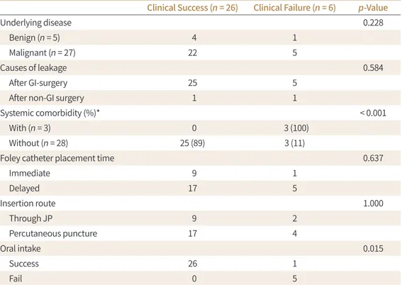
In statistical analysis, the statistically significant risk factors for clinical failure were sys- temic infectious condition (p < 0.001) and failed oral intake (p = 0.015). The characteristics of underlying disease (benign/malignant), causative surgery of leakage, Foley catheter place- ment time, and insertion route were not significantly related to clinical outcome (Table 2).
DISCUSSION
When post-operative complication of GI leakage occurs, early recognition and immediate resuscitation should be started. Many operative or non-operative approaches have been devel- oped to solve this problem, but it still remains an embarrassment to the surgeon, because it is often very challenging to manage bowel leakage and may finally have a potential to increase patient’s mortality rate (7).
Previous study revealed several clinical advantages of drainage through a Foley catheter.
One of the most important advantages is that it allows for early oral intake by preventing ad- ditional leakage through blockage of the opening via Foley catheter ballooning (6). In most studies, approximately 40% to 80% of patients with malignancy are presented as malnour-
Table 2. Clinical Results after Foley Catheter Placement
Clinical Success (n = 26) Clinical Failure (n = 6) p-Value
Underlying disease 0.228
Benign (n = 5) 4 1
Malignant (n = 27) 22 5
Causes of leakage 0.584
After GI-surgery 25 5
After non-GI surgery 1 1
Systemic comorbidity (%)* < 0.001
With (n = 3) 0 3 (100)
Without (n = 28) 25 (89) 3 (11)
Foley catheter placement time 0.637
Immediate 9 1
Delayed 17 5
Insertion route 1.000
Through JP 9 2
Percutaneous puncture 17 4
Oral intake 0.015
Success 26 1
Fail 0 5
*Crohn’s disease/Bechet’s disease, soft tissue sarcoma, carcinomatosis peritonei.
GI = gastrointestinal, JP = Jackson-Pratt
ished which increases their susceptibility to infection and overall tendency to delayed wound healing (8). It is widely known that enteral nutrition is superior to parenteral nutrition during the early phase of wound healing (9). Early enteral feeding can help maintain the barrier mechanism of the GI tract, and promotes mucosal growth and development (10). Eventually, it is very helpful to control the fistula. In our study, majority of patients started a regular diet at a median of 3 days (range, 1–26) after Foley catheter placement, which promoted early en- teral feeding. Interestingly, oral intake was a statistically significant factor related to clinical success in our study. This result is noticeable because it is consistent with the previous theo- ries, which emphasize the benefit of early enteral feeding. Our result demonstrated that per- cutaneous Foley catheter placement enables early oral diet, and oral diet itself can be strong- ly associated with successful clinical recovery of the patient.
Another important factor which affects the clinical success was systemic comorbidity that patients had when leakage was diagnosed. It is known that there are many risk factors both modifiable (such as stress, smoking, inappropriate alcohol consumption, malnutrition, obe- sity, diabetes, cardio-vascular disease, etc.) and non-modifiable (such as genetic diseases and ageing) strongly contributing to the impaired wound healing (10). To achieve proper tissue homeostasis during healing, the fine-tuned balance between a complex network of various leukocyte cell subsets and numerous pro- and anti-inflammatory mediators is crucial (11).
Dysregulation of critical parameters of these interactions results in pathologic and chronic inflammatory disease states that impair the quality of healing (11). Mawdsley et al. (7) dem- onstrated when the post-operative fistula tract is formed, the presence of comorbidity is the only factor to affect independently fistula-related mortality in the postoperative condition. In our study, patients with severe comorbidities such as carcinomatosis peritonei, soft tissue sarcoma, and inflammatory bowel diseases failed to achieve proper tract healing. A plausible reason for this result is that a poor general condition interrupted the closure of the stump opening. It is also reported that patients with multiple comorbidities have a significantly high- er rate of duodenal stump fistula after gastrectomy for gastric cancer (12). In the presence of poor prognostic factors, Foley catheter removal time should be determined carefully and other additional treatments should be considered (6).
In our study, the catheter indwelling time was median 35 days, and the clinical success rate was 81%. Gauderer and Stellato (13) described how the skin grows inward and the mucosa grows outward over time to line the tract with epithelium and create a chronic fistula. The emphasis has been on time controlling this process of tract epithelialization. Removal is not advised before the bowel is firmly attached to the abdominal wall (14). However, it is also known that the longer the tube is allowed to be in place, the greater the risk for persistence of the gastrocutaneous fistula (13). In addition, the longer catheter indwelling time, the more likely the risk of catheter mal-position and infection increases. Thus, proper catheter remov- al timing is an important factor for tract healing. Based on the results of our study, we sug- gest that optimal Foley catheter indwelling time is about 4-6 weeks. There was one patient with clinical failure despite having successfully started an oral diet. The point of note is that the patient had the shortest catheter indwelling time of 14 days. It can be explained that when the catheter is removed too early for the tract to be fully matured, the chance to un- complete disclosure of leakage point increases.
The size of Foley catheter that used in this study was mostly ten to twelve French and there was no technical or clinical problem associated the catheter size. Our result showed catheter malposition or migration was independent of catheter size. Proper tension and balloon shrinkage was important factor in complication. Therefore, we recommend the catheter size of ten to twelve for the procedure.
Of 32 patients, ten patients who showed matured fistula tract on the initial tubography un- derwent immediate catheter placement. The other twenty-two patients had sequential per- cutaneous drainage and delayed Foley catheter insertion. Two groups did not show statisti- cally different clinical success rate. In other words, there was no significant advantage of delayed catheter insertion for the clinical success. This result is very interesting, because it is widely known that it is favorable to delay any procedure until the inflammation is near total- ly subsides (15). Based on our study, the catheter insertion does not have to be delayed if the fistula tract is matured and active inflammatory phase is resolved.
This study has several limitations. First, the research was limited by a small sample size.
The use of a larger sample would improve future studies by analyzing the efficacy, safety of the procedure and more clinical factors affecting the successful outcome. Second, it is retro- spective in design. Third, the pigtail indwelling time and the Foley catheter placement time varied among the cases. The duration of the research was approximately five years. During the study, it was empirically known that the optimal catheter indwelling time is about four to six weeks, and that is the reason why the indwelling time of the Foley catheter was relatively shortened in recent cases
In conclusion, percutaneous Foley catheter insertion can be considered to be an effective approach to manage the post-operative bowel leakage. The presence of systemic comorbidity and successful oral diet after Foley catheter placement are significant factors for the clinical- ly successful recovery.
Author Contributions
Conceptualization, O.J.S., C.S.Y.; data curation, O.J.S., C.S.Y.; formal analysis, O.J.S., C.S.Y.; investiga- tion, O.J.S., C.S.Y.; methodology, all authors; project administration, all authors; resources, all authors;
software, O.J.S., C.S.Y.; supervision, O.J.S., L.H.G., C.B.G.; validation, O.J.S., C.S.Y.; visualization, O.J.S., C.S.Y.; writing—original draft, O.J.S., C.S.Y.; and writing—review & editing, O.J.S., C.S.Y.
Conflicts of Interest
The authors have no potential conflicts of interest to disclose.
REFERENCES
1. Fielding LP, Stewart-Brown S, Blesovsky L, Kearney G. Anastomotic integrity after operations for large- bowel cancer: a multicentre study. Br Med J 1980;281:411-414
2. Tuson JR, Everett WG. A retrospective study of colostomies, leaks and strictures after colorectal anastomo- sis. Int J Colorectal Dis 1990;5:44-48
3. Golub R, Golub RW, Cantu R Jr, Stein HD. A multivariate analysis of factors contributing to leakage of intes- tinal anastomoses. J Am Coll Surg 1997;184:364-372
4. McIntyre PB, Ritchie JK, Hawley PR, Bartram CI, Lennard-Jones JE. Management of enterocutaneous fistu- las: a review of 132 cases. Br J Surg 1984;71:293-296
5. Pickleman J, Watson W, Cunningham J, Fisher SG, Gamelli R. The failed gastrointestinal anastomosis: an inevitable catastrophe? J Am Coll Surg 1999;188:473-482
6. Oh JS, Lee HG, Chun HJ, Choi BG, Lee SH, Hahn ST, et al. Percutaneous management of postoperative du- odenal stump leakage with foley catheter. Cardiovasc Intervent Radiol 2013;36:1344-1349
7. Mawdsley JE, Hollington P, Bassett P, Windsor AJ, Forbes A, Gabe SM. An analysis of predictive factors for healing and mortality in patients with enterocutaneous fistulas. Aliment Pharmacol Ther 2008;28:1111- 1121
8. Payne WG, Naidu DK, Wheeler CK, Barkoe D, Mentis M, Salas RE, et al. Wound healing in patients with can- cer. Eplasty 2008;8:e9
9. Kiyama T, Witte MB, Thornton FJ, Barbul A. The route of nutrition support affects the early phase of wound healing. JPEN J Parenter Enteral Nutr 1998;22:276-279
10. Avishai E, Yeghiazaryan K, Golubnitschaja O. Impaired wound healing: facts and hypotheses for multi-pro- fessional considerations in predictive, preventive and personalised medicine. EPMA J 2017;8:23-33 11. Eming SA, Krieg T, Davidson JM. Inflammation in wound repair: molecular and cellular mechanisms. J In-
vest Dermatol 2007;127:514-525
12. Paik HJ, Lee SH, Choi CI, Kim DH, Jeon TY, Kim DH, et al. Duodenal stump fistula after gastrectomy for gas- tric cancer: risk factors, prevention, and management. Ann Surg Treat Res 2016;90:157-163
13. Gauderer MW, Stellato TA. Gastrostomies: evolution, techniques, indications, and complications. Curr Pro- bl Surg 1986;23:661-719
14. Tøttrup A. Foley catheter enterostomy for postoperative bowel perforation: an effective source control.
World J Surg 2010;34:2752-2754
15. Schecter WP. Management of enterocutaneous fistulas. Surg Clin North Am 2011;91:481-491
경피적 폴리 카테터 삽입을 이용하여 수술 후 장 누출을 성공적으로 관리하기 위한 기술적 및 임상적 요인들
조소영 · 오정석* · 이해규 · 최병길
목적 본 연구의 목적은 경피적 폴리 카테터 삽입을 이용하여 수술 후 장 누출을 성공적으로 관리하기 위한 기술적 및 임상적 요인을 분석함에 있다.
대상과 방법 이번 후향적 연구에는 32명의 환자가 포함되었다. 수술 후 위장 누출은 컴퓨터단 층촬영(CT)을 이용하여 진단되었고, 환자는 Jackson-Pratt 튜브 또는 이미지 유도 방법을 통 해 경피적 경로로 누출 위치에 폴리 카테터 삽입을 시행 받았다. 성공은 합병증 없이 폴리 카 테터를 성공적으로 제거한 것으로 정의하였고, 임상적 실패와 관련된 인자들에 대하여 분석 하였다.
결과 모든 환자에서 경피성 폴리 카테터 배치는 합병증 없이 성공적으로 시행되었다. 폴리 카 테터 삽입은 CT에서 누출이 확인된 후 중앙값 10일(범위, 1~68)째에 시행되었다. 임상적 성공 은 32명의 환자 중 26명(81%)에서 달성되었다. 통계학적 분석에서, 임상적 실패에 대한 통계 적으로 유의미한 위험 인자는 전신적 동반 질환(p < 0.001) 및 구강 섭취 실패(p = 0.015)였다.
결론 경피적 폴리 카테터 삽입은 수술 후 장 누출을 관리하기 위한 안전하고 효과적인 접근 방법으로 간주될 수 있다. 폴리 카테터 배치 후, 전신적 동반 질환의 유무 및 성공적인 구강 식이를 빠른 시일 내로 시작하는 것은 임상적으로 성공적인 회복에 중요한 요소이다.
가톨릭대학교 의과대학 서울성모병원 영상의학과