Correspondence to:Woo Hyun Park, Ph.D.
Department of Physiology, Chonbuk National University Medical School
1 Deokjin-dong, Deokjin-gu, Jeonju 561-756, Korea Tel: +82-63-270-3079, Fax: +82-63-274-9892 E-mail: parkwh71@yahoo.co.kr
접수:2005년 12월 18일, 수정:2006년 1월 20일 승인:2006년 1월 31일
교신저자:박우현, 전북 전주시 덕진구 덕진동 1가 664-14번지
561-756, 전북대학교 의과대학 심리학교실 Tel: 063-270-3079, Fax: 063-274-9892
E-mail: parkwh71@yahoo.co.kr
1
Overexpression of the Fanconi Anemia A Gene in Hela and MCF10A Cells
Woo Hyun Park, Ph.D.
Department of Physiology, Chonbuk National University Medical School, Jeonju, Korea
Background: Fanconi Anemia (FA) is an autosomal recessive inherited disease, which is characterized by developmental abnormalities, progressive bone marrow failure and a predisposition to cancer. The phenotypes of FA cells show extreme sensitivities towards oxygen and DNA cross linking agents, such as diepoxybutane and mitomycin C (MMC).
Methods: In the current study, retroviruses expressing the FANCA gene were prepared to create the stable cell lines, Hela (cervical carcinoma) and MCF10A (breast). The expression of FANCA protein in the Hela and MCF10A stable cells, following puromycin selection, was checked using Western blot.
The difference in the cell growth between the parent and FANCA expressing cells following MMC treatment was checked using the MTT assay.
Results: The expression of exogenous FANCA protein in the Hela and MCF10A stable cells was observed using Western blot. The MCF10A cells expressing exogenous FANCA were resistant to MMC concentrations with the range 0.01~1µM compared with the MCF10 parent cells. However, at an MMC concentration of 10µM, there was no difference in the susceptibility between the parent and FANCA expressing MCF10 cells. The Hela cells expressing FANCA showed no resistance at any MMC con- centration (0.01~10µM).
Conclusion: FANCA protein is an important factor for resistance to the cross linking agent, MMC, in MCF10A breast cells, but not in Hela cervical carcinoma cells. (Korean J Hematol 2006;41:1-7.)
Key Words: Fanconi anemia, FANCA, Hela cell, MCF10A cell
INTRODUCTION
Fanconi Anemia (FA) is an autosomal recessive disorder characterized by developmental abnor- malities and progressive bone marrow failure, and the incidences of aplastic anemia, myelodys- plastic syndrome (MDS) and acute myeloid leu- kemia (AML) are all greatly increased in homo-
zygotes.1-3) Cells from FA patients exhibit hyper- sensitivity to DNA crosslinking agents, such as mitomycin C (MMC), DEB, and cisplatin.2,4) Cu- rrently, more than eleven FA complementation groups are known to exist, and the nine have been identified and cloned, referred to as FA subtypes A, C, BRCA2/D1, D2, E, F, G, L and B.5-7) At present, most (60~80%) of FA patients fall into group A. The protein of FANCA (165
kDa) is the largest protein among other FA proteins, suggesting that FANCA gene plays an important role in FA pathogenesis. The encoded FA proteins cooperate in a common pathway: the FA/BRCA pathway.3) Six of the FA proteins, A, C, E, F, G, B and L, form a protein complex constitutively found in the nucleus of cells, the FA protein complex.3,6-8) The principal function of the FA complex is to activate FANCD2 follo- wing response to DNA damage.9) The activation of FANCD2 appears to occurs by the FANCL subunit of the FANC complex monoubiquitina- ting FANCD2 on lysine 516.6) The activated FANCD2 protein is subsequently targeted to sub- nuclear foci, which are thought to be the sites of DNA repair, and which contain BRCA1, FAN- CD2/BRCA2, and Rad51.3,10,11)
Despite these remarkable advances, little is known about the regulation and functional role of FA proteins, especially FANCA and FANCD2.
A number of recent findings have suggested that FA cells are defective in the repair of DNA dou- ble-strand breaks.12-14) Repair of double-strand DNA breaks can occur by non-homologous end joining (NHEJ) or by homologous recombination (HR).15,16) The repair of interstrand cross links by NHEJ, however, would be error-prone, suggesting that HR is important for the repair of that class of lesion. It is currently unclear which pathway, NHEJ or HR, is important in the FA phenotype.
To know the function of FANCA in the cancer cell lines, I expressed full length FANCA in the MCF10A breast cell and Hela cervical carcinoma cells. I found that the MCF10A cells which ex- pressed exogenous FANCA were resistant to the MMC, compared with the MCF10A parent cells.
But Hela cells which expressed FANCA did not show the resistance to the MMC.
MATERIALS AND METHODS
1. Cell cultures
MCF10A cells were grown in Dulbecco's modified Eagle's media-F12 (DMEM/F12, GIBCO-BRL), which
was supplemented with 5% fetal calf serum (GIB- CO-BRL), penicillin (10units/mL, Sigma), strepto- mycin (0.1mg/mL, Sigma), 10ul EGF (1mg/mL), 5ml sodium pyruvate (100mM), 5mL glutamine (200mM), 70uL hydrocortizone (10mM), 500uL insulin (1mg/mL) and 50uL cholera toxin (1mg/
mL). Hela and 293T cells were grown in Dul- becco's modified Eagle's Medium (DMEM), which was supplemented with 10% fetal calf serum (GI- BCO-BRL), penicillin (10units/mL, Sigma) and streptomycin (0.1mg/mL, Sigma).
2. Gene construct (plasmid)
The cDNA for human FANCA was from the D'Andrea lab (Dana-Farber Cancer institute, Har- vard Medical School). I subcloned the cDNA of FANCA in pcDNA3 vector into the pBabe retro- virus vector by PCR using the forward primer 5'-ATCGGGGGGCCGGCCAAATGTCCGACTC GTGGGTC-3' and the reverse primer 5'-ATCG- GGGGGCGCGCCTCAGAAGAGATGAGGCTCC- 3'. The forward primer contains the FseI restric- tion endonuclease site and the reverse primer contains the AscI restriction endonuclease site.
The pbabe puro vector contains a ZZ domain which shows the high affinity to IgG immuno- globulin. For purification of amino acids 1-232 of human FANCA, I subcloned the cDNA of FAN CA in pcDNA3 vector in the pET-21a(+) with a hexhistidine tag (Novagen), by PCR using the forward primer 5'-ATCGGGGGGATCCTCCGA- CTCGTGGGTCCCGAACTCCGC-3' and the re- verse primer 5'-AT CGGGGCTCGAGGGCCCT- GGCGACGTCAGCATGCTG-3'. The forward primer contains the BamHI restriction endonuclease site and the reverse primer contains the XhoI restric- tion endonuclease site. The fusion protein was expressed Rosetta cells (Novagen) using standard procedures. The fusion protein was purified on Ni2+-NTA agarose resin (Qiagen).
3. Making the stable cell lines which express FAN CA protein by retrovirus
The indicated pbabe construct was transfected
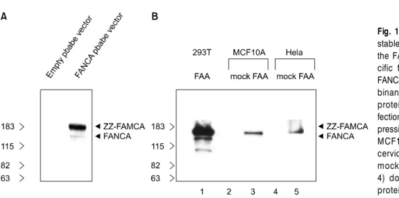
Fig. 1. FANCA expression in the stable cell lines. Western blot of the FANCA with antibodies spe- cific for amino acids 1-232 of FANCA is shown. (A) The recom- binant expression of zz-FANCA protein in 293T cells by trans- fection. (B) The recombinant ex- pression of zz-FANCA protein in MCF10A breast cell and Hela cervical carcinoma cells. The mock stable cells (lanes 2 and 4) do not express the FANCA protein.
by lipofection into 293T producer cells (human embryonic kidney cells) with pKAT plasmid (pa- ckaging construct) and pHCMV-G plasmid (ex- pressing the VSV-G envelope protein). Retroviral supernatants were collected on day 3 following lipofection. MCF10A and Hela cells were in- fected with the retrovirus for the 2 hour in the presence of 8ug/mL polybrene. Infected cells were washed free of viral supernatant and resuspended in growth media. After 2 days, cells were trans- ferred to media containing puromycin (1ug/mL).
Surviving cells were grown under continuous se- lection in puromycin.
4. Antibody and chemical
Anti-FANCA antiserum (polyclonal) was gene- rated in rabbits using the immunogen amino acids 1-232 of human FANCA. Mitomycin C (MMC) was purchased from Sigma.
5. SDS-PAGE and Western blot analysis
Cell lysates (50 ug per lane) were resolved on 7% SDS-polyacrylamide gels. Proteins were then transferred to polyvinylidene difluoride memb- rane, probed with an anti-FANCA antibody (a rabbit polyclonal IgG), followed by horseradish peroxidase-conjugated secondary antibody. Pro- teins were visualized by using the ECL system.
6. Growth Inhibition Assay
In vitro growth inhibition effect of MMC on MCF10A and Hela cells was determined by mea- suring MTT dye absorbance of living cells. Brie- fly, cells (2×10⁵ cells/well) were seeded in 96-well microtiter plates (Nunc, Roskilde, Denmark). After exposure to the drug for 72h, 50μL of MTT (Sigma) solution (2mg/mL in PBS) were added to each well, and the plates were incubated for additional 4h at 37°C. MTT solution in medium was aspirated off. To achieve solubilization of the formazan crystal formed in viable cells, 200μL of DMSO were added to each well before absor- bance at 570nm was measured.
RESULT
As shown in Fig. 1A, the human recombinant FANCA protein with ZZ domain was expressed well in 293T cell after transfection of pbabe- zz-FANCA vector (retrovirus vector). But the empty vector did not show FANCA Western band. Next, I made the retrovirus which ex- pressed the FANCA, infected the MCF10A and Hela cells with this retrovirus and finally ob- tained the stable MCF10A and Hela cells which expressed the zz-FANCA protein well (Fig. 1B).
I could not detect the expression of endogenous FANCA in Hela and MCF10A cells (Fig. 1B).
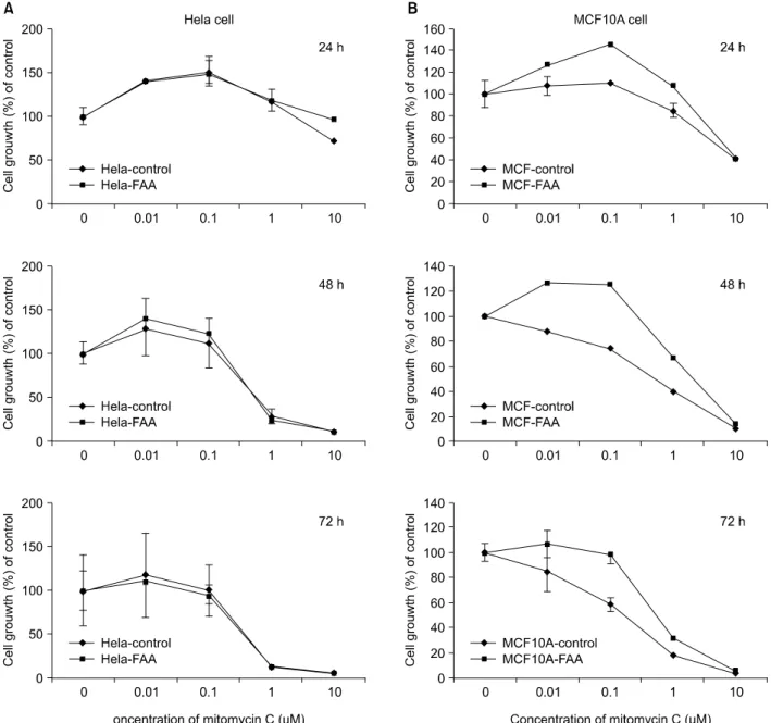
Fig. 2. (A) Effect of mitomycin C on the growth of normal Hela cells and FANCA stable Hela cells in vitro. (B) Effect of mitomycin C on the growth inhibition of normal MCF10A cells and FANCA stable MCF10A cells in vitro. Exponentially growing cells were treated with the indicated concentration of mitomycin C for 24, 48 and 72 h. Cell growth inhibition was assessed by MTT assay as described in "Materials and Methods". Results represent the mean of at least four independent experiments. bars, SE.
Because the exogenous level of FANCA expre- ssion is very higher than the endogenous FAN- CA, these stable cell lines are thought to be good for study on FANCA function only to crosslin- king agents, such as MMC. Therefore, I used these cells lines for knowing whether the FANCA has a resistant function against MMC.
As shown in Fig. 2A, the FANCA stable Hela cells did not show the resistance to MMC, com-
pared with parent cell at any doses in early and late time (24~72 hour). As for the MCF10A cell with exogenous FANCA protein, the FANCA showed a key function for resistance to MMC in the MCF10A breast cells (Fig. 2B). At the 24 hours, MCF10A cell with exogenous FANCA sho- wed the difference of susceptibility to MMC at the lower doses (from 0.01uM and 1uM), com- pared with parent cells. But at the high dose (10
uM), the difference of resistance to MMC between the parent cells and FANCA stable cells was very low. This result indicates that the FANCA protein is very closely involved in the susceptibility to low doses of MMC (from 0.01 uM to 1uM) in the MCF10A breast cell.
DISCUSSION
Fanconi Anemia (FA) is an autosomal recessive disorder characterized by birth defects, develop- ing pancytopenia and increased risk of mali- gnancy. It has been shown that BRCA1 (breast cancer tumor suppressor gene) regulates two of FA proteins, called FANCA and FANCD2, by a process called ubiquitination. However, exactly how the FA proteins and BRCA1 interact to regulate DNA damage repair remains obscure.
Therefore, in this experiment, I used the breast cell line (MCF10A) in which the BRCA1 and FA proteins work together for the DNA damage repair after intercross linker agents normally, and non-breast cell line (Hela cervical carcinoma cells) as a control cell line, which is not yet known whether the BRCA1 and FA proteins play a role for DNA damage repair triggered by MMC treatment.
I used the retrovirus system to make the stable cell lines expressing the FANCA protein. As shown in Fig. 1B, I successfully could detect the expression of exogenous FANCA in Hela and MCF10A cells. But I could not detect the expre- ssion of endogenous FANCA in Hela and MCF- 10A cells (Fig. 1B). Because I didn't use the RT- PCR to see the minimal expression of this pro- tein, I could not exclude the possibility that these cells have the endogenous FANCA protein very rare, However, because the exogenous level of FANCA expression is very higher than the endo- genous FANCA, these stable cell lines are thought to be good for study on FANCA function to crosslinking agents, such as MMC. Therefore, I used these cells lines for knowing whether the FANCA has a resistant function against MMC.
As shown in Fig. 2A, the FANCA stable Hela cells did not show the resistance to MMC, com- pared with parent cell at any doses in early and late time (24~72 hour). This result suggests that FANCA expression in Hela cervical carcinoma cells is not sufficient to confer resistance to MMC and Hela cells may have low expression of other FA pathway proteins such as FANCD2, FANCE, FANCL for the resistant to MMC. To be resistant to intercross linker agents such as MMC, the Hela cervical carcinoma needs more other pro- teins for DNA damage repair with BRCA1 than just overexpression of FANCA protein in our model.
As for the MCF10A cell with exogenous FANCA protein, the FANCA showed a key func- tion for resistance to MMC in the MCF10A breast cells (Fig. 2B). This result indicates that the FANCA protein is very closely involved in working with BRCA1 protein as well as the other FA proteins for DNA damage repair after the MMC treatment. But we need to clarify the direct or indirect interaction of FANCA and BRCA1 in this cell by ways of immunoprecipitation and Western method or other methods. It is of in- terest that the only breast cell having the exogenous FANCA can show the resistance to MMC, even though I need to use more FANCA stable cell lines.
FANCA as well as FANCD2 has been impli- cated as an important factor in the process of DNA damage repair in response to the MMC crosslink agents and the activated FANCD2 is required for DNA damage repair with BRCA1 and BRCA2.1-3,10,11) FANCA is directly bound to
BRCA1.17) BRCA2/FANCD1 has a well-defined
role in homology-directed DNA repair.10,14,18-20) These facts suggested that FANCA and FAN- CD2 are tightly related to the BRCA1 and BR- CA2 (breast tumor suppressor genes) and the FA pathway and BRCA pathway in cells is an impor- tant shared signaling pathways. Also, the BRCA proteins have an important role in breast cells.
These reports might explain the difference of sus-
ceptibility toward MMC between the Hela cer- vical carcinoma cells and MCF10A breast cells, which express the exogenous FANCA protein. Even though the role of FANCA is not clear now, this protein plays more important role in breast cell rather than other tissue cells including the cervical carcinoma cells. Probably, overexpressed FANCA protein can specifically interact with BRCA1 protein in the MCF10A breast cells, resulting in more resistant to MMC. The interaction of FA- NC proteins and BRCA proteins in breast cell and other cells after the intercross linker agent treatment or endogenous DNA damage will be studied further in view of the regulation of protein function by each other.
Taken together, I conclude that FANCA pro- tein is an important factor for resistance to MMC in the MCF10A breast cell, but not the Hela cervical carcinoma cells.
요 약
배경: 판코 빈혈(Fanconi anemia)은 열성으로 유전되
는 질환으로, 임상적인 특징으로는 성장에 장애가 오 며, 진행성 골수 이상과 암으로 대부분 사망하는 질환 으로 알려져 있다. 특히 환자 세포에서 나타나는 주요 특징은 DNA crosslink인 mitomycin C (MMC)나 die- poxybutane에 대해 민감하게 반응을 한다.
방법: 본 실험을 하기 위해서 Hela 세포주와 MCF 10A 세포주를 사용하였으며, FANCA 단백을 발현하 는 안정화 세포주를 만들기 위해서 retrovirus을 이용하 였다. FANCA 단백을 발현하는 세포주의 선택을 위해 서 puromycin을 사용하였으며, MMC에 대한 세포주들 의 성장 억제 변화를 보기 위해서 MTT assay를 이용하 였다.
결과: 성공적으로 FANCA 단백을 발현하는 HeLa 세 포주와 MCF10A 세포주를 얻었으며, 이들 세포에 MMC을 처리하여 세포의 성장 억제를 분석한 결과, MMC 농도에 따라(0.01~10uM) HeLa 세포주와 FAN- CA를 발현시킨 HeLa 세포주는 세포성장의 차이에 아 무런 변화를 보이지 않았다. MCF10A 세포주는 MMC 농도 0.01uM 부터 성적 억제가 관찰되었고(72 시간 배 양), FANCA를 발현시킨 MCF10A 세포주는 MMC 농 도(0.01~0.1uM)에서 성장이 확연히 덜 억제됨을 관찰
할 수 있었다.
결론: FANCA 단백은 breast 세포주인 MCF10A에서 MMC에 대한 감수성을 억제하는데 중요하게 작용하 는 하나의 인자임을 확인하였다.
ACKNOWLEDGEMENT
This paper was supported by research funds of Chonbuk National University in 2005 and a postdoctoral fellowship from the Department of Defense Breast Cancer Research Program (BC 031671). I appreciate the professor, Jeff Parvin, who helped me to do this work.
REFERENCES
1) Bagby GC Jr. Genetic basis of Fanconi anemia. Curr Opin Hematol 2003;10:68-76.
2) Joenje H, Patel KJ. The emerging genetic and mo- lecular basis of Fanconi anaemia. Nat Rev Genet 2001;2:446-57.
3) D'Andrea AD, Grompe M. The Fanconi anaemia/BR- CA pathway. Nat Rev Cancer 2003;3:23-34.
4) Kwee ML, Poll EH, van de Kamp JJ, de Koning H, Eriksson AW, Joenje H. Unusual response to bifunc- tional alkylating agents in a case of Fanconi anaemia.
Hum Genet 1983;64:384-7.
5) Joenje H, Oostra AB, Wijker M, et al. Evidence for at least eight Fanconi anemia genes. Am J Hum Genet 1997;61:940-4.
6) Meetei AR, de Winter JP, Medhurst AL, et al. A no- vel ubiquitin ligase is deficient in Fanconi anemia.
Nat Genet 2003;35:165-70.
7) Meetei AR, Levitus M, Xue Y, et al. X-linked inhe ritance of Fanconi anemia complementation group B. Nat Genet 204;36:1219-24.
8) Medhurst AL, Huber PA, Waisfisz Q, de Winter JP, Mathew CG. Direct interactions of the five known Fanconi anaemia proteins suggest a common func- tional pathway. Hum Mol Genet 2001;10:423-4.
9) Garcia-Higuera I, Taniguchi T, Ganesan S, et al. In- teraction of the Fanconi anemia proteins and BRCA1 in a common pathway. Mol Cell 2001;7:249-62.
10) Moynahan ME, Pierce AJ, Jasin M. BRCA2 is requir- ed for homology-directed repair of chromosomal breaks. Mol Cell 2001;7:263-72.
11) Moynahan ME, Chiu JW, Koller BH, Jasin M. Brca1 controls homology-directed DNA repair. Mol Cell
1999;4:511-8.
12) Lundberg R, Mavinakere M, Campbell C. Deficient DNA end joining activity in extracts from fanconi anemia fibroblasts. J Biol Chem 2001;276:9543-9.
13) Donahue SL, Campbell C. A DNA double strand break repair defect in Fanconi anemia fibroblasts. J Biol Chem 2002;277:46243-7.
14) Donahue SL, Campbell C. A Rad50-dependent path- way of DNA repair is deficient in Fanconi anemia fibroblasts. Nucleic Acids Res 2004;32:3248-57.
15) Pfeiffer P, Goedecke W, Kuhfittig-Kulle S, Obe G.
Pathways of DNA double-strand break repair and their impact on the prevention and formation of chromosomal aberrations. Cytogenet Genome Res 2004;104:7-13.
16) van den Bosch M, Lohman PH, Pastink A. DNA
double-strand break repair by homologous recombi- nation. Biol Chem 2002;383:873-92.
17) Folias A, Matkovic M, Bruun D, et al. BRCA1 inte- racts directly with the Fanconi anemia protein FAN- CA. Hum Mol Genet 2002;11:2591-7.
18) Howlett NG, Taniguchi T, Olson S, et al. Biallelic in- activation of BRCA2 in Fanconi anemia. Science 2002;
297:606-9.
19) Hirsch B, Shimamura A, Moreau L, et al. Association of biallelic BRCA2/FANCD1 mutations with sponta- neous chromosomal instability and solid tumors of childhood. Blood 2004;103:2554-9.
20) West SC. Molecular views of recombination proteins and their control. Nat Rev Mol Cell Biol 2003;4:435- 45.