Imaging Science in Dentistry 2015; 45: 95-101 http://dx.doi.org/10.5624/isd.2015.45.2.95
Introduction
The main goal of endodontic treatment is the thorough cleaning and shaping of the root canal system and its complete obturation with an inert filling material.1 How- ever, in order to ensure successful treatment, prior knowl- edge of the root canal anatomy is necessary, based on the careful interpretation of angled radiographs, proper access preparation, and a detailed exploration of the interior of the tooth, as incomplete or untreated canals may cause the
treatment to fail.2
Mandibular molars are the teeth most often affected by decay, and may therefore require endodontic treatment, even at a young age.3 The mandibular first molar is one of the most frequently treated teeth, accounting for 17.4%
of all endodontic procedures.4,5 Among endodontically treated teeth, mandibular molars are most frequently ex- tracted, and one of the main reasons for extraction is the failure of endodontic treatment (19.3%).6 Therefore, prior knowledge of the anatomy of the root canal system, in- formed by a knowledge of the anatomical variations that may occur in various populations, is likely to increase the probability that the treatment will be successful.
The staining and clearing technique is the most common
Characterization of mandibular molar root and canal morphology using cone beam computed tomography and its variability in Belgian and Chilean population samples
Andres Torres1,*, Reinhilde Jacobs1, Paul Lambrechts2, Claudia Brizuela3, Carolina Cabrera3, Guillermo Concha4, María Eugenia Pedemonte3
1Department of Imaging and Pathology, OMFS-IMPATH Research Group, Katholieke Universiteit Leuven, Leuven, Belgium
2Department of Conservative Dentistry, Katholieke Universiteit Leuven, Leuven, Belgium
3Department of Endodontics, Universidad de los Andes, Santiago, Chile
4Department of Oral and Maxillofacial Radiology, Universidad de los Andes, Santiago, Chile
ABstrACt
Purpose: This study used cone-beam computed tomography (CBCT) to characterize mandibular molar root and canal morphology and its variability in Belgian and Chilean population samples.
Materials and Methods: We analyzed the CBCT images of 515 mandibular molars (257 from Belgium and 258 from Chile). Molars meeting the inclusion criteria were analyzed to determine (1) the number of roots; (2) the root canal configuration; (3) the presence of a curved canal in the cross-sectional image of the distal root in the mandibular first molar and (4) the presence of a C-shaped canal in the second mandibular molar. A descriptive analysis was performed. The association between national origin and the presence of a curved or C-shaped canal was evaluated using the chi-squared test.
results: The most common configurations in the mesial root of both molars were type V and type III. In the distal root, type I canal configuration was the most common. Curvature in the cross-sectional image was found in 25%
of the distal canals of the mandibular first molars in the Belgian population, compared to 11% in the Chilean population. The prevalence of C-shaped canals was 10% or less in both populations.
Conclusion: In cases of unclear or complex root and canal morphology in the mandibular molars, CBCT imaging might assist endodontic specialists in making an accurate diagnosis and in treatment planning.(Imaging Sci Dent 2015; 45: 95-101)
Key words: Anatomy; Cone-Beam Computed Tomography; Molar; Dental Pulp Cavity
Copyright ⓒ 2015 by Korean Academy of Oral and Maxillofacial Radiology
This is an Open Access article distributed under the terms of the Creative Commons Attribution Non-Commercial License(http://creativecommons.org/licenses/by-nc/3.0) which permits unrestricted non-commercial use, distribution, and reproduction in any medium, provided the original work is properly cited.
Imaging Science in Dentistry·pISSN 2233-7822 eISSN 2233-7830 Received November 18, 2014; Revised January 1, 2015; Accepted February 12, 2015
*Correspondence to : Dr. Andres Torres
Department of Imaging and Pathology, Katholieke Universiteit Leuven, Campus Sint-Rafaël, Kapucijnenvoer 33, B-3000, Leuven, Belgium
Tel) 32-16-33-2951, Fax) 32-16-332410, E-mail) [email protected]
*표 선 두께
=
1/0.3ptmethod used to assess the root canal system.1,7 However, recent studies have used cone-beam computed tomogra- phy (CBCT) because it allows the visualization of fine details without visual noise as well as the overlapping vi- sualization of adjacent structures.8,9 CBCT technology uses isotropic voxels, which are equal dimensions in all three planes of space, enabling accurate linear geometric and three-dimensional measurements of the data acquired.8,10-13 CBCT is also a non-invasive method that allows accurate information to be obtained without requiring the tooth to be extracted for analysis.14
Although numerous studies have used CBCT to study root and canal anatomy,7,15-18 most previous studies were conducted in Asian populations, which are characterized by different features than those observed in Caucasian pop- ulations.7,16 Moreover, to date, no comparative study has assessed cross-population variability in mandibular molar root and canal morphology.
The aim of this study was to use CBCT to characterize mandibular molar root and canal morphology and its vari- ability in Belgian and Chilean population samples.
Materials and Methods
Patients
CBCT images were obtained from the database of the Oral Imaging Center of the Department of Oral Health Sci- ence at the University Hospital of the Katholieke Univer- siteit Leuven, Leuven, Belgium, between November 2010 and January 2012, and from the database of a private dento- maxillofacial radiology practice in Santiago, Chile, between February 2007 and May 2012. Patients were referred to these radiological centers for CBCT scanning in accordance with various clinical indications. Informed consent was ob- tained from the patients who were treated at the University Hospitals of the Katholieke Universiteit Leuven, Leuven, Belgium. For patients evaluated in the private dentomaxil- lofacial radiology practice in Santiago, Chile, the authori- zation of the director of the center was obtained to use the information in the database for research purposes. All image data sets were anonymized.
The following selection criteria were applied: (1) per- manent mandibular molars without any type of resto- ration; (2) the absence of periapical lesions or periapical ligament widening; (3) no previous endodontic treatment;
(4) a fully developed root (Nolla stage 10); (5) the absence of root resorption, and (6) the availability of a high-quality CBCT image. A total of 515 mandibular molars (257 from
the Belgian population and 258 from the Chilean popula- tion) from 270 patients (100 Belgian and 170 Chilean) met these criteria and were further analyzed.
Radiographic technique
CBCT images were taken using a 3D Accuitomo 170® machine (Morita, Kyoto, Japan) operating at 90 kV and 87.5 mAs. The size of the field of view was selected de- pending on the requirements of each case. The voxel size was 0.25 mm. All CBCT imaging was performed by an appropriately licensed medical imaging technologist and diagnosed by a dentomaxillofacial radiologist, with the min- imum exposure necessary for adequate image quality.
Image evaluation
All images were evaluated by a dentist, who was pre- viously calibrated under the supervision of a specialist in
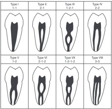
Fig. 1. Classification of root canal configuration.1 Type I: a single canal extends from the pulp chamber to the apex. Type II: two separate canals leave the pulp chamber and join short of the apex to form one canal. Type III: one canal leaves the pulp chamber, divides into two within the root, and then merges to exit as one canal. Type IV: Two separate and distinct canals extend from the pulp chamber to the apex. Type V: one canal leaves the pulp chamber and divides short of the apex into two separate and dis- tinct canals with separate apical foramina. Type VI: two separate canals leave the pulp chamber, merge in the body of the root, and re-divide short of the apex to exit as two distinct canals. Type VII:
one canal leaves the pulp chamber, divides and then rejoins within the body of the root, and finally re-divides into two distinct canals short of the apex. Type VIII: three separate and distinct canals ex- tend from the pulp chamber to the apex.
Type I 1-1
Type V 1-2
Type II 2-1
Type VI 2-1-2
Type III 1-2-1
Type VII 1-2-1-2
Type IV 2-2
Type VIII 3-3
dental and maxillofacial radiology. The calibration was performed by having the examiner dentist and a dento- maxillofacial radiologist evaluate a randomized set of CBCT images. A total of 80 mandibular molars were eval- uated, and complex cases were discussed and analyzed until a consensus was reached. Cohen’s kappa coefficient for interobserver agreement was 0.776.
All CBCT slices were analyzed using i-Dixel One Data Viewer Plus software (version 1.5.4.5, Morita, Kyoto, Ja- pan) under standardized conditions in a quiet and dimmed room with a diagnostic display (Barco® MDRC-2120, Bar- co, Kortrijk, Belgium). Axial, coronal, and sagittal two-di- mensional sections were displayed on the monitor and the complete data set was assessed. When necessary, image contrast and brightness were adjusted using the software image processing tool in order to ensure optimal visualiza- tion.
The teeth that met the inclusion criteria were evaluated to determine (1) the number of roots, (2) the root canal configuration according to Vertucci’s classification1 (Fig.
1), (3) the presence of a curved canal in the cross-sectional image of the distal root in the mandibular first molar, and (4) the presence of a C-shaped canal in the mandibular second molar. The age and gender of the patients were also recorded.
In the mandibular first molars, a curved distal canal was only recorded in the distal root when the canal exhibited the following features in a cross-sectional image (Fig. 2):
(1) the presence of a ribbon-shaped canal with (2) a curved
shape, as verified by the ability to draw a straight line in- tersecting only the ends of the buccal and lingual sides of the canal.
A B
Fig. 2. Curved canal in the cross-sectional image of the distal root in the mandibular first molar. A. Left: a mandibular first molar with (M) mesial and (D) distal roots. Right: cross-sectional image of a mandibular first molar as seen in an axial-plane CBCT view. (M) mesial root, (D) distal root, (*), cortical and trabecular bone structure. When a straight line is drawn in the distal ribbon-shaped canal, it only comes in contact with the (a) buccal and (b) lingual ends of the canal. B. Cone-beam computed tomography cross-sectional images. 1a-c: curved distal canals in cross-sectional images, 1d: an oval canal, 2a and b: curved distal canals in cross-sectional images, 2c: a canal starting to divide, 2d: a fully divided canal, 3a-d: the absence of a curved distal canal in cross-sectional images.
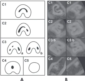
Fig. 3. Classification of C-shaped canal configurations.19 A. C1:
an uninterrupted “C” with no separation or division, C2: a canal shape resembling a semi-column resulting from the discontinu- ation of the “C” outline, C3: three (a) or two (b) separate canals, C4: only one round or oval canal in the cross-sectional image, C5:
no observable canal lumen (usually only found near the apex). B.
Cross-sectional CBCT images of different types of C-shaped canal configurations, at different root levels.
A B
In the mandibular second molars, the presence of a C- shaped canal was recorded. The definition of a C-shaped canal system developed by Fan et al.19 requires a tooth to exhibit the following three features: (1) fused roots, (2) a longitudinal groove on the lingual or buccal surface of the root, and (3) at least one cross-section of the canal exhibit- ing the C1, C2, or C3 configuration (Fig. 3).
A descriptive analysis of the variables was performed. For quantitative variables (age), central tendency, dispersion, and position were measured for each population group.
Nominal variables (country of origin, evaluated molar, root canal configuration, presence of a curved canal in the cross-sectional image of the distal root in mandibular first molar, and presence of a C-shaped canal in the mandibu- lar second molar) and the number of roots were tabulated according to absolute frequencies and percentages. The association between country of origin and the presence of a curved canal in the cross-sectional image of the distal root in the mandibular first molar and the presence of a C-shaped canal in the mandibular second molar was evaluated via the chi-squared test, using the RStudio® version 0.98.501 soft- ware (RStudio Inc., Boston, Massachusetts, USA).
results
The median age of the Belgian population sample ex- amined was 19.5 years, and that of the Chilean population sample was 19.0 years, with an interquartile range of 11 years in both samples. The minimum age in both popu- lations was 11 years, and the maximum age was 47 and 60 years in the Belgian and Chilean population samples, respectively.
In the Belgian sample consisting of a total of 100 pa- tients, 48% were female and 52% were male. In the Chil- ean sample with a total of 170 patients, 55.88% were fe- male and 44.12% were male.
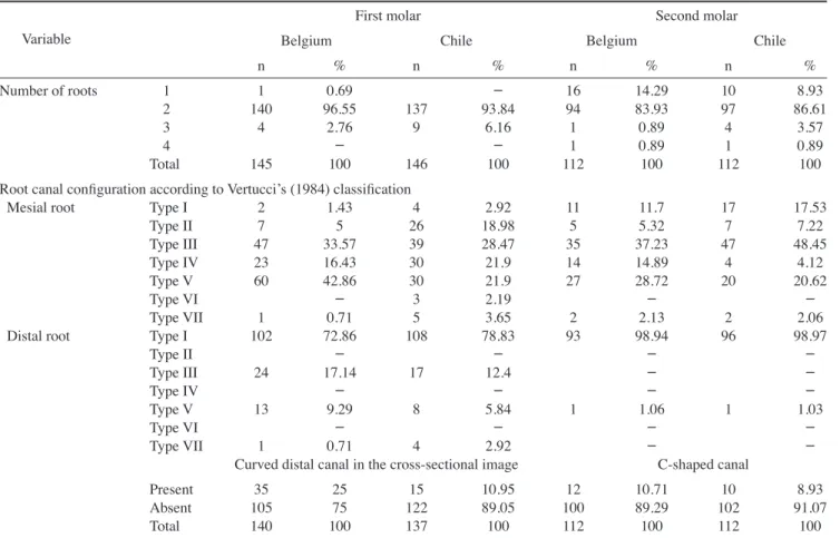
All the variables analyzed for the mandibular first and second molars are shown in Table 1: the number of roots, the root canal configuration of each root, the presence of a curved canal in the cross-sectional image of the distal root (first molar), and the presence of a C-shaped ca- nal (second molar).
Both groups were compared regarding the presence of a curved canal in the cross-sectional image of the dis- tal root (first molar) and the presence of a C-shaped ca-
Table 1. Variables analyzed regarding the mandibular first and second molars by population.
Variable
First molar Second molar
Belgium Chile Belgium Chile n % n % n % n %
Number of roots 1 1 0.69 - 16 14.29 10 8.93
2 140 96.55 137 93.84 94 83.93 97 86.61
3 4 2.76 9 6.16 1 0.89 4 3.57
4 - - 1 0.89 1 0.89
Total 145 100 146 100 112 100 112 100 Root canal configuration according to Vertucci’s (1984) classification
Mesial root Type I 2 1.43 4 2.92 11 11.7 17 17.53
Type II 7 5 26 18.98 5 5.32 7 7.22
Type III 47 33.57 39 28.47 35 37.23 47 48.45
Type IV 23 16.43 30 21.9 14 14.89 4 4.12
Type V 60 42.86 30 21.9 27 28.72 20 20.62
Type VI - 3 2.19 - -
Type VII 1 0.71 5 3.65 2 2.13 2 2.06
Distal root Type I 102 72.86 108 78.83 93 98.94 96 98.97
Type II - - - -
Type III 24 17.14 17 12.4 - -
Type IV - - - -
Type V 13 9.29 8 5.84 1 1.06 1 1.03
Type VI - - - -
Type VII 1 0.71 4 2.92 - -
Curved distal canal in the cross-sectional image C-shaped canal
Present 35 25 15 10.95 12 10.71 10 8.93 Absent 105 75 122 89.05 100 89.29 102 91.07 Total 140 100 137 100 112 100 112 100
nal (second molars). A significant positive association was found between the Belgian group and the presence of a curved canal in the cross-sectional image of the distal root (first molar) in comparison to the Chilean group (rel- ative risk=2.28, 95% confidence interval=1.3-3.9) No significant association was found between the two popula- tion groups and the prevalence of C-shaped canals.
discussion
All CBCT images were obtained using the same scan- ner in both countries, from patients who required a CBCT examination as an integral part of their diagnosis and treatment planning. It was difficult to establish and imple- ment a standard protocol for obtaining the images, as the protocol varied on a case-by-case basis in order to ensure the lowest radiation dose possible for a given patient. In general, variability in the size of the field of view, im- age resolution, slice thickness, and interval may prevent the collection of standardized images, which potentially would allow a more accurate assessment. In future re- search, a standard image acquisition protocol should be defined, to the extent possible, in order to ensure an opti- mally accurate comparison.
The ethnic background of each patient was not recorded and was not taken into consideration as an inclusion crite- rion. Assessing specific ethnic groups may be technically challenging, in light of factors such as migration rates, the increased prevalence of individuals with multiethnic backgrounds, and globalization, which can make it diffi- cult to define appropriate inclusion criteria. Therefore, the individuals analyzed represent only a sample of the popu- lation that attended the radiological centers in both coun- tries. However, when our results are compared to those reported in studies analyzing other populations,7,15-18,20-28
it can be concluded that our population samples showed features corresponding to groups of predominantly Cau- casian background.
The mandibular first molar generally has one mesial root and one distal root.7 The most frequent variation in the number of roots is the presence of a third root, which is located in the distolingual position and is found with a prevalence >30% in Asian populations.7,15 In contrast, in the present study, a third root was found in both groups much less frequently (in approximately 3% of the Belgian sample and 6% of the Chilean sample). Other studies, such as that by Curzon et al.20 in an English population or Schäfer et al.21 in a German population, have reported
frequencies of 3.3% and 0.7%, respectively. It is consid- ered normal in Caucasian populations for this anatomical variation to be present with a frequency <10%.7
The mesial root generally contains two canals (types II-VII) in 94.4% of cases,7 corresponding to the results obtained in the Belgian and Chilean populations, in which two canals were found in most cases in a certain third of the root. The most common configurations in the Belgian sample were types V and III, whereas types III, IV, and V were the frequent in the Chilean sample. Vertucci,1 using the staining and clearing technique, found that the most common configurations were types IV and II, with preva- lence rates of 43% and 28%, respectively.
However, type I was the most common configuration of the distal root in both populations. This result is similar to that obtained by Vertucci,1 who reported a prevalence of 70% for the type I configuration.
Canals have traditionally been described as having oval or round shapes when seen in a cross-sectional image.
They may even appear to have a ribbon-like shape when the root is flattened in a mesiodistal manner. However, when visualized with CBCT, this description may not be completely accurate, as is the case for the distal canal of the mandibular first molar. This canal presents, at times, a curved ribbon-like shape at the coronal third of the root, resembling the shape of an open C or parenthesis facing the mesial side of the root, which can subsequently take another shape or split along the root (Fig. 2).
When comparing both population samples, a signifi- cant positive association of this feature with the Belgian sample was found. The probability of observing a curved canal in the cross-sectional image of the distal root in the mandibular first molar for the Belgian group was 2.28 times the probability of the Chilean group (95% confi- dence interval=1.3-3.9). The complex morphology of this type of canals may make cleaning and shaping proce- dures more difficult. In addition, being aware of this fea- ture may prevent perforation or stripping when the canal morphology is not preserved.
In a significant percentage of individuals (22%16-25%23), the mandibular second molar may have a single root. This type of anatomy is most frequently found in Asian pop- ulations.24 In the present study, lower percentages were found, with a higher frequency of this feature found in the Belgian population sample. In rare cases (approximately 2%), this molar presents with three roots.25 A similarly low prevalence of this feature was found in both population samples, although the prevalence was greater in the Chil-
ean population.
In the mesial root of the mandibular second molar, types III and V were the most frequent configurations in both population samples. In Vertucci’s1 study, the most com- mon configurations were types II, I, and IV, with frequen- cies of 38%, 27%, and 26%, respectively.
Type I was also the most common configuration in the distal root of the second mandibular molar in both popu- lations, with a prevalence higher than that observed in the first molar. This finding is consistent with other studies, where this type of configuration was reported in over 80%
of cases.1,16,22,25-28
The prevalence of C-shaped canals was approximately 10% in both population samples. No statistically signif- icant association was found between national origin and the presence of a C-shaped canal in the mandibular sec- ond molar. Other studies, in Korean and Chinese popula- tions,16,18,29 have reported prevalence rates of C-shaped ca- nals of 29% to 44.5%, respectively. However, as observed in this study, the prevalence of this feature decreases to approximately 10% or less when analyzing populations with a Caucasian background.24
The mandibular molars are the teeth most often affected by decay and may therefore require endodontic treatment, even at a young age.3 Nonetheless, among endodontically treated teeth, the mandibular molars are extracted most frequently, and one of the main reasons for extraction is endodontic treatment failure (19.3%).6 This may occur due to the complex root canal anatomy that poses a chal- lenge for endodontic specialists during treatment. The shape of the root canals can vary considerably. In the first molar, the distal canal may present curvature in the axial plane along with curvature in other planes, and second molars can present C-shaped canals with different con- figurations along the root, among other variations. These aspects can complicate cleaning and shaping procedures, compromising the treatment and reducing the likelihood of a successful outcome. Further research is needed to develop new cleaning and shaping strategies in order to manage the complex anatomy of these teeth in a way that would lead to more predictable outcomes.
Periapical radiography is an indispensable tool for end- odontic treatment. However, one of its limitations is that it represents a two-dimensional view of three-dimensional structures, revealing only limited aspects of the three-di- mensional anatomy.9 This technique can also present problems such as the geometric distortion of anatomical structures and anatomical noise.30 In contrast, CBCT was
specifically designed to reproduce the three-dimensional anatomy of the maxillofacial skeleton without distortion.
Using CBCT, it is possible to completely analyze the anat- omy of root canals, to detect periapical pathologies more quickly than with conventional radiographs, and to evaluate both root fractures and the shape and size of the alveolar bone.9
However, although the clinical use of CBCT is increas- ing, current technologies have certain limitations related to the geometry of the cone-beam projection, sensor sensi- tivity, and image clarity that may be affected by artifacts, noise, and poor contrast in soft tissue.30
In conclusion, the root and canal anatomy of 515 man- dibular molars was analyzed using CBCT. A low preva- lence of three-rooted mandibular first molars and C-shaped canals in the second mandibular molar were found in both populations. Most of the molars were found to have two canals in the mesial root and one canal in the distal root.
The Belgian population showed a higher prevalence of curved canals in the cross-sectional image of the distal root in the mandibular first molar. CBCT imaging is a useful tool for assessing root and canal anatomy and may assist endodontic specialists in making a diagnosis and planning further treatment. Additional improvements in cleaning and shaping procedures may be needed in order to handle anatomical variations properly and achieve more predict- able outcomes.
Acknowledgments
The authors would like to kindly thank Dr. Andrés Bri- ner for his contribution and support during this research.
Without his help this research would not have been possi- ble.
references
1. Vertucci FJ. Root canal anatomy of the human permanent teeth. Oral Surg Oral Med Oral Pathol 1984; 58: 589-99.
2. Vertucci FJ. Root canal morphology and its relationship to endodontic procedures. Endod Topics 2005; 10: 3-29.
3. Mărgărit R, Andrei OC. Anatomical variations of mandibular first molar and their implications in endodontic treatment.
Rom J Morphol Embryol 2011; 52: 1389-92.
4. Zaatar EI, al-Kandari AM, Alhomaidah S, al-Yasin IM. Fre- quency of endodontic treatment in Kuwait: radiographic eval- uation of 846 endodontically treated teeth. J Endod 1997; 23:
453-6.
5. Scavo R, Martinez Lalis R, Zmener O, Dipietro S, Grana D, Pameijer CH. Frequency and distribution of teeth requiring endodontic therapy in an Argentine population attending a
specialty clinic in endodontics. Int Dent J 2011; 61: 257-60.
6. Touré B, Faye B, Kane AW, Lo CM, Niang B, Boucher Y.
Analysis of reasons for extraction of endodontically treated teeth: a prospective study. J Endod 2011; 37: 1512-5.
7. de Pablo OV, Estevez R, Peix Sanchez M, Heilborn C, Cohen- ca N. Root anatomy and canal configuration of the permanent mandibular first molar: a systematic review. J Endod 2010;
36: 1919-31.
8. Cotton TP, Geisler TM, Holden DT, Schwartz SA, Schindler WG. Endodontic applications of cone-beam volumetric to- mography. J Endod 2007; 33: 1121-32.
9. Patel S. New dimensions in endodontic imaging: part 2. Cone beam computed tomography. Int Endod J 2009; 42: 463-75.
10. Damstra J, Fourie Z, Huddleston Slater JJ, Ren Y. Accuracy of linear measurements from cone-beam computed tomog- raphy-derived surface models of different voxel sizes. Am J Orthod Dentofacial Orthop 2010; 137: 16.e1-6.
11. Stratemann SA, Huang JC, Maki K, Miller AJ, Hatcher DC.
Comparison of cone beam computed tomography imaging with physical measures. Dentomaxillofac Radiol 2008; 37:
80-93.
12. Ludlow JB, Laster WS, See M, Bailey LJ, Hershey HG. Ac- curacy of measurements of mandibular anatomy in cone beam computed tomography images. Oral Surg Oral Med Oral Pathol Oral Radiol Endod 2007; 103: 534-42.
13. Pinsky HM, Dyda S, Pinsky RW, Misch KA, Sarment DP. Ac- curacy of three-dimensional measurements using cone-beam CT. Dentomaxillofac Radiol 2006; 35: 410-6.
14. Neelakantan P, Subbarao C, Subbarao CV. Comparative evalu- ation of modified canal staining and clearing technique, cone- beam computed tomography, peripheral quantitative computed tomography, spiral computed tomography, and plain and con- trast medium-enhanced digital radiography in studying root canal morphology. J Endod 2010; 36: 1547-51.
15. Wang Y, Zheng QH, Zhou XD, Tang L, Wang Q, Zheng GN, et al. Evaluation of the root and canal morphology of mandib- ular first permanent molars in a western Chinese population by cone-beam computed tomography. J Endod 2010; 36: 1786-9.
16. Zhang R, Wang H, Tian YY, Yu X, Hu T, Dummer PM. Use of cone-beam computed tomography to evaluate root and canal morphology of mandibular molars in Chinese individuals. Int Endod J 2011; 44: 990-9.
17. Zheng QH, Wang Y, Zhou XD, Wang Q, Zheng GN, Huang DM. A cone-beam computed tomography study of maxillary first permanent molar root and canal morphology in a Chinese population. J Endod 2010; 36: 1480-4.
18. Zheng Q, Zhang L, Zhou X, Wang Q, Wang Y, Tang L, et al.
C-shaped root canal system in mandibular second molars in a Chinese population evaluated by cone-beam computed to- mography. Int Endod J 2011; 44: 857-62.
19. Fan B, Cheung GS, Fan M, Gutmann JL, Bian Z. C-shaped canal system in mandibular second molars: part I - anatomical features. J Endod 2004; 30: 899-903.
20. Curzon ME. Three-rooted mandibular permanent molars in English Caucasians. J Dent Res 1973; 52: 181.
21. Schafer E, Breuer D, Janzen S. The prevalence of three-rooted mandibular permanent first molars in a German population. J Endod 2009; 35: 202-5.
22. Gulabivala K, Aung TH, Alavi A, Ng YL. Root and canal mor- phology of Burmese mandibular molars. Int Endod J 2001; 34:
359-70.
23. Gulabivala K, Opasanon A, Ng YL, Alavi A. Root and canal morphology of Thai mandibular molars. Int Endod J 2002; 35:
56-62.
24. Jafarzadeh H, Wu YN. The C-shaped root canal configuration:
a review. J Endod 2007; 33: 517-23.
25. Manning SA. Root canal anatomy of mandibular second mo- lars. Part I. Int Endod J 1990; 23: 34-9.
26. Peiris R, Takahashi M, Sasaki K, Kanazawa E. Root and canal morphology of permanent mandibular molars in a Sri Lankan population. Odontology 2007; 95: 16-23.
27. Al-Qudah AA, Awawdeh LA. Root and canal morphology of mandibular first and second molar teeth in a Jordanian popula- tion. Int Endod J 2009; 42: 775-84.
28. Rwenyonyi CM, Kutesa A, Muwazi LM, Buwembo W. Root and canal morphology of mandibular first and second perma- nent molar teeth in a Ugandan population. Odontology 2009;
97: 92-6.
29. Jin GC, Lee SJ, Roh BD. Anatomical study of C-shaped ca- nals in mandibular second molars by analysis of computed tomography. J Endod 2006; 32: 10-3.
30. Scarfe WC, Farman AG. What is cone-beam CT and how does it work? Dent Clin North Am 2008; 52: 707-30.