https://doi.org/10.5624/isd.2018.48.2.79
Introduction
Periodontal diagnosis requires a thorough clinical exam
ination, involving a consideration of the patient’s signs, symptoms, medical history, and dental history. Radiogra
phic evaluation, in turn, plays a decisive role in confirm
ing and establishing diagnoses by providing information on the type and severity of damage to the alveolar bone.1 For this purpose, a series of conventional radiographic techniques have been used, the most common of which are bitewing, periapical, and panoramic radiographs.2
The establishment of sensitive radiographic techniques and 3dimensional methods for assessing dentoalveolar structures led to the development of new techniques such as digital imaging, which enabled the manipulation of con
trast and density levels using specialized software. In addi
tion, a smaller dose of radiation is required to sensitize digi
tal sensors than is needed for conventional films. Never
theless, digital radiographs do not overcome certain limita
tions of conventional 2dimensional imaging techniques, such as the risk of overestimating or underestimating the amount of alveolar bone loss.36
The advent of conebeam computed tomography(CBCT), in turn, offered solutions to some of the abovementioned limitations. CBCT involves a conically shaped Xray beam, directed at a region of interest, which sensitizes a 2dimen
sional array of image detectors.7 This modality has brought
Comparison of conventional imaging techniques and CBCT for periodontal evaluation:
A systematic review
Isabela Goulart Gil Choi1,*, Arthur Rodriguez Gonzalez Cortes1, Emiko Saito Arita1, Marco Antonio Paupério Georgetti2
1Department of Oral Radiology, School of Dentistry, University of São Paulo, São Paulo, SP, Brazil
2Department of Periodontics, School of Dentistry, University of São Paulo, São Paulo, SP, Brazil
ABsTrACT
Purpose: This study aimed to carry out a systematic review of studies in the literature comparing conventional imag
ing techniques with conebeam computed tomography in terms of the role of these techniques for assessing any of the following periodontal conditions and parameters: infrabony defects, furcation involvement, height of the alveolar bone crest, and the periodontal ligament space.
Materials and Methods: Interventional and observational studies comparing conventional imaging techniques with conebeam computed tomography were considered eligible for inclusion. The MEDLINE and Embase databases were searched for articles published through 2017. The PRISMA statement was followed during data assessment and extraction.
results: The search strategy yielded 351 publications. An initial screening of the publications was performed using abstracts and key words, and after the application of exclusion criteria, 13 studies were finally identified as eligible for review.
Conclusion: These studies revealed conebeam computed tomography to be the best imaging technique to assess infrabony defects, furcation lesions, the height of the alveolar bone crest, and the periodontal ligament space.
(Imaging Sci Dent 2018; 48: 79-86)
Key words: ConeBeam Computed Tomography; Radiology; Diagnosis, Oral; Periodontics
Copyright ⓒ 2018 by Korean Academy of Oral and Maxillofacial Radiology
This is an Open Access article distributed under the terms of the Creative Commons Attribution NonCommercial License(http://creativecommons.org/licenses/bync/3.0) which permits unrestricted noncommercial use, distribution, and reproduction in any medium, provided the original work is properly cited.
Imaging Science in Dentistry·pISSN 22337822 eISSN 22337830 Received January 23, 2018; Revised May 24, 2018; Accepted May 29, 2018
*Correspondence to : Dr. Isabela Goulart Gil Choi
Department of Oral Radiology, School of Dentistry, University of São Paulo, SP, Brazil.
Av. Prof. Lineu Prestes, 2227. São Paulo, SP 05508000, Brazil
Tel) 551130917831, Fax) 551130917831, Email) isabelaggilchoi@gmail.com
a series of benefits to the field of diagnostic imaging, in
cluding the elimination of distortions and the ability to visualize structures in all 3 orthogonal planes.8,9 Although CBCT is a promising technique in the field of periodontal diagnosis, it is still more expensive than conventional tech
niques and involves a higher radiation dose.10
A few studies using CBCT for periodontal diagnosis have been conducted, and have shown potential benefits.11 However, little is known regarding comparisons between conventional and 3dimensional imaging techniques in terms of diagnostic performance, precision, accuracy, and advantages and disadvantages in specific circumstances.
Therefore, the objective of this study was to carry out a systematic review of studies in the literature comparing conventional imaging techniques and CBCT for the assess
ment of infrabony defects, furcation involvement, height of the alveolar bone crest, and the periodontal ligament space.
Materials and Methods
In this study, a systematic review design was adopted to compare the precision and accuracy of conventional imag
ing techniques and CBCT for infrabony defects, furcation involvement, height of the alveolar bone crest, and the peri
odontal ligament space.
The MEDLINE(via PubMed) and Embase databases were searched for Englishlanguage articles published thro
ugh 2017 using the following search strategy developed for MEDLINE:((CBCT OR cone beam computed tomo
graphy) AND(digital radiography OR digital radiology) AND(periodontal OR periodontitits) AND(diagnosis OR diagnostic accuracy)). The reference lists of articles con
sidered for inclusion and the OpenGrey12 database were screened for relevant unpublished studies and papers not identified by the electronic search.
Original articles, systematic reviews, and case reports were considered for inclusion. Interventional studies were included if they reported clinical outcomes from adult pati
ents with infrabony defects, furcation involvement, or mar
ginal bone loss. Book chapters and conference abstracts were excluded from the study. To be considered eligible for inclusion, studies must have reported the advantages and disadvantages of visualizing some of the above peri
odontal conditions with more than 1 diagnostic imaging technique. Studies that did not compare CBCT findings with results obtained using other diagnostic modalities were not considered eligible for inclusion. The review text was structured in accordance with the PRISMA(Preferred
Reporting Items for Systematic Reviews and MetaAnaly
sis) guidelines.13 Data extraction
Two independent reviewers with expertise in periodon
tal research screened the titles, abstracts, and full texts of the articles that were identified. When considered neces
sary, attempts to contact the authors were made. The fol
lowing data points were extracted and recorded: year of publication, location of the study, characteristics of the groups or sample, methodological characteristics, outcome measures, conclusions, and source of funding.
results
A total of 351 potentially eligible papers were screened.
Of these, 328 were excluded after title, key word, and/or abstract assessment, yielding 23 papers that potentially met the inclusion criteria. Ten additional studies were exclud
ed because their full texts did not present any direct com
parisons between a conventional imaging technique and CBCT. Thus, 13 studies were ultimately identified as eligi
ble for inclusion in this systematic review. The character
istics of the included studies are summarized in Table 1.
Comparison of clinical findings and CBCT data Among the included publications, 4 discussed infrabony defects, 3 discussed the height of the alveolar bone crest, 3 discussed furcation involvement, 2 discussed the peri
odontal ligament space, and 1 discussed infrabony defects, furcation involvement, and the periodontal ligament space.
In 2 of the studies that analyzed infrabony defects, the findings were obtained from naturally occurring defects in human skulls,14,15 while the other 2 evaluated simulated infrabony defects.16,17
Of the studies that evaluated the height of the alveolar bone crest, 1 imaged bone defects in human skulls,18 an
other was an in vivo study that evaluated correlations bet
ween CBCT and direct surgical measurements after bone
replacement graft procedures,19 and the third analyzed images selected from a secondary database containing images of patients referred for periodontal evaluation.20
Two of the three studies that were included because they discussed furcation involvement utilized intraoperative findings as the gold standard,21,22 while the third analyzed simulated lesions in 15 macerated pig mandibles.24
The 13th study was a human ex vivo study that used histological evaluations as the gold standard and confirm
ed close agreement between the histological findings and
CBCT in terms of the identification of infrabony defects, furcation involvement, and the periodontal ligament space.23 Only 4 of the 13 publications were in vivo studies, and all of them used different scanning parameters, making it dif
ficult to compare their findings directly.
Infrabony defects
The studies performed to evaluate artificially created
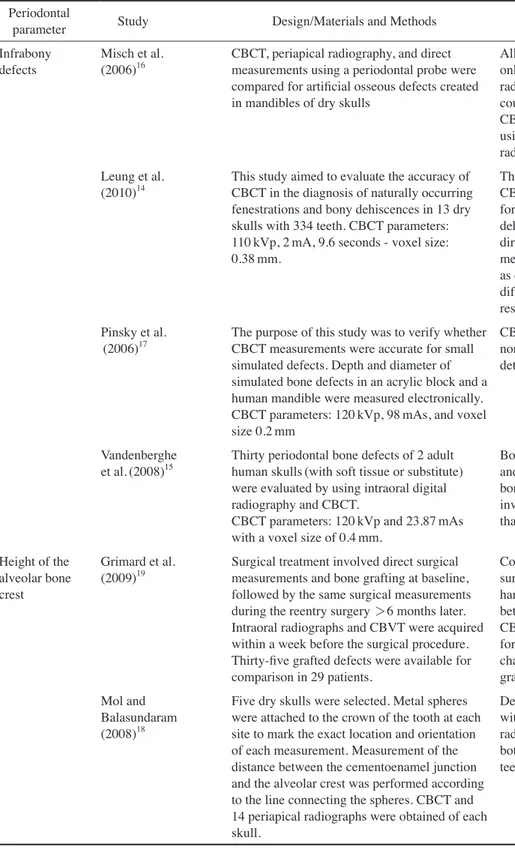
Table 1. Characteristics of the 13 studies included in this systematic review Periodontal
parameter Study Design/Materials and Methods Results/Conclusion Infrabony
defects Misch et al.
(2006)16 CBCT, periapical radiography, and direct measurements using a periodontal probe were compared for artificial osseous defects created in mandibles of dry skulls
All defects were detected using CBCT and only 67% of them were diagnosed using radiography. Buccal and lingual defects could not be diagnosed with radiography.
CBCT is as accurate as direct measurements using a periodontal probe and as reliable as radiographs for interproximal areas.
Leung et al.
(2010)14 This study aimed to evaluate the accuracy of CBCT in the diagnosis of naturally occurring fenestrations and bony dehiscences in 13 dry skulls with 334 teeth. CBCT parameters:
110kVp, 2mA, 9.6 seconds voxel size:
0.38mm.
The number of fenestrations detected by CBCT was more than 3 times higher than for direct examination and the number of dehiscences was less for CBCT than for direct examination. The study showed that measurements on CBCT were not as accurate as direct measurements on skulls and these differences were due to limitations in spatial resolution of the CBCT images.
Pinsky et al.
(2006)17 The purpose of this study was to verify whether CBCT measurements were accurate for small simulated defects. Depth and diameter of simulated bone defects in an acrylic block and a human mandible were measured electronically.
CBCT parameters: 120kVp, 98mAs, and voxel size 0.2mm
CBCT showed potential to be an accurate, noninvasive, practical method to reliably determine osseous lesion size and volume.
Vandenberghe
et al.(2008)15 Thirty periodontal bone defects of 2 adult human skulls(with soft tissue or substitute) were evaluated by using intraoral digital radiography and CBCT.
CBCT parameters: 120kVp and 23.87mAs with a voxel size of 0.4mm.
Both imaging modalities had same over and underestimation rates for periodontal bone defects. Bone craters and furcation involvements were better depicted on CBCT than on intraoral images.
Height of the alveolar bone crest
Grimard et al.
(2009)19 Surgical treatment involved direct surgical measurements and bone grafting at baseline, followed by the same surgical measurements during the reentry surgery >6 months later.
Intraoral radiographs and CBVT were acquired within a week before the surgical procedure.
Thirty-five grafted defects were available for comparison in 29 patients.
Correlations between CBCT and direct surgical measurements were higher for all hard tissue parameters than were correlations between intraoral and surgical measurements.
CBCT may be an equivalent substitution for direct surgical measurements of bony changes occurring after bonereplacement graft procedures
Mol and Balasundaram (2008)18
Five dry skulls were selected. Metal spheres were attached to the crown of the tooth at each site to mark the exact location and orientation of each measurement. Measurement of the distance between the cementoenamel junction and the alveolar crest was performed according to the line connecting the spheres. CBCT and 14 periapical radiographs were obtained of each skull.
Detecting bone loss was significantly better with CBCT than with conventional intraoral radiographs, but the diagnostic accuracy of both imaging modalities was low for anterior teeth.
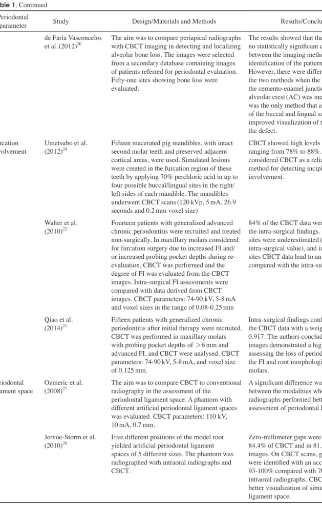
Table 1. Characteristics of the 13 studies included in this systematic review Periodontal
parameter Study Design/Materials and Methods Results/Conclusion de Faria Vasconcelos
et al.(2012)20 The aim was to compare periapical radiographs with CBCT imaging in detecting and localizing alveolar bone loss. The images were selected from a secondary database containing images of patients referred for periodontal evaluation.
Fiftyone sites showing bone loss were evaluated.
The results showed that there were no statistically significant differences between the imaging methods in terms of identification of the pattern of bone loss.
However, there were differences between the two methods when the distance between the cementoenamel junction(CEJ) and the alveolar crest(AC) was measured, as CBCT was the only method that allowed an analysis of the buccal and lingual surfaces and an improved visualization of the morphology of the defect.
Furcation
involvement Umetsubo et al.
(2012)24 Fifteen macerated pig mandibles, with intact second molar teeth and preserved adjacent cortical areas, were used. Simulated lesions were created in the furcation region of these teeth by applying 70% perchloric acid in up to four possible buccal/lingual sites in the right/
left sides of each mandible. The mandibles underwent CBCT scans(120kVp, 5mA, 26.9 seconds and 0.2mm voxel size)
CBCT showed high levels of accuracy, ranging from 78% to 88%. The authors considered CBCT as a reliable and accurate method for detecting incipient furcation involvement.
Walter et al.
(2010)22 Fourteen patients with generalized advanced chronic periodontitis were recruited and treated nonsurgically. In maxillary molars considered for furcation surgery due to increased FI and/
or increased probing pocket depths during re
evaluation, CBCT was performed and the degree of FI was evaluated from the CBCT images. Intrasurgical FI assessments were compared with data derived from CBCT images. CBCT parameters: 7490 kV, 58mA and voxel sizes in the range of 0.080.25mm
84% of the CBCT data were confirmed by the intra-surgical findings. 14.7% of the sites were underestimated(CBCT less than intrasurgical value), and in 1.3% of the sites CBCT data lead to an overestimation compared with the intrasurgical analysis.
Qiao et al.
(2014)21 Fifteen patients with generalized chronic periodontitis after initial therapy were recruited.
CBCT was performed in maxillary molars with probing pocket depths of >6mm and advanced FI, and CBCT were analysed. CBCT parameters: 7490kV, 58mA, and voxel size of 0.125mm.
Intra-surgical findings confirmed 82.4% of the CBCT data with a weighted kappa of 0.917. The authors concluded that CBCT images demonstrated a high accuracy assessing the loss of periodontal tissue of the FI and root morphologies in maxillary molars.
Periodontal
ligament space Ozmeric et al.
(2008)27 The aim was to compare CBCT to conventional radiography in the assessment of the
periodontal ligament space. A phantom with different artificial periodontal ligament spaces was evaluated. CBCT parameters: 110 kV, 10mA, 0.7mm.
A significant difference was observed between the modalities where conventional radiographs performed better than CBCT for assessment of periodontal ligament space.
JervoeStorm et al.
(2010)26 Five different positions of the model root yielded artificial periodontal ligament spaces of 5 different sizes. The phantom was radiographed with intraoral radiographs and CBCT.
Zeromillimeter gaps were recognized in 84.4% of CBCT and in 81.6% of intraoral images. On CBCT scans, gaps of 0.19mm were identified with an accuracy of 93100% compared with 70.2%81.7% in intraoral radiographs. CBCT provided a better visualization of simulated periodontal ligament space.
Table 1. Continued
infrabony defects, comparing measurements made using periapical radiographs and CBCT with control measure
ments obtained directly from dry skulls, reported that high
er precision and accuracy were obtained using CBCT. The greater precision of CBCT measurements can be attributed to the advantage of identifying defects present in both the buccal and lingual aspects of the teeth. Such defects cannot be fully identified by conventional radiography.14,16,17 As a result, all artificially created defects were detected with CBCT, whereas only 67% of the defects were identified in periapical radiographs.16 Studies have demonstrated that CBCT has the potential to be more accurate17 than conven
tional techniques in assessing artificially created peri
odontal defects.15,24
Only 1 study evaluated the accuracy of CBCT in identi
fying clinical bone defects, such as fenestrations and dehis
cences.18 The authors of that study compared observations made using tomographic images with direct observations of the defects in the skulls. The number of fenestrations detected by CBCT was 3 times higher than the number detected by direct visualization, thus leading to several false positive results(104 observed versus 32 actual fen
estrations). In contrast, fewer dehiscences were found in the tomographic images than by direct visualization(43 observed versus 52 actual dehiscences).14 The authors sug
gest that the low accuracy could be attributed to certain natural aspects of clinical bone defects, which present with less welldefined margins than artificially created defects, or to limitations in the spatial resolution of the CBCT de
vice used.
Another study comparing measurements of periodontal defects(fenestrations, dehiscences, and furcations) made using periapical radiographs, panoramic films, CT, and CBCT with the corresponding histological specimens show
ed that image quality(contrast, brightness, distortion, over
lay, and clarity of structures) was superior using CBCT.
CBCT scans showed smaller deviations in the dimensions of the periodontal defects than were obtained using the histological results.23
Height of the alveolar bone crest
CBCT was found to be more accurate than convention
al radiographs for assessing alveolar crest height, because it led to less underestimation of horizontal bone loss.18,20 One of the studies found that the mean alveolar bone crest was 0.23mm higher in the CBCT images than the actual crest height, whereas the corresponding deviation was 1.17 mm in periapical radiographs.18 Similar results were also obtained by de Faria Vasconcelos et al.(2012),20 who eval
uated 51 dental sites.
Nevertheless, the diagnostic accuracy of CBCT images achieved in the study by Mol and Balasundaram(2008)18 for horizontal bone loss in the anterior teeth was low. Both the underestimation of bone loss and the poor accuracy of measurements of crest height in the anterior maxilla could be attributed to the very thin cortical bone present in this region and the low sharpness of the images produced by the CBCT device.
One of the few in vivo studies comparing bone height measurements from digital intraoral radiographs and CBCT images to direct surgical measurements for the evaluation of regenerative treatment outcomes confirmed that CBCT yielded satisfactory results. In that study, digital IR and CBCT images were taken prior to initial bone grafting and after a 6month followup. A total of 35 intrabony defects were analyzed. The results suggested that CBCT reliably detected graft resorption based on a comparison with di
rect surgical measurements.19
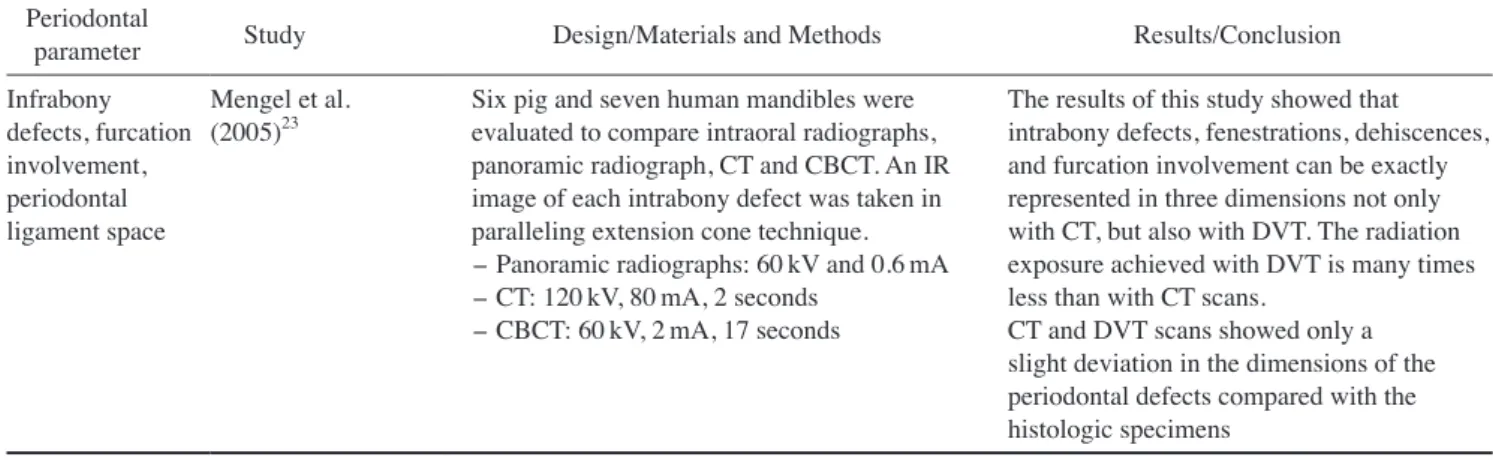
Table 1. Characteristics of the 13 studies included in this systematic review Periodontal
parameter Study Design/Materials and Methods Results/Conclusion Infrabony
defects, furcation involvement, periodontal ligament space
Mengel et al.
(2005)23 Six pig and seven human mandibles were evaluated to compare intraoral radiographs, panoramic radiograph, CT and CBCT. An IR image of each intrabony defect was taken in paralleling extension cone technique.
Panoramic radiographs: 60kV and 0.6mA
CT: 120kV, 80mA, 2 seconds
CBCT: 60kV, 2mA, 17 seconds
The results of this study showed that intrabony defects, fenestrations, dehiscences, and furcation involvement can be exactly represented in three dimensions not only with CT, but also with DVT. The radiation exposure achieved with DVT is many times less than with CT scans.
CT and DVT scans showed only a slight deviation in the dimensions of the periodontal defects compared with the histologic specimens
CBCT: conebeam computed tomography, CT: computed tomography, FI: furcation involvement, DVT: digital volumetric tomography.
Table 1. Continued
Furcation involvement
CBCT images were highly accurate(7888%) in detect
ing artificially created furcation involvement in 15 mace
rated pig mandibles in the in vitro study conducted by Umetsubo et al.(2012). Furthermore, furcation defects were also identified more accurately in CBCT images than in intraoral digital radiography in the study by Vandenber
ghe et al.(2008).15,24 Some in vivo studies also evaluated furcation involvement in tomographic images that were compared with direct intrasurgical assessments as the gold standard. The CBCT and surgical measurements agreed in 84% of cases, which is in accord with previous studies that found similar results.21,22 In the consensus report of the AAP Regeneration Workshop, the use of 3dimensional radiographic modalities was encouraged for the assessment of furcation defect treatment outcomes.25
Periodontal ligament space
Few studies have evaluated the accuracy of imaging tech
niques for evaluating the periodontal ligament space, and the existing studies have reported different results. Some studies have shown that CBCT was more accurate than conventional radiography in detecting marginal widening of the periodontal space.23,26 For instance, JervoeStorm et al.(2010) found almost 100% accuracy in identifying periodontal ligament space widening on CBCT images.26 Ozmeric et al.(2008), in contrast, found that periapical radiography was superior to CBCT for periodontal spaces smaller than or equal to 200 micrometers.27 The authors of that study suggested that the observers’ lack of experience with CBCT may have favored the results from 2dimen
sional conventional images.
discussion
Some CBCT parameters are known to lead to higher or lower spatial resolution. In the study of Leung et al.
(2010),14 the authors reported that the low accuracy of CBCT images in identifying clinical bone defects was due to limitations in the spatial resolution of the CBCT device used. The smallest thickness of bone measured in the axi
al and coronal slices was 0.6mm, which may explain the large number of falsepositive fenestrations found in the maxilla, where the cortical bone is less dense. In addition, the aforementioned study also had image quality limita
tions. Image quality is also influenced by the scanning parameters of the CBCT device. In this context, lower mil
liamperage(mA) leads to lower contrast resolution.28 Misch et al.16 (2006) chose to use high milliamperage in their re
search(47.7mA, 120kVp, and 20s) and obtained satisfac
tory results for identifying artificially created defects. In contrast, Leung et al.14 (2010) chose to use lower milliam
perage(2mA, 110kVp, and 9.6s) and obtained lower accu
racy and sensitivity in their measurements and classifica
tions.
In the study of Mol and Balasundaram(2008),18 bone loss detection was significantly better with CBCT than with conventional intraoral radiographs, but the diagnostic accu
racy of both imaging modalities was low for the anterior teeth. These results could have been better, as the authors reported image quality limitations because of the obsolete CBCT device used in their study. Advances in spatial reso
lution and other parameters have been incorporated into most modern devices, improving the resolution and preci
sion of tomographic images.22 Thus, further investigations with modern devices using optimal parameters can be re
commended to address the feasibility of CBCT.
The studies performed of CBCT data for furcation invol
vement not only showed high accuracy in identifying dif
ferent levels of furcation involvement, but also demonstrat
ed that the CBCT images provided additional important information about the root morphology and residual attach
ment of maxillary molars, which is a significant advantage of CBCT over conventional presurgical clinical assessment methods.21 Furthermore, the differences reported in the studies of furcation involvement may have been due to dif
ferent levels of expertise among the observers, who should be also trained to properly manipulate the CBCT software.
In the study conducted by Umetsubo et al.(2012),24 for instance, moderate interobserver agreement was found(κ=
0.400.59), indicating a lack of reproducibility.
Another example of how CBCT parameters can interfere with the final diagnosis of various periodontal conditions was provided by the study of JervoeStorm et al.(2010).26 The authors found an accuracy of almost 100% in identi
fying periodontal ligament space widening, and that result was probably obtained due to the high resolution used for imaging, as reflected by the voxel size of 0.15mm. This finding is in contrast with that of Ozmeric et al.(2008),27 who reported that periapical radiography was superior to CBCT for periodontal spaces smaller than or equal to 200 micrometers, using a voxel size of 0.7mm.
A limitation of the articles reviewed herein is that most of them were based on in vitro experiments. Only 4 of the 13 articles were in vivo studies. Another limitation was variation in the levels of expertise of the observers who participated in the research. Observers should be very well trained in manipulating the CBCT software, but several
authors reported that observers experienced difficulties in manipulating the CBCT software to visualize the different periodontal parameters.
The lack of studies that qualitatively or quantitatively compared conventional imaging techniques and CBCT was another limitation. The most recent in vitro/in vivo research found was from 2014.
CBCT is significantly more accurate and reliable than 2dimensional conventional imaging techniques for assess
ing infrabony defects, furcation lesions, the height of the alveolar bone crest, and the periodontal ligament space.
However, differences in imaging protocol parameters can affect the reproducibility and reliability of CBCT measure
ments.
references
1. Songa VM, Jampani ND, Babu V, Buggapati L, Mittapally S.
Accuracy of cone beam computed tomography in diagnosis and treatment planning of periodontal bone defects: a case report.
J Clin Diagn Res 2014; 8: ZD235.
2. Noujeim M, Prihoda T, Langlais R, Nummikoski P. Evalua
tion of highresolution cone beam computed tomography in the detection of simulated interradicular bone lesions. Dento
maxillofac Radiol 2009; 38: 15662.
3. Akesson L, Håkansson J, Rohlin M. Comparison of panoramic and intraoral radiography and pocket probing for the measure
ment of the marginal bone level. J Clin Periodontol 1992; 19:
32632.
4. Renvert S, Badersten A, Nilvéus R, Egelberg J. Healing after treatment of periodontal intraosseous defects. I. Comparative study of clinical methods. J Clin Periodontol 1981; 8: 38799.
5. Suomi JD, Plumbo J, Barbano JP. A comparative study of radio
graphs and pocket measurements in periodontal disease evalu
ation. J Periodontol 1968; 39: 3115.
6. Tugnait A, Clerehugh V, Hirschmann PN. The usefulness of radiographs in diagnosis and management of periodontal dis
eases: a review. J Dent 2000; 28: 21926.
7. Arai Y, Tammisalo E, Iwai K, Hashimoto K, Shinoda K. Devel
opment of a compact computed tomographic apparatus for den
tal use. Dentomaxillofac Radiol 1999; 28: 2458.
8. Ludlow JB, DaviesLudlow LE, Brooks SL. Dosimetry of two extraoral direct digital imaging devices: NewTom cone beam CT and Orthophos Plus DS panoramic unit. Dentomaxillofac Radiol 2003; 32: 22934.
9. Mah JK, Danforth RA, Bumann A, Hatcher D. Radiation ab
sorbed in maxillofacial imaging with a new dental computed tomography device. Oral Surg Oral Med Oral Pathol Oral Ra
diol Endod 2003; 96: 50813.
10. Batista WO, Navarro MV, Maia AF. Effective doses in pano
ramic images from conventional and CBCT equipment. Radiat Prot Dosimetry 2012; 151: 6775.
11. Aljehani YA. Diagnostic applications of conebeam CT for peri
odontal diseases. Int J Dent 2014; 2014: 865079.
12. OpenGrey.eu[Internet]. VandoeuvrelèsNancy Cedex: System for information on Grey Literature in Europe.[cited 2018 Jan
uary 23] Available from: http://www.opengrey.eu.
13. Moher D, Liberati A, Tetzlaff J, Altman DG, PRISMA group.
Preferred reporting items for systematic reviews and metaanal
yses: the PRISMA statement. PLoS Med 2009; 6: e1000097.
14. Leung CC, Palomo L, Griffith R, Hans MG. Accuracy and reli
ability of conebeam computed tomography for measuring al
veolar bone height and detecting bony dehiscences and fenes
trations. Am J Orthod Dentofacial Orthop 2010; 137(4 Suppl):
S10919.
15. Vandenberghe B, Jacobs R, Yang J. Detection of periodontal bone loss using digital intraoral and cone beam computed tomo
graphy images: an in vitro assessment of bony and/or infrabo
ny defects. Dentomaxillofac Radiol 2008; 37: 25260.
16. Misch KA, Yi ES, Sarment DP. Accuracy of cone beam com
puted tomography for periodontal defect measurements. J Peri
odontol 2006; 77: 12616.
17. Pinsky HM, Dyda S, Pinsky RW, Misch KA, Sarment DP. Ac
curacy of threedimensional measurements using conebeam CT. Dentomaxillofac Radiol 2006; 35: 4106.
18. Mol A, Balasundaram A. In vitro cone beam computed tomo
graphy imaging of periodontal bone. Dentomaxillofac Radiol 2008; 37: 31924.
19. Grimard BA, Hoidal MJ, Mills MP, Mellonig JT, Nummikoski PV, Mealey BL. Comparison of clinical, periapical radiograph, and conebeam volume tomography measurement techniques for assessing bone level changes following regenerative peri
odontal therapy. J Periodontol 2009; 80: 4855.
20. de Faria Vasconcelos K, Evangelista KM, Rodrigues CD, Estre
la C, de Sousa TO, Silva MA. Detection of periodontal bone loss using cone beam CT and intraoral radiography. Dento
maxillofac Radiol 2012; 41: 649.
21. Qiao J, Wang S, Duan J, Zhang Y, Qiu Y, Sun C, et al. The accu
racy of conebeam computed tomography in assessing maxil
lary molar furcation involvement. J Clin Periodontol 2014; 41:
26974.
22. Walter C, Weiger R, Zitzmann NU. Accuracy of threedimen
sional imaging in assessing maxillary molar furcation involve
ment. J Clin Periodontol 2010; 37: 43641.
23. Mengel R, Candir M, Shiratori K, FloresdeJacoby L. Digital volume tomography in the diagnosis of periodontal defects: an in vitro study on native pig and human mandibles. J Periodon
tol 2005; 76: 66573.
24. Umetsubo OS, Gaia BF, Costa FF, Cavalcanti MG. Detection of simulated incipient furcation involvement by CBCT: an in vitro study using pig mandibles. Braz Oral Res 2012; 26: 341
25. Reddy MS, AichelmannReidy ME, AvilaOrtiz G, Klokkevold 7.
PR, Murphy KG, Rosen PS, et al. Periodontal regeneration furcation defects: a consensus report from the AAP Regenera
tion Workshop. J Periodontol 2015; 86(2 Suppl): S1313.
26. JervoeStorm PM, Hagner M, Neugebauer J, Ritter L, Zoller JE, Jepsen S, et al. Comparison of conebeam computerized tomography and intraoral radiographs for determination of the periodontal ligament in a variable phantom. Oral Surg Oral Med Oral Pathol Oral Radiol Endod 2010; 109: e95101.
27. Ozmeric N, Kostioutchenko I, Hagler G, Frentzen M, Jervoe
Storm PM. Conebeam computed tomography in assessment of periodontal ligament space: in vitro study on artificial tooth model. Clin Oral Investig 2008; 12: 2339.
28. Kim JH, Arita ES, Pinheiro LR, Yoshimoto M, Watanabe PC, Cortes AR. Computed tomographic artifacts in maxillofacial surgery. J Craniofac Surg 2018; 29: e7880.