Introduction
Artificial intelligence (AI) has evolved from the concept of strong AI, which imitates human intelligence, to the im- plementation of weak AI that can solve certain problems.
1Studies of weak AI explore ways to construct algorithms that can learn from data and make predictions. Machine learning is a branch of computer science that builds al- gorithms guided by data.
2Among them, neural networks (NNs), which consist of nodes and weights, were one of the first types of AI algorithms to be developed. The com- putational power of these networks relies on the quality and quantity of training data, which allow these networks to update the weights of the connections. Simple network structures with only a few layers are known as “shallow”
learning neural networks, whereas network structures that employ numerous and large layers are referred to as “deep”
learning neural networks.
3Deep learning structures re- ferred to as convolutional neural networks (CNNs), which can extract many features from abstracted layers of filters, are mainly used for processing large and complex images.
Deep learning is being accelerated by the development of self-learning back-propagation algorithms that progressive- ly refine the results from the data, as well as by increases in computational power. Due to these rapid technological advances, AI, represented by deep learning, can be used for real-life problems and is applied across all sectors of soci- ety.
4The diagnostic accuracy of deep learning algorithms in the medical field is approaching levels of human exper- tise, changing the role of computer-assisted diagnosis from a ‘second-opinion’ tool to a more collaborative one.
3The development of AI applications in the dental field is also remarkable.
1,2In this article, papers about deep learning ap- plied to the field of oral and maxillofacial radiology will be reviewed.
Materials and Methods
Search strategy
In PubMed, Scopus, and the IEEE Xplore Digital Library,
An overview of deep learning in the field of dentistry
Jae-Joon Hwang
1, Yun-Hoa Jung
1, Bong-Hae Cho
1, Min-Suk Heo
2,*
1Department of Oral and Maxillofacial Radiology, School of Dentistry, Pusan National University, Dental Research Institute, Yangsan, Korea
2Department of Oral and Maxillofacial Radiology and Dental Research Institute, School of Dentistry, Seoul National University, Seoul, Korea
ABSTRACT
Purpose: Artificial intelligence(AI), represented by deep learning, can be used for real-life problems and is applied across all sectors of society including medical and dental field. The purpose of this study is to review articles about deep learning that were applied to the field of oral and maxillofacial radiology.
Materials and Methods: A systematic review was performed using Pubmed, Scopus, and IEEE explore databases to identify articles using deep learning in English literature. The variables from 25 articles included network architecture, number of training data, evaluation result, pros and cons, study object and imaging modality.
Results: Convolutional Neural network(CNN) was used as a main network component. The number of published paper and training datasets tended to increase, dealing with various field of dentistry.
Conclusion: Dental public datasets need to be constructed and data standardization is necessary for clinical application of deep learning in dental field.(Imaging Sci Dent 2019; 49: 1-7)
KEY WORDS: Artificial Intelligence; Deep Learning; Dentistry; Radiology
Copyright ⓒ 2019 by Korean Academy of Oral and Maxillofacial Radiology
This is an Open Access article distributed under the terms of the Creative Commons Attribution Non-Commercial License(http://creativecommons.org/licenses/by-nc/3.0) which permits unrestricted non-commercial use, distribution, and reproduction in any medium, provided the original work is properly cited.
Imaging Science in Dentistry·pISSN 2233-7822 eISSN 2233-7830
*This study was supported by 2017 Clinical Research Grant from Pusan National University Dental Hospital.
Received November 27, 2018; Revised December 15, 2018; Accepted December 17, 2018
*Correspondence to: Prof. Min-Suk Heo. Department of Oral and Maxillofacial Radiology, School of Dentistry, Seoul National University, 101 Daehak-ro, Jongno-gu, Seoul 03080, Korea
Tel) 82-2-2072-3016, E-mail) [email protected]
a search was performed for ‘deep learning OR neural net- work’ and ‘dental AND (diagnosis OR detection OR clas- sification OR segmentation)’ extending through December 2018, and 144, 33, and 32 search results were obtained, re- spectively. A total of 25 peer-reviewed papers were obtained by removing articles not written in English, those focusing on non-dental fields, papers not related to imaging dentistry, as well as reviews, editorials, and in-press papers. The mul- tilayer perceptron emerged as an early field of deep learn- ing, and papers on this topic were excluded from this study because it is not a true end-to-end learning method-it learns features extracted from images using existing ma- chine learning algorithms-and it has shallow networks and limited accuracy when the number of layers is increased.
5Data extraction
Study-specific data describing deep learning architecture, the size of the training datasets, evaluation results, advan- tages and disadvantages, the object of the study, and imag- ing modality were collected, in addition to other variables such as author and publication year.
Results
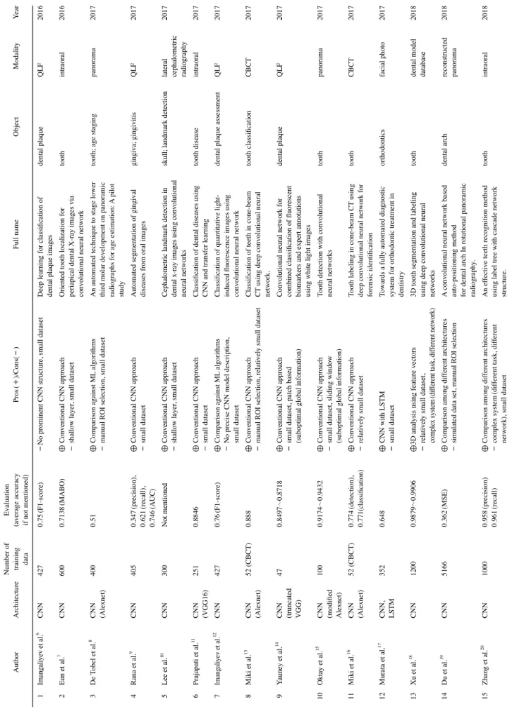
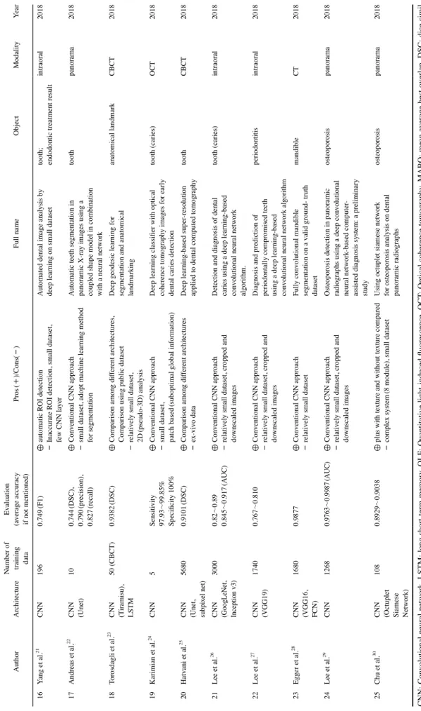
The data extracted from the selected papers are summa- rized in Table 1.
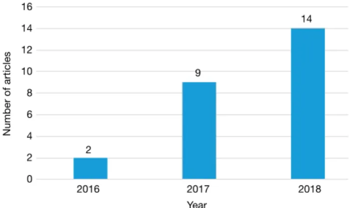
In all studies, CNN was used as a main network compo- nent, and there were also studies using various other types of networks, such as long short-term memory and siamese networks, in addition to CNNs. CNN-based papers have appeared in the field of dentistry since 2016, and subse- quently, more and more dentistry papers using CNN have been published (Fig. 1).
The median size of the datasets used for training also tended to increase, from 100 units to 1000 units (Fig. 2).
Many papers that used pretrained networks such as Alex- net, VGG, GoogLeNet, and Inception v3 showed good re- sults for general purposes.
31However, the structure of CNN networks tends to change from networks with shallow lay- ers to deeper or problem-specific home-made or complex networks.
These studies dealt with various field of dentistry. Most of them were related to teeth, but other subjects such as the gingiva and periodontium, the dental arch, osteoporosis, and anatomical landmarks were also studied using deep learning (Table 2).
Various imaging modalities have been studied in con- junction with the abovementioned subjects. Efforts are underway to diagnose dental disease using traditional 2-di-
mensional radiographs (intraoral and panoramic), as well as using 3-dimensional cone-beam computed tomography (CBCT). Other studies have investigated new modalities in dental applications, such as quantitative light-induced fluorescence, optical coherence tomography, and the use of intra-oral laser scanners.
Discussion
Computer assisted diagnosis (CAD) software in the med- ical field has been used to obtain second opinions, but the design and tuning of conventional CAD tends to be very arduous. Recently, deep learning techniques have been in- tegrated into CAD, with promising results for various med- ical applications.
32,33The qualitative and quantitative ap- plications of deep learning in dentistry are also expanding, but certain areas need to be complemented to promote the continued development of deep learning research in oral and maxillofacial radiology.
However, because all the data sets used in the research analyzed herein were in-house, objective comparison of the studies was difficult. Only a single study tried to evalu- ate the accuracy of developed networks using other public datasets.
23Efforts are needed to develop a public dataset, such as in the medical field,
34to develop algorithms that can be used in clinical applications. In order to achieve this, researchers need to release the data used in their pa- pers with appropriate removal of personal information, and legal and institutional support from each country is also necessary.
35,36There is also a need to build a common, free repository that can reliably collect, catalog, and archive publicly available data in the dental field.
The overall increase in the size of training datasets is
desirable for clinical applications of deep learning to the
dental field. However, most studies used relatively small
data sets (fewer than 1000 units per group), and the accu-
racy of most studies was less than 90%. This is below the
clinically expected accuracy of 98%-99%.
37Deep learning
requires a large amount of data because it learns features
directly from the data via an end-to-end process. In an an-
atomical classification study of CT data, at least 1,000 data
sets per group were required to achieve 98% validation
accuracy with deep learning, and 4,092 data sets per group
were required to reach the desired accuracy of 99.5%.
38CBCT, which is the most popular 3D imaging modality in
the dental field, does not utilize defined Hounsfield unit
values like medical CT, and the pixel values of the acquired
images change at every exposure.
39The image quality and
magnification of panoramic radiographs, which are com-
Table 1. Summary of deep learning articles in the field of dentistry AuthorArchitecture
Number of training data
Evaluation (average accuracy if not mentioned) Pros(+)/Cons(-) Full name ObjectModalityYear 1Imangaliyev et al.6CNN4270.75(F1-score)-No prominent CNN structure, small dataset
Deep learning for classification of dental plaque images
dental plaqueQLF2016 2Eun et al.7CNN6000.7138(MABO)⊕ Conventional CNN approach - shallow layer, small dataset
Oriented tooth localization for periapical dental X-ray images via convolutional neural network
toothintraoral2016 3De Tobel et al.8CNN (Alexnet)4000.51 ⊕ Comparison against ML algorithms - manual ROI selection, small dataset
An automated technique to stage lower third molar development on panoramic radiographs for age estimation:
A pilot study
tooth; age stagingpanorama2017 4Rana et al.9CNN4050.347(precision), 0.621(recall), 0.746(AUC)
⊕ Conventional CNN approach - small dataset
Automated segmentation of gingival diseases from oral images
gingiva; gingivitisQLF2017 5Lee et al.10CNN300Not mentioned⊕ Conventional CNN approach - shallow layer, small dataset
Cephalometric landmark detection in dental x-ray images using convolutional neural networks
skull; landmark detection
lateral cephalometric radiography
2017 6Prajapati et al.11CNN (VGG16)2510.8846⊕ Conventional CNN approach - small dataset
Classification of dental diseases using CNN and transfer learning
tooth diseaseintraoral2017 7Imangaliyev et al.12CNN4270.76(F1-score)⊕ Comparison against ML algorithms - No precise CNN model description, small dataset
Classification of quantitative light- induced fluorescence images using convolutional neural network
dental plaque assessmentQLF2017 8Miki et al.13CNN (Alexnet)52 (CBCT)0.888⊕ Conventional CNN approach - manual ROI selection, relatively small dataset
Classification of teeth in cone-beam CT using deep convolutional neural network.
tooth classificationCBCT2017 9Yauney et al.14CNN (truncated VGG)
470.8497~0.8718⊕ Conventional CNN approach - small dataset, patch based (suboptimal global information)
Convolutional neural network for combined classification of fluorescent biomarkers and expert annotations using white light images
dental plaqueQLF2017 10Oktay et al.15CNN (modified Alexnet)
1000.9174~0.9432⊕ Conventional CNN approach - small dataset, sliding window (suboptimal global information)
Tooth detection with convolutional neural networks
toothpanorama2017 11Miki et al.16CNN (Alexnet)52 (CBCT)0.774(detection), 0.771(classification)⊕ Conventional CNN approach - relatively small datasetTooth labeling in cone-beam CT using
deep convolutional neural network for forensic identification
toothCBCT2017 12Murata et al.17CNN, LSTM3520.648 ⊕ CNN with LSTM - small dataset
Towards a fully automated diagnostic system for orthodontic treatment in dentistry
orthodonticsfacial photo2017 13Xu et al.18CNN12000.9879~0.9906⊕3D analysis using feature vectors - relatively small dataset, complex system(different task, different network)
3D tooth segmentation and labeling using deep convolutional neural networks
tooth
dental model database
2018 14Du et al.19CNN51660.362(MSE)⊕ Comparison among different architectures - simulated data set, manual ROI selection
A convolutional neural network based auto-positioning method for dental arch In rotational panoramic radiography
.
dental arch
reconstructed panorama
2018 15Zhang et al.20CNN10000.958(precision) 0.961(recall)⊕ Comparison among different architectures - complex system(different task, different network), small dataset
An effective teeth recognition method
using label tree with cascade network structure.
toothintraoral2018
Table 1. Summary of deep learning articles in the field of dentistry AuthorArchitecture
Number of training data
Evaluation (average accuracy if not mentioned) Pros(+)/Cons(-) Full name ObjectModalityYear 16Yang et al.21CNN1960.749(F1)⊕ automatic ROI detection - Inaccurate ROI detection, small dataset, few CNN layer
Automated dental image analysis by deep learning on small dataset
tooth; endodontic treatment resultintraoral2018 17Andreas et al.22CNN (Unet)100.744(DSC), 0.790(precision), 0.827(recall)
⊕ Conventional CNN approach - small dataset, adopt machine learning method for segmentation
Automatic teeth segmentation in panoramic X-ray images using a coupled shape model in combination with a neural network
toothpanorama2018 18Torosdagli et al.23
CNN (Tiramisu), LSTM
50 (CBCT)0.9382(DSC)⊕ Comparison among different architectures, Comparison using public dataset - relatively small dataset, 2D(pseudo-3D) analysis
Deep geodesic learning for segmentation and anatomical landmarking
anatomical landmarkCBCT2018 19Karimian et al.24CNN5
Sensitivity 97.93
~99.85% Specificity 100%
⊕ Conventional CNN approach - small dataset, patch based(suboptimal global information)
Deep learning classifier with optical coherence tomography images for early dental caries detection
tooth (caries)OCT2018 20Hatvani et al.25CNN (Unet, subpixel net)
56800.9101(DSC)⊕ Comparison among different architectures - ex-vivo dataDeep learning-based super-resolution applied to dental computed tomographytoothCBCT2018 21Lee et al.26
CNN (GoogLeNet, Inception v3)
30000.82~0.89 0.845~0.917(AUC)⊕ Conventional CNN approach - relatively small dataset, cropped and downscaled images
Detection and diagnosis of dental caries using a deep learning-based convolutional neural network algorithm.
tooth (caries)intraoral2018 22Lee et al.27CNN (VGG19)17400.767~0.810⊕ Conventional CNN approach - relatively small dataset, cropped and downscaled images
Diagnosis and prediction of periodontally compromised teeth using a deep learning-based coritgo alrktwoneeural l nnaioutolnvhm
periodontitisintraoral2018 23Egger et al.28CNN (VGG16, FCN)
16800.9877 ⊕ Conventional CNN approach - relatively small dataset
Fully convolutional mandible segmentation on a valid ground- truth dataset
mandibleCT2018 24Lee et al.29CNN1268 0.9763~0.9987(AUC)⊕ Conventional CNN approach - relatively small dataset, cropped and downscaled images
Osteoporosis detection in panoramic radiographs using a deep convolutional neural network-based computer
- assisted diagnosis system: a preliminary study
osteoporosispanorama2018 25Chu et al.30CNN (Octuplet Siamese Network)
1080.8929~0.9038⊕ plus with texture and without texture compared - complex system(8 module), small dataset Using octuplet siamese network for osteoporosis analysis on dental panoramic radiographs
osteoporosispanorama2018 CNN: Convolutional neural network, LSTM: long short-term memory, QLF: Quantitative light-induced fluorescence, OCT: Optical coherence tomography, MABO: mean average best overlap, DSC: dice simil arity coefficient, AUC: area under the curve
Table1. Continued
monly used in dental practice, depend on the positioning of the patient.
40Therefore, to achieve clinically meaningful high accuracy, trans-hospital or hybrid data sets from mul- tiple machines and conditions are likely to be needed due to the nature of dental images. For this reason, it is espe- cially important to emphasize the need to construct a large- scale dental public dataset to make the clinical application of deep learning possible.
It is also necessary to emphasize the need for data stan- dardization in the dental field, as well as for standardization of data set construction. In particular, CBCT exhibits large image variation according to brand, machine, and exposure conditions, which can be an obstacle to deep learning re-
search. For example, collecting and learning data on a ma- chine-by-machine basis is difficult because models learned on one machine do not apply to other machines. Although attempts have been made to develop guidelines in Europe, Germany, and England regarding the image quality of CBCT, no international standard has yet been established.
41Therefore, in order for 3-dimensional diagnosis using deep learning to be practical, an international standard for the quality of CBCT images needs to be established in the near future.
Many papers have used preprocessed images via manual cropping of the region of interest. This makes it difficult to analyze and compare results accurately due to errors in the manual process. Some papers
9,10,19have described networks that learned by dividing images into patches of a certain size. However, this method is limited because the network cannot learn the whole image, and instead only fo- cuses on a small part of the image. Some papers
21,22,24used downsampling, which might delete important details of the image. These choices seem to have been made due to limitations in the amount of data or computational power, as indicated in the limitations sections of some papers.
21,22However, as computing power per cost increases, it is nec- essary to use entire images to learn, without any artificial manipulation in the preprocessing stage, in order to obtain more accurate and general results.
Currently, the use of AI is expanding in the medical field.
For example, Watson, developed by IBM, has been used to support doctors’ clinical decisions.
42However, the clinical accuracy of AI in the dental field must be verified with a variety of cases and imaging modalities due to the difficul- ty of standardizing dental radiology before AI can take on a more important role in making diagnostic recommenda- tions. Furthermore, current AI algorithms function as black boxes, making it difficult for humans to identify or adjust the criteria used for diagnoses.
43Therefore, in order to in- crease the reliability of AI, it is necessary to develop a visu- alization and modification tool for deep learning networks that can be easily understood and edited by humans.
References
1. Park WJ, Park JB. History and application of artificial neural networks in dentistry. Eur J Dent 2018; 12: 594-601.
2. Mupparapu M, Wu CW, Chen YC. Artificial intelligence, ma- chine learning, neural networks, and deep learning: futuristic con- cepts for new dental diagnosis. Quintessence Int 2018; 49: 687-8.
3. Burt JR, Torosdagli N, Khosravan N, RaviPrakash H, Mortazi A, Tissavirasingham F, et al. Deep learning beyond cats and dogs:
recent advances in diagnosing breast cancer with deep neural
2016 2017 2018
Year 1400
1200 1000 800 600 400 200 0
Number of training data
Fig. 2. Median size of training datasets from 2016 to 2018.
Table 2. Frequency of subjects in deep learning articles
Subject Frequency
Tooth related 12
Dental plaque 3
Gingiva or periodontium 2
Osteoporosis 2
etc. 5
16 14 12 10 8 6 4 2 0
Number of articles
2016 2017 2018
Year Fig. 1. Number of articles from 2016 to 2018.
14
2
9
600.5
175.5
1200
networks. Br J Radiol 2018; 91: 20170545.
4. Rabuñal JR, Dorado J. Artificial neural networks in real-life ap- plications. IGI Global: Hershey; 2005. p. 166-346.
5. Panchal G, Ganatra A, Kosta YP, Panchal D. Behaviour analy- sis of multilayer perceptrons with multiple hidden neurons and hidden layers. Int J Comput Theory Eng 2011; 3: 332-7.
6. Imangaliyev S, van der Veen MH, Volgenant CM, Keijser BJ, Crielaard W, Levin E. Deep learning for classification of dental plaque images. In: Conca PP, Nicosia GG. Machine learning, optimization, and Big data. Second International Workshop, MOD 2016, Volterra, Italy, August 26-29, 2016, Revised Se- lected Papers. Heidelberg: Springer. 2016. p. 407-10.
7. Eun H, Kim C. Oriented tooth localization for periapical dental X-ray images via convolutional neural network. 2016 Asia-Pa- cific Signal and Information Processing Association Annual Summit and Conference (APSIPA 2016); 2016 Dec 13-16; Jeju, Korea. Red Hook, NY: IEEE; p. 33-9.
8. De Tobel J, Radesh P, Vandermeulen D, Thevissen PW. An automated technique to stage lower third molar development on panoramic radiographs for age estimation: a pilot study. J Forensic Odontostomatol 2017; 2: 42-54.
9. Rana A, Yauney G, Wong LC, Gupta O, Muftu A, Shah P. Au- tomated segmentation of gingival diseases from oral images.
IEEE-NIH 2017 Special Topics Conference on Healthcare Inno- vations and Point-of-Care Technologies. Bethesda, MD: Nation- al Institutes of Health. p. 144-7.
10. Lee H, Park M, Kim J. Cephalometric landmark detection in dental X-ray images using convolutional neural networks. Proc.
SPIE 10134, Medical Imaging 2017: Computer-Aided Diag- nosis, 101341W (3 March 2017). Available from: https://doi.
org/10.1117/12.2255870.
11. Prajapati SA, Nagaraj R, Mitra S. Classification of dental diseases using CNN and transfer learning. 2017 5th Interna- tional Symposium on Computational and Business Intelligence (ISCBI), Dubai, 2017, p. 70-4. Available from: https://doi.
org/10.1109/ISCBI.2017.8053547.
12. Imangaliyev S, van der Veen MH, Volgenant CM, Loos BG, Keijser BJ, Crielaard W, et al. Classification of quantitative light-induced fluorescence images using convolutional neural network. arXiv:1705.09193. 2017 [cited 2018 November 20].
Available from: https://arxiv.org/pdf/1705.09193.pdf
13. Miki Y, Muramatsu C, Hayashi T, Zhou X, Hara T, Katsumata A, et al. Classification of teeth in cone-beam CT using deep con- volutional neural network. Comput Biol Med 2017; 80: 24-9.
14. Yauney G, AngelinoK, Edlund DA, Shah P. Convolutional neural network for combined classification of fluorescent bio- markers and expert annotations using white light images. 2017 IEEE 17th International Conference on Bioinformatics and Bio- engineering (BIBE), Washington, DC, 2017, p. 303-9. Available from: https://doi.org/10.1109/BIBE.2017.00-37
15. Oktay AB. Tooth detection with Convolutional Neural Net- works. 2017 Medical Technologies National Congress (TIPTE- KNO), Trabzon, 2017, p. 1-4. Available from: https://doi.
org/10.1109/TIPTEKNO.2017.8238075
16. Miki Y, Muramatsu C, Hayashi T, Zhou X, Hara T, Katsumata A, et al. Tooth labeling in cone-beam CT using deep convolutional neural network for forensic identification. In: Armato SG, Pet- rick NA. Medical Imaging 2017: Computer-Aided Diagnosis.
Proceedings Volume 10134, SPIE Medical Imaging; 2017 Feb 11-16; Orlando, USA: SPIE; 2017. Available from: https://doi.
org/10.1117/12.2254332
17. Murata S, Lee C, Tanikawa C, Date S. Towards a fully auto- mated diagnostic system for orthodontic treatment in dentistry.
2017 IEEE 13th International Conference on e-Science (e-Sci- ence), Auckland, 2017. p. 1-8. Available from: https://doi.
org/10.1109/eScience.2017.12
18. Xu X, Liu C, Zheng Y. 3D tooth segmentation and labeling us- ing deep convolutional neural networks. IEEE Trans Vis Com- put Graph (in press).
19. Du X, Chen Y, Zhao J, Xi Y. A convolutional neural network based auto-positioning method for dental arch in rotational panoramic radiography. Conf Proc IEEE Eng Med Biol Soc 2018; 2018: 2615-8.
20. Zhang K, Wu J, Chen H, Lyu P. An effective teeth recognition method using label tree with cascade network structure. Com- put Med Imaging Graph 2018; 68: 61-70.
21. Yang J, Xie Y, Liu L, Xia B, Cao Z, Guo C. Automated dental image analysis by deep learning on small dataset. 2018 IEEE 42nd Annual Computer Software and Applications Conference (COMPSAC), Tokyo, 2018. 492-7. Available from: https://doi.
org/10.1109/COMPSAC.2018.00076
22. Andreas W, Sudesh G, Stefan W. Automatic teeth segmenta- tion in panoramic X-ray images using a coupled shape model in combination with a neural network. Proceedings from the 21st International Conference on Medical Image Computing and Computer Assisted Intervention, Part IV; 2018 Sep 16-20;
Granada, Spain. Basel: Springer; 2018. Available from: https://
doi.org/10.1007/978-3-030-00937-3_81.
23. Torosdagli N, Liberton DK, Verma P, Sincan M, Lee JS, Bagci U.
Deep geodesic learning for segmentation and anatomical land- marking. IEEE Trans Med Imaging (in press).
24. Karimian N, Salehi HS, Mahdian M, Alnajjar H, Tadinada A.
Deep learning classifier with optical coherence tomography im- ages for early dental caries detection. Proc. SPIE 10473, Lasers in Dentistry XXIV, 1047304 (8 February 2018); Available from:
https://doi.org/10.1117/12.2291088
25. Hatvani J, Horváth A, Michetti J, Basarab A, Kouamé D, Gyöngy M. Deep learning-based super-resolution applied to dental computed tomography. IEEE Transactions on Radia- tion and Plasma Medical Sciences. Available from: https://doi.
org/10.1109/TRPMS.2018.2827239
26. Lee JH, Kim DH, Jeong SN, Choi SH. Detection and diagnosis of dental caries using a deep learning-based convolutional neu- ral network algorithm. J Dent 2018; 77: 106-111.
27. Lee JH, Kim DH, Jeong SN, Choi SH. Diagnosis and prediction of periodontally compromised teeth using a deep learning-based convolutional neural network algorithm. J Periodontal Implant Sci 2018; 48: 114-23.
28. Egger J, Pfarrkirchner B, Gsaxner C, Lindner L, Schmalstieg D, Wallner J. Fully convolutional mandible segmentation on a valid ground - truth dataset. Conf Proc IEEE Eng Med Biol Soc 2018; 2018: 656-60.
29. Lee JS, Adhikari S, Liu L, Jeong HG, Kim H, Yoon SJ. Osteo- porosis detection in panoramic radiographs using a deep con- volutional neural network-based computer-assisted diagnosis system: a preliminary study. Dentomaxillofac Radiol 2018:
20170344 (in press).
30. Chu P, Bo C, Liang X, Yang J, Megalooikonomou V, Yang F, et al. Using octuplet siamese network for osteoporosis analysis on dental panoramic radiographs. Conf Proc IEEE Eng Med Biol Soc 2018; 2018: 2579-82.
31. Kim YD, Jang TW, Han BY, Choi, SJ. Learning to select pre- trained deep representations with bayesian evidence framework.
2016 IEEE Conference on Computer Vision and Pattern Recog- nition (CVPR), Las Vegas, NV, 2016, pp. 5318-5326. p. 5318- 26. Available from: https://doi.org/10.1109/CVPR.2016.574 32. Cheng JZ, Ni D, Chou YH, Qin J, Tiu CM, Chang YC, et al.
Computer-aided diagnosis with deep learning architecture:
applications to breast lesions in US images and pulmonary nod- ules in CT scans. Sci Rep 2016; 6: 24454.
33. Suzuki K. Overview of deep learning in medical imaging. Ra- diol Phys Technol 2017; 10: 257-73.
34. Greenspan H, Ginneken BV, Summers RM. Guest editorial:
deep learning in medical imaging: overview and future promise of an exciting new technique. IEEE Trans Med Imaging 2016;
35: 1153-9.
35. Berman JJ. Confidentiality issues for medical data miners. Artif Intell Med 2002; 26: 25-36.
36. Cooper T, Collman J. Managing information security and priva- cy in healthcare data mining. In: Chen H, Fuller SS, Friedman CH. Medical informatics: knowledge management and data mining in biomedicine. New York: Springer; 2005. p. 95-137.
37. Weese J, Lorenz C. Four challenges in medical image analysis from an industrial perspective. Med Image Anal 2016; 33: 44-9.
38. Cho J, Lee K, Shin E, Choy G, Do S. How much data is needed to train a medical image deep learning system to achieve nec- essary high accuracy? arXiv:1511.06348 [Preprint]. 2016 [cited 2018 Nov 20] Available from: https://arxiv.org/abs/1511.06348 39. Pauwels R, Araki K, Siewerdsen JH, Thongvigitmanee SS.
Technical aspects of dental CBCT: state of the art. Dentomaxil- lofac Radiol 2015; 44: 20140224.
40. Devlin H, Yuan J. Object position and image magnification in dental panoramic radiography: a theoretical analysis. Dento- maxillofac Radiol 2013; 42: 29951683.
41. de Las Heras Gala H, Torresin A, Dasu A, Rampado O, Delis H, Hernández Girón I, et al. Quality control in cone-beam com- puted tomography (CBCT) EFOMP-ESTRO-IAEA protocol (summary report). Phys Med 2017; 39: 67-72.
42. Chen Y, Elenee Argentinis JD, Weber G. IBM Watson: how cognitive computing can be applied to big data challenges in life sciences research. Clin Ther 2016; 38: 688-701.
43. Anifowose FA. Artificial intelligence application in reservoir characterization and modeling: whitening the black Box. Pro- ceedings of the SPE Saudi Arabia section Young Professionals Technical Symposium; 2011 Mar 14-16; Dharan, Saudi Arabia.
Dharan: Society of Petroleum Engineers; 2011. Available from:
https://doi.org/10.2118/155413-MS.v