Chemotherapy
Infect Chemother 2013;45(3):292-298 pISSN 2093-2340 · eISSN 2092-6448
Received: April 23, 2013 Revised: July 17, 2013 Accepted: July 18, 2013 Corresponding Author : Sang-Oh Lee, MD, PhD
Department of Infectious Diseases, Asan Medical Center, University of Ulsan College of Medicine, 88 Olympic-ro 43-gil, Songpa-gu, Seoul 138-736, Korea Tel: +82-2-3010-3301, Fax: +82-2-3010-6970
E-mail: [email protected]
This is an Open Access article distributed under the terms of the Creative Commons Attribution Non-Commercial License (http://creativecommons.org/licenses/by-nc/3.0) which permits unrestricted non-commercial use, distribution, and repro- duction in any medium, provided the original work is properly cited.
Copyrights © 2013 by The Korean Society of Infectious Diseases | Korean Society for Chemotherapy
www.icjournal.org
Risk Factors for Mortality in Patients with Invasive Mucormycosis
Hyo-Lim Hong
1, Yu-Mi Lee
1, Tark Kim
1, Joo-Young Lee
1, Yoo-Sam Chung
2, Mi-Na Kim
3, Sung-Han Kim
1, Sang-Ho Choi
1, Yang Soo Kim
1, Jun Hee Woo
1, and Sang-Oh Lee
1Departments of 1Infectious Diseases, 2Otolaryngology, and 3Laboratory Medicine, Asan Medical Center, University of Ulsan College of Medicine, Seoul, Korea
Background: Mucormycosis is an uncommon and life-threatening fungal infection. The clinical predictors of outcome were evaluated in patients with invasive mucormycosis.
Materials and Methods: We retrospectively reviewed histologically proven cases of invasive mucormycosis in our institution from 1996 to 2012.
Results: A total of 64 patients were analyzed. The median age was 59 years (interquartile range [IQR], 50–67), and 32 patients (50%) were male. The most common underlying diseases were diabetes mellitus (67%), hematologic malignancy (22%), and solid cancer (19%). The most common infection sites were the rhino-orbito-cerebral area (56%) and the lungs (31%). The 180-day all-cause mor- tality was 33%. Disseminated infection was associated with increased mortality (hazard ratio [HR]: 169.74, 95% confidence interval [CI]: 6.41 to 4492.64; P = 0.002). Pulmonary infection (HR: 0.08, 95% CI: 0.01 to 0.66; P = 0.02) and complete surgical removal of infected tissue (HR: 0.12, 95% CI: 0.02 to 0.64; P = 0.01) were associated with decreased mortality.
Conclusions: These results suggest that patients with mucormycosis had a lower risk of mortality if they developed a pulmonary infection, rather than a disseminated infection and with complete debridement of infected tissue.
Key Words: Risk factor, Mortality, Mucormycosis
Introduction
Mucormycosis is an uncommon, life-threatening infection caused by filamentous fungi of the order Mucorales and class Zygomycetes. These ubiquitous organisms can be found in bread mold, soil, manure, and decaying vegetation. Along with candidiasis and aspergillosis, mucormycosis is one of the
primarily opportunistic invasive mycoses that frequently de- velop in immunocompromised patients who have received hematopoietic stem cell or solid organ transplantations or who have hematologic malignancies [1-4]. Moreover, mucor- mycosis can occur in immunocompetent patients with diabe- tes mellitus, subcutaneous tissue injury, and iron overload [5], and who are undergoing deferoxamine therapy [6]. The site of
infection varies, and can occur in the lungs, the skin and soft tissue, the rhino-orbito-cerebral region, and the gastrointesti- nal tract. Mucormycosis infection may also appear as a dis- seminated disease that infects more than one noncontiguous site [4].
The mortality rate depends on the patient’s underlying dis- eases and the infection site [7]. Treatment strategy involves timely diagnosis, aggressive surgical debridement combined with high-dose amphotericin B, and reversal of underlying predisposing factors whenever possible [8]. Factors associated with poor outcome have not been well defined. In addition, the only published data available on mucormycosis in Korea consists of case reports. Therefore, we conducted a retrospec- tive study to specifically evaluate clinical predictors of out- come in patients with mucormycosis.
Materials and Methods
This retrospective study was performed at the Asan Medical Center, a 2,700-bed tertiary care teaching hospital in Seoul, Korea. The pathology database was electronically searched to identify all cases of mucormycosis from patients admitted be- tween April 1996 and November 2012. Data was collected on patients’ demographics, underlying conditions, concomitant immunosuppressive medications, laboratory data, radiologic findings, clinical features, antifungal treatment, surgical pro- cedures, and outcomes.
Patients were included in the study if they met the criteria for proven invasive mucormycosis based on the revised defi- nitions of invasive fungal disease of the European Organiza- tion for Research and Treatment of Cancer/Mycosis Study Group (EORTC/MSG) [9]. A diagnosis of mucormycosis was based on histopathological demonstration of broad, ribbon- like, wide-angled branching, non-septate hyphae even in the absence of positive cultures, and accompanying tissue inva- sion by fungal hyphae [10, 11]. The mucormycosis genus was determined by morphological examination of conidia, hy- phae, and whole colonies.
Infection sites were classified as rhino-orbito-cerebral, pul- monary, gastrointestinal, cutaneous, or disseminated mucor- mycosis. The rhino-orbito-cerebral involvements were classi- fied as rhinocerebral, sino-orbital, or isolated sinusitis [4], while the pulmonary infections were subcategorized into po- tentially resectable cases and definitively non-resectable cas- es. The day of diagnosis was defined as the day on which a procedure (biopsy, surgery, or culture) leading to a mucormy-
cosis diagnosis was conducted. The outcome was assessed by 180-day all-cause mortality after a diagnosis of invasive mu- cormycosis.
Data were analyzed using IBM SPSS for Windows (version 19.0; IBM Corp., Armonk, NY, USA). Categorical variables were analyzed using either the Chi-square test or Fisher’s ex- act test. Continuous variables were analyzed using the Stu- dent’s t-test or Mann-Whitney U-test, as appropriate. The pri- mary objective of the study was to identify independent predictors of 180-day mortality in invasive mucormycosis.
Therefore, a Cox proportional hazards regression model for multivariate analyses was built using a forward stepwise method. Covariates with a P < 0.2 were used in the stepwise method. All tests were 2-tailed and differences were consid- ered significant when P < 0.05.
Results
1. Patient characteristics and clinical outcomes During the study period, 64 patients with histologically proven invasive mucormycosis were identified. Mycological diagnosis was performed in only 7 patients (11%); 4 patients were infected with Rhizopus species (6%) and 3 patients were infected with Mucor species (5%). The median age was 59 years (nterquartile range [IQR], 50–67), and 32 patients (50%) were male. The majority of patients had an underlying disease (63/64, 98%), the most common of which was diabetes melli- tus (67%), followed by hematologic malignancy (22%) and solid cancer (19%). The most common infection site was the rhino-orbito-cerebral area (36 cases, 56%), followed by the lung (20 cases, 31%) and the gastrointestinal tract (4 cases, 6%). All 64 mucormycosis patients received antifungal thera- py; the majority of patients (57/64, 89%) received amphoteri- cin B (conventional or liposomal) as the first-line antifungal agent. Surgery was performed on 46 patients (46/64, 72%), with complete removal of the infected site in 22 cases (22/46, 48%). In cases of rhino-orbito-cerebral mucormycosis, surgi- cal debridement was performed in 89% (32/36) of the pa- tients, and the majority was for rhino-cerebral lesions (17/19), followed by sinusitis (12/14) and sino-orbital lesions (3/3).
Eight patients (8/20, 40%) received surgical resection for pul- monary mucormycosis. The 180-day all-cause mortality after diagnosis of invasive mucormycosis was 33% (21/64).
2. Risk factors for mortality
Demographic and clinical characteristics of the 43 surviving
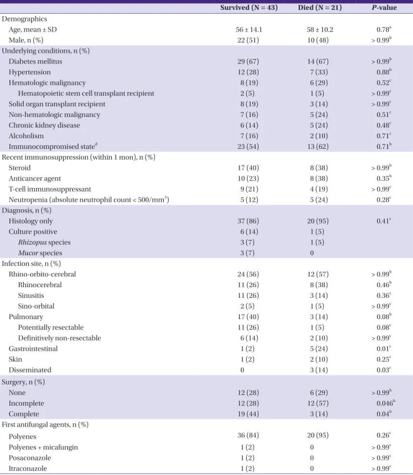
Table 1. Comparison of clinical characteristics between patients with invasive mucormycosis who survived and died within 180 days after diagnosis Survived (N = 43) Died (N = 21) P-value Demographics
Age, mean ± SD 56 ± 14.1 58 ± 10.2 0.78a
Male, n (%) 22 (51) 10 (48) > 0.99b
Underlying conditions, n (%)
Diabetes mellitus 29 (67) 14 (67) > 0.99b
Hypertension 12 (28) 7 (33) 0.88b
Hematologic malignancy 8 (19) 6 (29) 0.52c
Hematopoietic stem cell transplant recipient 2 (5) 1 (5) > 0.99c
Solid organ transplant recipient 8 (19) 3 (14) > 0.99c
Non-hematologic malignancy 7 (16) 5 (24) 0.51c
Chronic kidney disease 6 (14) 5 (24) 0.48c
Alcoholism 7 (16) 2 (10) 0.71c
Immunocompromised stated 23 (54) 13 (62) 0.71b
Recent immunosuppression (within 1 mon), n (%)
Steroid 17 (40) 8 (38) > 0.99b
Anticancer agent 10 (23) 8 (38) 0.35b
T-cell immunosuppressant 9 (21) 4 (19) > 0.99c
Neutropenia (absolute neutrophil count < 500/mm3) 5 (12) 5 (24) 0.28c
Diagnosis, n (%)
Histology only 37 (86) 20 (95) 0.41c
Culture positive 6 (14) 1 (5)
Rhizopus species 3 (7) 1 (5)
Mucor species 3 (7) 0
Infection site, n (%)
Rhino-orbito-cerebral 24 (56) 12 (57) > 0.99b
Rhinocerebral 11 (26) 8 (38) 0.46b
Sinusitis 11 (26) 3 (14) 0.36c
Sino-orbital 2 (5) 1 (5) > 0.99c
Pulmonary 17 (40) 3 (14) 0.08b
Potentially resectable 11 (26) 1 (5) 0.08c
Definitively non-resectable 6 (14) 2 (10) > 0.99c
Gastrointestinal 1 (2) 5 (24) 0.01c
Skin 1 (2) 2 (10) 0.25c
Disseminated 0 3 (14) 0.03c
Surgery, n (%)
None 12 (28) 6 (29) > 0.99b
Incomplete 12 (28) 12 (57) 0.046b
Complete 19 (44) 3 (14) 0.04b
First antifungal agents, n (%)
Polyenes 36 (84) 20 (95) 0.26c
Polyenes + micafungin 1 (2) 0 > 0.99c
Posaconazole 1 (2) 0 > 0.99c
Itraconazole 1 (2) 0 > 0.99c
IQR, interquartile range.
a Student’s t-test was used.
b Chi-square test was used.
c Fisher’s exact test was used.
d The immunocompromised state was verified if the patients (i) had daily administration of corticosteroids, (ii) were solid organ or hematopoietic stem cell transplant recipients, and (iii) had received treatment with chemotherapy for an underlying malignancy during the 6 months prior to hospital admission.
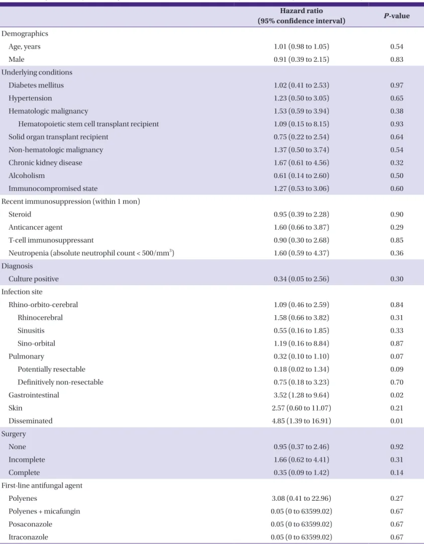
Table 2. Cox regression analysis with single clinical variable associated with 180-day all-cause mortality from invasive mucormycosis Hazard ratio
(95% confidence interval) P-value Demographics
Age, years 1.01 (0.98 to 1.05) 0.54
Male 0.91 (0.39 to 2.15) 0.83
Underlying conditions
Diabetes mellitus 1.02 (0.41 to 2.53) 0.97
Hypertension 1.23 (0.50 to 3.05) 0.65
Hematologic malignancy 1.53 (0.59 to 3.94) 0.38
Hematopoietic stem cell transplant recipient 1.09 (0.15 to 8.15) 0.93
Solid organ transplant recipient 0.75 (0.22 to 2.54) 0.64
Non-hematologic malignancy 1.37 (0.50 to 3.74) 0.54
Chronic kidney disease 1.67 (0.61 to 4.56) 0.32
Alcoholism 0.61 (0.14 to 2.60) 0.50
Immunocompromised state 1.27 (0.53 to 3.06) 0.60
Recent immunosuppression (within 1 mon)
Steroid 0.95 (0.39 to 2.28) 0.90
Anticancer agent 1.60 (0.66 to 3.87) 0.29
T-cell immunosuppressant 0.90 (0.30 to 2.68) 0.85
Neutropenia (absolute neutrophil count < 500/mm3) 1.60 (0.59 to 4.37) 0.36
Diagnosis
Culture positive 0.34 (0.05 to 2.56) 0.30
Infection site
Rhino-orbito-cerebral 1.09 (0.46 to 2.59) 0.84
Rhinocerebral 1.58 (0.66 to 3.82) 0.31
Sinusitis 0.55 (0.16 to 1.85) 0.33
Sino-orbital 1.19 (0.16 to 8.84) 0.87
Pulmonary 0.32 (0.10 to 1.10) 0.07
Potentially resectable 0.18 (0.02 to 1.34) 0.09
Definitively non-resectable 0.75 (0.18 to 3.23) 0.70
Gastrointestinal 3.52 (1.28 to 9.64) 0.02
Skin 2.57 (0.60 to 11.07) 0.21
Disseminated 4.85 (1.39 to 16.91) 0.01
Surgery
None 0.95 (0.37 to 2.46) 0.92
Incomplete 1.66 (0.62 to 4.41) 0.31
Complete 0.35 (0.09 to 1.42) 0.14
First-line antifungal agent
Polyenes 3.08 (0.41 to 22.96) 0.27
Polyenes + micafungin 0.05 (0 to 63599.02) 0.67
Posaconazole 0.05 (0 to 63599.02) 0.67
Itraconazole 0.05 (0 to 63599.02) 0.67
patients and the 21 patients who died within 180 days after di- agnosis of invasive mucormycosis are listed in Table 1. Gastro- intestinal (2% vs. 24%, P = 0.01) and disseminated mucormy- cosis (0% vs. 14%, P = 0.03) were less common in surviving patients than in patients who did not survive. Complete sur- gery was done more commonly in surviving patients than in patients who did not survive (44% vs. 14%, P = 0.04), while in- complete surgical removal (28% vs. 57%, P = 0.046) was more common in patients who did not survive. Cases with pulmo- nary mucormycosis were also more common in survived pa- tients than in patients who died (40% vs. 14%, P = 0.08).
Univariate analysis was performed using a Cox proportional hazards model to determine clinical variables significantly as- sociated with 180-day all-cause mortality (Table 2). Our analy- ses revealed that gastrointestinal mucormycosis (P = 0.02) and disseminated mucormycosis (P = 0.01) were significantly as- sociated with mortality. In subsequent multivariate analysis (Table 3), disseminated infection was independently associat- ed with an increased risk of 180-day mortality (hazard ratio [HR]: 169.74, 95% confidence interval [CI]: 6.41 to 4492.64;
P = 0.002). Pulmonary infection (HR: 0.08, 95% CI: 0.01 to 0.66;
P = 0.02) and complete surgical removal of lesions (HR: 0.12, 95% CI: 0.02 to 0.64; P = 0.01) were associated with improved survival at 180 days post-diagnosis.
Discussion
This study suggests that the greatest risk for mortality from invasive mucormycosis was associated with disseminated in- fection, and complete surgical removal of the infection site or pulmonary infection was associated with decreased mortality.
Mucormycosis is rare but often highly fatal. Our results show the mucormycosis mortality rate to be 33%, which is lower
than the previously reported rates in mucormycosis (44–80%) [4, 7, 12-14]. This may be explained the differences in patients’
characteristics, resectable lesions of infection site, and thera- peutic strategies. In the present study, the proportion of pa- tients with diabetes mellitus as an underlying disease was much higher than in other recent reports (67% vs. 17–23%) [12, 13, 15], whereas the frequency of patients with underlying hematologic malignancy was lower (22% vs. 44–50%). Since rapid correction of underlying predisposing factors is critical to outcomes, a relatively higher proportion of patients with more easily controlled diabetes mellitus than hematologic malignancy, could have a favorable effect on outcomes in our cases. Additionally, compared to the previous epidemiologic studies (44–61%), a large proportion of patients in this study (72%) underwent surgery as part of therapy [4, 12, 13], which may have contributed to relatively high survival rates in our patients. Many patients had more resectable lesions and tend- ed to undergo aggressive surgical debridement despite deep or multiple lesions. Among 36 patients with rhino-orbito-ce- rebral mucormycosis, 14 patients had isolated sinusitis with more resectable lesions; 89% of the patients (32/36) received surgical management. Twelve patients out of 20 with pulmo- nary mucormycosis had potentially resectable lesions and 2 patients out of 8 patients with definitively non-resectable le- sions received surgical management.
Several previous studies have investigated risk factors for mucormycosis [4, 7, 12, 13]. In agreement with our results, Roden et al. [4] reported disseminated infection to be an inde- pendent risk factor for increased mortality (odds ratio [OR]:
11.2, 95% CI: 5.79 to 21.73). They found, however, that pulmo- nary infection presented a relatively lower 180-day mortality rate (15%, 3/20) and was associated with greater survival (HR:
0.08, 95% CI: 0.01 to 0.66). Spellberg et al. [7] reported that the 90-day mortality rate for patients with pulmonary infection Table 3. Cox regression with analysis multiple clinical variables associated with 180-day all-cause mortality from invasive mucormycosis
Adjusted hazard ratio 95% confidence interval P-value Pulmonary infection
No 1
Yes 0.08 0.01 to 0.66 0.02
Disseminated infection
No 1
Yes 169.74 6.41 to 492.64 0.002
Complete surgery
No 1
Yes 0.12 0.02 to 0.64 0.01
was 75% (6/8). Pulmonary involvement was associated with lower Kaplan-Meier survival compared to non-pulmonary in- fection (pulmonary [n = 8] vs. non-pulmonary [n =12]). The re- ported survival differences could be explained by more re- sectable lesions in our study population, which may have con- tributed to the favorable outcomes of pulmonary involvement in our study (180-day mortality rate of potentially resectable, 8% [1/12] vs. definitively non-resectable, 25% [2/8]; P = 0.54).
Except for the frequency of surgical intervention, no underly- ing medical conditions were significantly different between potentially resectable lesions and definitively non-resectable lesions. Considering the relatively small number of patients with pulmonary mucormycosis in our study, further investiga- tion with larger study populations is required to delineate the relationship between pulmonary involvement and mortality in mucormycosis.
Effective mucormycosis treatment is challenging, involving extensive debridement, high-dose amphotericin B, and cor- rection of any underlying diseases [4, 13, 16]. Our study indi- cates that complete removal of infected lesions significantly improves outcomes (HR: 0.18, 95% CI: 0.03 to 1.05). Roden et al. [4] also reported surgery to be associated with a decreased risk of mortality (OR: 0.24, 95% CI: 0.15 to 0.37). Similarly, the mortality rate in pulmonary mucormycosis patients treated with both surgery and antifungal agents was 11%, significantly lower than the 68% mortality rate for patients treated with only antifungal agents (P = 0.0004) [14]. These results suggest that extensive debridement of infected tissue may be neces- sary to minimize mortality. In contrast to previous reports, we did not find amphotericin B to be associated with survival (HR: 3.08, 95% CI: 0.41 to 22.96). Further, because amphoteri- cin B was used in the majority of patients that received an an- tifungal agent (89%), we could not evaluate the comparative effectiveness of various antifungal agents.
Our study has several limitations. First, the study was per- formed at a single center. Second, our study was based on ret- rospective observation, which limits the amount and type of information that could be gathered. Third, the number of cas- es of mucormycosis identified by culture was small (7/64;
11%), so associations between mucormycosis genus/species and mortality could not be evaluated.
Despite these limitations, our results suggest that patients with mucormycosis had reduced mortality risk if they had a pulmonary infection, if their infection was not disseminated, and if extensive and complete surgical debridement of the in- fected tissue was performed. With the increasing incidence of mucormycosis [17], further investigation based on data gath-
ered at multiple centers will be necessary to characterize the prevalence and risk factors for mortality in mucormycosis.
References
1. Kontoyiannis DP, Wessel VC, Bodey GP, Rolston KV. Zygo- mycosis in the 1990s in a tertiary-care cancer center. Clin Infect Dis 2000;30:851-6.
2. Marr KA, Carter RA, Crippa F, Wald A, Corey L. Epidemi- ology and outcome of mould infections in hematopoietic stem cell transplant recipients. Clin Infect Dis 2002;34:
909-17.
3. Ribes JA, Vanover-Sams CL, Baker DJ. Zygomycetes in hu- man disease. Clin Microbiol Rev 2000;13:236-301.
4. Roden MM, Zaoutis TE, Buchanan WL, Knudsen TA, Sarkisova TA, Schaufele RL, Sein M, Sein T, Chiou CC, Chu JH, Kontoyiannis DP, Walsh TJ. Epidemiology and out- come of zygomycosis: a review of 929 reported cases. Clin Infect Dis 2005;41:634-53.
5. Spellberg B, Edwards J Jr, Ibrahim A. Novel perspectives on mucormycosis: pathophysiology, presentation, and management. Clin Microbiol Rev 2005;18:556-69.
6. Boelaert JR, de Locht M, Van Cutsem J, Kerrels V, Canti- nieaux B, Verdonck A, Van Landuyt HW, Schneider YJ.
Mucormycosis during deferoxamine therapy is a sidero- phore-mediated infection. In vitro and in vivo animal studies. J Clin Invest 1993;91:1979-86.
7. Spellberg B, Kontoyiannis DP, Fredricks D, Morris MI, Per- fect JR, Chin-Hong PV, Ibrahim AS, Brass EP. Risk factors for mortality in patients with mucormycosis. Med Mycol 2012;50:611-8.
8. Skiada A, Lanternier F, Groll AH, Pagano L, Zimmerli S, Herbrecht R, Lortholary O, Petrikkos GL; European Con- ference on Infections in Leukemia. Diagnosis and treat- ment of mucormycosis in patients with haematological malignancies: guidelines from the 3rd European Confer- ence on Infections in Leukemia (ECIL 3). Haematologica 2013;98:492-504.
9. De Pauw B, Walsh TJ, Donnelly JP, Stevens DA, Edwards JE, Calandra T, Pappas PG, Maertens J, Lortholary O, Kauffman CA, Denning DW, Patterson TF, Maschmeyer G, Bille J, Dismukes WE, Herbrecht R, Hope WW, Kibbler CC, Kullberg BJ, Marr KA, Muñoz P, Odds FC, Perfect JR, Re- strepo A, Ruhnke M, Segal BH, Sobel JD, Sorrell TC, Viscoli C, Wingard JR, Zaoutis T, Bennett JE; European Organiza- tion for Research and Treatment of Cancer/Invasive Fun-
gal Infections Cooperative Group; National Institute of Al- lergy and Infectious Diseases Mycoses Study Group (EORTC/MSG) Consensus Group. Revised definitions of invasive fungal disease from the European Organization for Research and Treatment of Cancer/Invasive Fungal Infections Cooperative Group and the National Institute of Allergy and Infectious Diseases Mycoses Study Group (EORTC/MSG) Consensus Group. Clin Infect Dis 2008;46:
1813-21.
10. Frater JL, Hall GS, Procop GW. Histologic features of zygo- mycosis: emphasis on perineural invasion and fungal morphology. Arch Pathol Lab Med 2001;125:375-8.
11. Parfrey NA. Improved diagnosis and prognosis of mucor- mycosis. A clinicopathologic study of 33 cases. Medicine (Baltimore) 1986;65:113-23.
12. Lanternier F, Dannaoui E, Morizot G, Elie C, Garcia-Her- moso D, Huerre M, Bitar D, Dromer F, Lortholary O;
French Mycosis Study Group. A global analysis of mucor- mycosis in France: the RetroZygo Study (2005-2007). Clin Infect Dis 2012;54 (Suppl 1):S35-43.
13. Skiada A, Pagano L, Groll A, Zimmerli S, Dupont B, Lagrou K, Lass-Florl C, Bouza E, Klimko N, Gaustad P, Richardson M, Hamal P, Akova M, Meis JF, Rodriguez-Tudela JL, Roi- lides E, Mitrousia-Ziouva A, Petrikkos G; European Con- federation of Medical Mycology Working Group on Zygo- mycosis. Zygomycosis in Europe: analysis of 230 cases
accrued by the registry of the European Confederation of Medical Mycology (ECMM) Working Group on Zygomy- cosis between 2005 and 2007. Clin Microbiol Infect 2011;17:1859-67.
14. Cinque P, Bossolasco S, Vago L, Fornara C, Lipari S, Racca S, Lazzarin A, Linde A. Varicella-zoster virus (VZV) DNA in cerebrospinal fluid of patients infected with human im- munodeficiency virus: VZV disease of the central nervous system or subclinical reactivation of VZV infection? Clin Infect Dis 1997;25:634-9.
15. Rüping MJ, Heinz WJ, Kindo AJ, Rickerts V, Lass-Flörl C, Beisel C, Herbrecht R, Roth Y, Silling G, Ullmann AJ, Borchert K, Egerer G, Maertens J, Maschmeyer G, Simon A, Wattad M, Fischer G, Vehreschild JJ, Cornely OA. Forty- one recent cases of invasive zygomycosis from a global clinical registry. J Antimicrob Chemother 2010;65:296- 302.
16. Spellberg B, Walsh TJ, Kontoyiannis DP, Edwards J Jr, Ibra- him AS. Recent advances in the management of mucor- mycosis: from bench to bedside. Clin Infect Dis 2009;48:1743-51.
17. Bitar D, Van Cauteren D, Lanternier F, Dannaoui E, Che D, Dromer F, Desenclos JC, Lortholary O. Increasing inci- dence of zygomycosis (mucormycosis), France, 1997- 2006. Emerg Infect Dis 2009;15:1395-401.