26
Immune Network
생성과 ERK 활성화
1연세대학교 의과대학 미생물학교실, 2건양대학교 의과대학 미생물학교실
안혜정1․조상래1․백태현2․이정림2․최인홍1
Production of TNF-α and IL-6 in Macrophages by Mycobacterial Protein Antigens
Hae-Jeong Ahn1, Sang-Nae Cho1, Tae-Hyun Paik2, Junglim Lee2 and In-Hong Choi1
1Department of Microbiology, Yonsei University College of Medicine, Seoul, 2Department of Microbiology, College of Medicine, Konyang University, Daejeon, Korea
ABSTRACT
Background: Mycobacterial antigens released as PIM, LM, LAM, lipoproteins and other cellular factors may contribute to macrophage and dendritic cell activation through pattern recognition receptors such as TLRs. In this study, we assessed cytokine pro- duction and ERK activation with stimulation of several major mycobacterial antigens.
Methods: Purified mycobacterial antigens (10, 22, 30, 38kDa) and recombinant antigens (6, 16, 19, 38kDa, Ag85A antigen) were studied. The production of cytokines (TNF-α, IL-12, IL-6) was measured by ELISA. The ERK activation was detected by western blotting. The expression of TLR2 or TLR4 was measured by flow cytometry. Results:
Among purified antigens only 30kDa antigen induced production of IL-6 or TNF-α in THP-1 macrophage cells. When THP-1 macrophage cells were treated with 30kDa antigen, phosphorylation of ERK was detected. ERK activation also occurred in TLR2 transfectant HEK293 cells with 30kDa antigen stimulation. Conclusion: 30kDa antigen is one of the major mycobacterial antigens inducing cytokine production and MAP kinases phosphorylation in macrophages. (Immune Network 2007;7(1):26-30)
Key Words: Macrophage, M. tuberculosis, 30kDa antigen, TNF-α, IL-6, ERK
책임저자:최인홍, 연세대학교 의과대학 미생물학교실
}120-752, 서울시 서대문구 신촌동 134번지 Tel: 02-2228-1821, Fax: 02-392-7088
E-mail: inhong@yumc.yonsei.ac.kr
본 연구는 한국학술진흥재단 기초과학연구 (KRF-2004-E00115)에 의하여 이루어진 것임.
서 론
결핵균으로 인한 감염을 숙주가 방어하기 위해 선천 면역반응과 적응면역반응이 필요하게 되는데(1), 대식세 포는 체내에 들어온 병원균에 대해 가장 먼저 면역반응 을 유발하는 면역세포로서 선천면역을 담당한다. 활성 화된 대식세포는 다양한 세포독성 단백을 분비하고 병 원균, 바이러스 감염 세포, 암 세포, 세포 내 세균을 제거 하며, MHC class II 분자를 다량 발현하여 효과적인 항원 전달세포 역할을 한다(2-4).
뿐만 아니라 폐결핵 환자에서 분리한 균에 감염된 폐 포 대식세포는 몇몇의 cytokine과 chemokine을 분비하여 빠른 시간 내에 단핵구를 병변부위로 모여들게 한다 (5,6). 이러한 단핵구에서 분화된 수상돌기세포는 결핵균 세포벽 성분이나 결핵균 항원 자극에 의해 성숙과정을 거쳐 동시자극분자의 발현도 증가되고 항원전달 능력이 향상되어 숫(naïve) T세포를 활성화시킴으로써 적응면역 반응을 유도한다(7). 생체 내에서 수상돌기세포가 결핵 균에 대한 선천면역과 적응면역을 유도하는 숙주의 주 요세포이고(8), 수상돌기세포의 성숙과정이 선천면역반 응에 관여하는 세포에 발현되는 toll-like receptor (TLR) 을 매개하여 일어남이 보고되었다(9,10).
대식세포를 활성화시키는 항산균 항원은 여러 종류가 있다. 항산균 세포벽 glycolipid인 lipoarabinomannan (LAM) 과 mannosylated phosphatidylinositol (PIM)은 TLR2를 통 하여 대식세포를 활성화시키며(3) 마우스 대식세포주인
RAW 264.7 세포에서 결핵균 항원인 LAM, STF, PIM 등 이 NF-κB, AP1 및 MAP kinase를 급속하게 활성화시킨 다는 보고가 있다(4). 또한 항산균 19kDa 지질단백은 사 람 단핵세포에서 IL-12p40를 더 많이 유도하는 반면 20kDa 및 38kDa 단백항원은 TNF-α와 IL-6을 생성을 주 로 유도하였다(11).
본 연구에서는 결핵균 단백항원 중에서 대식세포를 활성화시키는 항원이 어떠한 것인지 확인하고 이들 항 원의 대식세포 활성화 기전을 파악하고자 하였다.
재료 및 방법
세포주 배양. 사람 대식세포주인 THP-1은 American Type Culture Collection에서 분양받았다. 세포는 10% 우태아 혈청(fetal bovine serum: FBS, Jeil Biotechservices Inc., Daegu, Korea)이 포함된 RPMI 1640배지(0.1 M sodium pyruvate, 2 mM penicillin, 50μg/ml streptomycin; Life tech- nologies 포함) 및 5% CO2, 37oC incubator에서 배양하였 다. 대식세포로 분화시키기 위해 100 ng/ml의 PMA (Sigma-Aldrich, St. Louis, MO)를 24시간 처리하였다.
TLR2 transfectant cell은 human embryonic kidney (HEK) 293 cell에 사람 TLR2 (HEK/TLR2/CD14) 유전자를 trans- fection한 세포(Invivogen, San Diego, CA)를 구입하여 사 용하였다. HEK293-TLR2 transfectant cell 배지로는 Dul- becco’s modified Eagle’s medium에 blasticidin (10μg/ ml), normocin (100μg/ml), 10% 우태아혈청을 첨가하여 사용 하였다.
결핵균 단백항원. 정제단백인 10kDa, 22kDa, 30kDa, 38kDa 항원과 합성된 6kDa, 16kDa, 19kDa, 38kDa, Ag85A 항원을 사용하였다. Salmonella minnesota에서 분리한 50 ng/ml의 LPS (lipopolysaccharide; 지다당류, Sigma, St.
Louis, MO, USA)와 Bacillus subtillis에서 분리한 10μg/
ml의 LTA (lipoteichoic acid, Sigma, St. Louis, MO, USA) 는 각각 TLR4와 TLR2를 자극하기 위한 positive control 로 사용하였다.
유세포 분석. 세포를 인산완충액으로 2회 세척한 후 세 포표면에 FITC-TLR4 (clone HTA125; Santa Cruz) 및 PE-TLR2 (clone TL2.1; Santa Cruz) 단클론항체(20μl/1 × 106 cell)로 얼음에서 30분간 반응시켜서 직접형광염색을 하였다. 결과는 FacsCalibur (BD Bioscience)를 사용하여 분 석하였으며 총 20,000개의 세포를 측정한 후 WinMDI 2.8 (Joseph Trotter, Scripps Research Institute, San Diego, CA, USA)을 이용하여 결과를 해석하였다.
ELISA. 대식세포 배양액에 포함된 cytokine을 측정하기 위해서 ELISA를 수행하였다. 항-cytokine 포획항체(BD Sciences)를 coating buffer (0.1M carbonate, pH 9.5)에 250 배 희석하여 microtiter plate (Costar, Acton, MA, USA)에 분주하고 4oC에서 하룻밤 동안 부착시킨 후, 0.05%
tween 20이 함유된 인산완충액으로 3회 세척하고 10%
소혈청알부민이 포함된 인산완충액으로 차단시켰다.
10% 소혈청알부민-인산완충액으로 2배로 희석시킨 세 포배양액은 포획항체가 부착된 평판의 각 well에 분주한 후 37oC에서 2시간 반응시킨 후 0.05% Tween 20이 함유 된 인산완충액으로 5회 세척하였다. 검출항체에 biotin이 부착된 항-사람 IL-6 항체, 항-TNF-α 항체 또는 항-IL-12 항체와 horseradish peroxidase (HRP)-conjugated streptavidin (1:1의 비율)을 10% 소혈청알부민-인산완충액으로 250 배 희석하여 각 well에 첨가한 후 37oC에서 1시간 반응시 켰다. 그 후 0.05% Tween 20이 함유된 인산완충액으로 7회 세척한 후에 tetramethyl benzidine와 hydrogen pe- roxide의 혼합액을 각 well에 첨가한 후에 어두운 상온에 서 20분간 반응시키고 2N H2SO4용액으로 발색반응을 정 지시켰다. Optical density는 microplate reader (Molecular Devices, Sunnyvale, CA, USA)를 이용하여 450 nm 파장에 서 측정하였다. 분비된 cytokine의 양은 사람의 재조합형 IL-6, TNF-α의 표준곡선으로부터 유추하여 계산하였다.
Western blotting. 사람 대식세포주인 THP-1과 HEK293- TLR2 transfactant cell에 15, 30, 40분간 결핵균 항원 자극 을 준 후 ERK (extracellular signal-regulated kinase) 인산 화를 측정하였다. 인산완충액으로 세척한 세포를 NP-40 lysis buffer (protease inhibitor가 포함된 25 mM Tris-HCl, 137 mM NaCl, 3 mM KCl, 1%NP-40)로 깨서 13,000 rpm 에서 15분간 원심 분리하여 상청액의 단백을 얻었다. 단 백의 농도는 Bradford protein assay (Bio-Rad, Hercules, CA, USA)로 정량하였고 총단백에서 40~50μg을 취하여 12%
SDS-PAGE를 통하여 전기영동 하였다. 그 후 nitrocel- lulose membrane (Amersham Pharmacia Biotech, Piscataway, NJ, USA)에 옮겨서 5% 탈지분유로 실온에서 2시간 동안 차단시킨 후 0.05% tween-20이 함유된 인산완충액으로 2회 세척하였다. 항-phospho-ERK 다클론항체(Cell Signa- ling Technology, Beverly, MA, USA)를 사용하여 5% 탈지 분유에 1:1,000의 비율로 항체를 붙여서 4oC에서 하룻 밤 동안 반응시켰다. 그 후 0.05% tween-20이 함유된 인 산완충액으로 5분씩 3회 세척한 후에 5% 탈지분유에 1:5,000의 비율로 희석한 이차항체인 HRP-conjugated goat anti- rabbit IgG (Jackson Immuno Research, Baltimore, MD, USA)와 4oC에서 1시간 동안 반응시키고 PBST로 10 분간 3회 세척한 후 west save (Lab Frontier, Seoul, Korea) 를 사용하여 발광반응을 측정하여 x-ray film에 노출시켜 서 현상하였다.
결 과
사람 대식세포주 THP-1에서 TLR 분자의 발현. THP-1 세포는 미성숙 단핵세포에서 유래된 세포로서 대식세포 로 분화시키기 위하여 PMA를 처리한 후 사용하였다. 단
0 2,000 4,000 6,000 8,000 10,000 12,000 14,000 16,000
Control 10kDa
22kDa 30kDa
38kDa 6kDa
16kDa 19kDa
38kDa Ag85A LTA
(pg/ mL)
A. TNF-α
0 1,000 2,000 3,000 4,000 5,000 (pg/ mL)
B. IL-12
Antigen
Purified Recombinant Control
10kDa 22kDa
30kDa 38kDa
6kDa 16kDa
19kDa 38kDa
Ag85A LTA
Antigen
Purified Recombinant 0
2,000 4,000 6,000 8,000 10,000 12,000 14,000 16,000
Control 10kDa
22kDa 30kDa
38kDa 6kDa
16kDa 19kDa
38kDa Ag85A LTA
(pg/ mL)
A. TNF-α
0 1,000 2,000 3,000 4,000 5,000 (pg/ mL)
B. IL-12
Antigen
Purified Recombinant Control
10kDa 22kDa
30kDa 38kDa
6kDa 16kDa
19kDa 38kDa
Ag85A LTA
0 1,000 2,000 3,000 4,000 5,000 (pg/ mL)
B. IL-12
Antigen
Purified Recombinant Control
10kDa 22kDa
30kDa 38kDa
6kDa 16kDa
19kDa 38kDa
Ag85A LTA
Antigen
Purified Recombinant
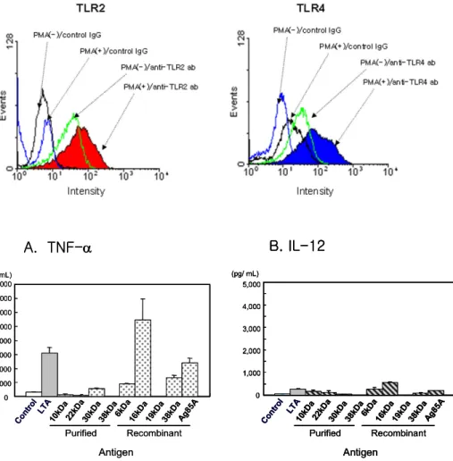
Figure 2. Cytokine production in THP-1 cells by mycobacterial anti- gens. THP-1 cells were treated with 10μg/ml of purified mycobacterial antigens (10, 22, 30, 38kDa) or re- combinant mycobacterial antigens (6, 16, 19, 38, Ag85A) for 48 hrs.
TNF-α and IL-12 in the culture supernatants were measured by ELI- SA. LTA (10μg/ml) was used as positive control. (A) TNF-α, (B) IL-12.
Figure 1. TLR expression on THP-1 cells. THP-1 cells were treated with 100 ng/ml of PMA for 24 hrs. The expression of TLR2 or TLR4 was measured by direct staining by anti-TLR2 antibody or anti-TLR4 antibody. Total 20,000 cells were analyzed. The result is the representative result from three independent experiments. Control IgG, isotype control antibody.
핵세포 상태의 THP-1 세포와 대식세포로 분화시킨 THP-1 세포 표면의 TLR2 및 TLR4 분자 발현을 비교하 였다. 그 결과 PMA로 분화시킨 THP-1 세포에서 TLR2 및 TLR4 분자가 더 많이 발현하였다(Fig. 1).
사람 대식세포주 THP-1에서 결핵균 항원에 의한 cyto- kine 생성. 결핵균 항원으로 자극된 대식세포에서 cytokine 생성을 관찰하기 위하여 THP-1 세포를 결핵균 정제단백 항원(10, 22, 30, 38kDa)과 재조합항원(6, 16, 19, 38kDa 항원, Ag85A)으로 자극하였다. 그 결과 결핵균 정제단백 항원 중에서는 30kDa항원만이 TNF-α 생성을 유도하였 다(Fig. 2A). 재조합항원의 경우 19kDa을 제외한 4종류의 항원이 TNF-α 생성을 유도하였다(Fig. 2A). IL-12는 정 제단백항원과 재조합항원 모두에서 유도되지 않았다 (Fig. 2B).
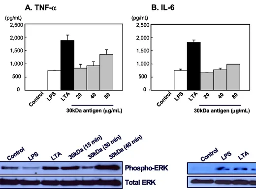
사람 대식세포주 THP-1에서 결핵균 30kDa항원 자극 에 따른 cytokine 생성. THP-1 세포의 cytokine 생성 실험 에서 사용한 4종류의 결핵균 정제단백항원 중에서 유일 하게 THP-1 세포를 자극한 30kDa항원을 대상으로 항원 농도에 따른 TNF-α와 IL-6 생성을 관찰하였다. ELISA 를 통하여 THP-1 세포 배양 상청액의 cytokine 생성을 측 정한 결과, 30kDa항원 양이 증가함에 따라 TNF-α와
IL-6의 생성이 증가하였다(Fig. 3).
결핵균 30kDa항원이 ERK 활성화에 미치는 영향. 30kDa 항원 자극에 의하여 THP-1세포에서 ERK의 인산화가 일 어나는지를 확인하기 위해 항-phosphor-ERK항체를 이용 하여 western blotting을 실시하였다. Positive control인 LPS, LTA 및 30kDa항원을 처리한 후 15, 30, 40분 후 총 단백을 얻었다. ERK 인산화를 측정한 결과 30kDa항원 을 자극하였을 때 40분 후 THP-1 세포의 ERK 인산화가 증가되었다(Fig. 4). HEK293-TLR2 transfactant 세포주에 서도 LPS, LTA 및 30kDa항원을 처리한 후 ERK의 인산 화를 측정하였는데 30kDa항원을 처리한 15분 후에 ERK 의 인산화가 관찰되었다가 30분에 감소되었다(Fig. 5).
고 찰
항산균 항원은 면역반응을 활성화시키기도 하고 억제 하기도 하는 다양한 항원의 집합체로 구성되어 있다. 항 산균 19kDa 지질단백은 TLR2 ligand로 알려져 있으며 숙 주 세포의 apoptosis를 유도하고 nitric oxide를 생성하여 면역반응을 증가시키는 반면 MHC class II 분자의 down- regulation시키기 때문에 항원전달을 감소시킨다(12).
19kDa 지질단백은 사람 단핵세포주에서 IL-12p40를 더
0 500 1,000 1,500 2,000 2,500
A. TNF-α
Control LPS
LTA 20 40 80
(pg/mL)
30kDa antigen (µg/mL) 0 500 1,000 1,500 2,000 2,500 (pg/mL)
B. IL-6
Control LPS
LTA 20 40 80
30kDa antigen (µg/mL) 0
500 1,000 1,500 2,000 2,500
A. TNF-α
Control LPS
LTA 20 40 80
(pg/mL)
30kDa antigen (µg/mL) 0 500 1,000 1,500 2,000 2,500 (pg/mL)
B. IL-6
Control LPS
LTA 20 40 80
30kDa antigen (µg/mL)
Figure 3. Dose-dependent cytokine production in THP-1 cells by 30kDa antigen. THP-1 cells were treated with 20, 40, 80μg/ml of 30kDa antigen for 48 hrs. TNF-α and IL-6 in the culture supernatants were measured by ELISA. LPS (50 ng/
ml) and LTA (10μg/ml) was used as positive control. The result is the representative result from three in- dependent experiments. (A) TNF- α, (B) IL-6.
Phospho-ERK Total ERK
LPS Control
LTA 30kDa (15 min) 30kDa (30 min)
30kDa (40 min)
Phospho-ERK Total ERK
LPS Control
LTA 30kDa (15 min) 30kDa (30 min)
30kDa (40 min)
Phospho-ERK Total ERK
LPS Control
LTA 30kDa (15 min) 30kDa (30 min)
30kDa (40 min)
Figure 4. ERK activation in THP-1 cells by 30kDa antigen.
THP-1 cells were treated with 10μg/ml of 30kDa antigen for 15, 30, 40 min. Total protein was prepared and western blot was done with anti-phospho-ERK antibody. LPS (50 ng/ml) and LTA (10μg/ml) was used as positive control.
Phospho-ERK Total ERK Control
LPS LTA
30kDa (15 min) 30kDa (30 min)
30kDa (40 min)
Phospho-ERK Total ERK Control
LPS LTA
30kDa (15 min) 30kDa (30 min)
30kDa (40 min)
Phospho-ERK Total ERK Control
LPS LTA
30kDa (15 min) 30kDa (30 min)
30kDa (40 min)
Figure 5. ERK activation in TLR2 transfectant cells by 30kDa antigen. HEK293 cells expressing TLR2 were treated with 10μg/
ml of 30kDa antigen for 15, 30, 40 min. Total protein was prepared and western blot was done with anti-phospho-ERK antibody. LPS (50 ng/ml) and LTA (10μg/ml) was used as positive control.
많이 유도하는 반면 20kDa 및 38kDa 단백항원은 TNF-α 와 IL-6을 생성을 유도한다(11). 결핵에 대한 면역반응을 이해하고 나아가서 백신 개발 등을 전제로 하여 여러 종 류의 항산균 항원에 의한 면역조절 연구가 활발하다.
본 연구에서는 사람 대식세포주인 THP-1 세포를 대상 으로 결핵균 단백항원들에 대한 cytokine 생성을 관찰한 결과 정제단백항원 중에서는 30kDa 항원이 TNF-α 생성 을 유도하였으며 재조합항원 중에서는 6, 16, 38kDa 항 원과 Ag85A가 TNF-α 생성을 유도하였다. 재조합항원 의 경우 대장균에서 생성하기 때문에 단백 분리 과정에 서 포함될 수 있는 대장균 유래 LPS의 가능성을 배제하 기가 용이하지 않았다. 그리하여 이후의 실험에서는 30kDa 항원을 중심으로 전개하였다. 30kDa 결핵균 항원 은 IL-12 또는 IL-18 등의 cytokine을 유도하는데 결핵에 감염된 환자에서는 30kDa 항원에 대한 반응이 저하되어 있다가 결핵 치료 후에는 정상으로 회복된다(13,14). 사 람 단핵구를 30kDa 항원으로 자극하면 TNF-α를 생성하 는데 TNF-α는 항산균 육아종(granuloma)의 형성에 중요 한 역할을 하고 대식세포 활성화인자로 작용하여 결핵 균을 포함한 세균 성장을 저해하기 때문에 30kDa 항원 (Ag85B)이 면역력을 보완하는 기능이 있는 것으로 제시 되었다(15).
본 연구에서 대식세포주인 THP-1 세포를 결핵균 30kDa
항원으로 자극하였을 때 MAP kinase member인 ERK가 TLR2의 배위자인 LTA나 TLR4의 배위자인 LPS에 대한 반응만큼 강하게 활성화되는 것으로 보아 30kDa항원도 MAP kinase 경로를 통하여 대식세포를 강하게 활성화시 킴으로써 숙주의 면역반응 유도에 관여하는 것으로 추 정된다. 결핵균 30kDa 항원은 Ag85 complex로도 알려져 있는데 결핵균 배양액에서 검출되는 단백으로 Ag85A, Ag85B, Ag85C의 세 가지 종류로 구분되고, 결핵균뿐 아 니라 모든 항산균에서 관찰된다(16). Ag85A, Ag85B, Ag- 85C간의 일부 상이한 유전자 sequence 차이는 Ag85 gene family가 조상 유전자의 duplication에 의해서 mycobacterial species로 나누어지기 전에 생성된 것으로 알려져 있다 (17). 또한 Ag85A는 M. tuberculosis나 Mycobacterium leprae (18), Mycobacterium bovis BCG-vaccinated mice (19)에 감 염된 후 T 세포 증식을 강력하게 유도하며 IFN-γ생성을 유도시키는 항원으로 보고되어 있고 그로 인하여 Ag85A 와 Ag85B가 현재 결핵균의 subunit 백신의 가장 강력한 후보분자로 제시되어 있다. 본 연구에서 TLR2를 발현하 는 HEK293 세포에서 30kDa 항원이 ERK 활성화를 유도 하였다. PMA로 자극한 THP-1 세포에서는 TLR2 발현이 높게 검출되기 때문에 THP-1 세포의 결과와 TLR2를 발 현하는 HEK293 세포의 결과를 비교하면 30kDa 항원이
TLR2를 통하여 ERK 활성화 등의 신호전달을 보낼 가능 성이 추정되지만 이는 추후 연구가 더 필요하다고 생각 한다.
결론적으로 본 연구에서는 결핵균 정제단백항원인 30kDa 항원이 대식세포에서 ERK의 활성화를 유도하고 TNF-α 및 IL-6 생성을 유도하는 것을 확인함으로써 숙 주 면역반응 유발과 결핵 백신 개발에서 중요한 역할을 할 것으로 제시한다.
참 고 문 헌
1. Flynn JL: Immunology of tuberculosis and implications in vaccine development. Tuberculosis (Edinburgh) 84;93-101, 2004
2. Fenton MJ, Vermeulen MW: Immunopathology of tuber- culosis: Roles of macrophages and monocytes. Infect Immun 64;683-690, 1996
3. Jones BW, Heldwein KA, Means TK, Saukkonen JJ, Fenton MJ: Differential roles of Toll-like receptors in the elicitation of proinflammatory responses by macrophages. Ann Rheum Dis 60;iii6-iii12, 2001
4. Jones BW, Means TK, Heldwein KA, Keen MA, Hill PJ, Belisle JT, Fenton MJ: Different Toll-like receptor agonists induce distinct macrophage responses. J Leukoc Biol 69;
1036-1044, 2001
5. Schluger NW, Rom WN: The host immune response to tuberculosis. Am J Respir Crit Care Med 157;679-691, 1998 6. Sadek MI, Sada E, Toossi Z, Schwander SK, Rich EA:
Chemokines induced by infection of mononuclear phagocytes with mycobacteria and present in lung alveoli during active pulmonary tuberculosis. Am J Respir Cell Mol Biol 19;
513-521,1998
7. Banchereau J, Steinman RM: Dendritic cells and the control of immunity. Nature 392;245-252, 1998
8. Jiao X, Lo-Man R, Guermonprez P, Fiette L, Burgaud S, Gicquel B, Winter N, Leclerc C: Dendritic cells are host cells for mycobacteria in vivo that trigger innate and acquired immunity. J Immunol 168;1294-1301, 2002
9. Tsuji S, Matsumoto M, Takeuchi O, Akira S, Azuma I, Hayashi A, Toyoshima K: Maturation of human dendritic cells by cell wall skeleton of Mycobacterium bovis bacillus Calmette-Guerin: involvement of toll-like receptors. Infect Immun 68;6883-6890, 2000
10. Hertz CJ, Kiertscher SM, Godowski PJ, Bouis DA, Norgard MV, Roth MD, Modlin RL: Microbial lipopeptides stimulate dendritic cell maturation via Toll-like receptor 2. J Immunol 166;2444-2450, 2001
11. Jung SB, Yang CS, Lee JS, Shin AR, Jung SS, Son JW, Hading CV, Kim HJ, Park JK, PAIK TH, Song CH, Cho EK: The mycobacterial 38kDa glycolipoprotein antigen activates the mitogen-activated protein kinase pathway and release of proinflammatory cytokine through toll-like receptors 2 and 4 in human monocytes. Infect Immun 74;2686-2696, 2006
12. Pennini ME, Pai RK, Schultz DC, Boom WH, Harding CV:
Mycobacterium tuberculosis 19-kDa lipoprotein inhibits IFN-gamma- induced chromatin remodeling of MHC2TA by TLR2 and MAPK signaling. J Immunol 176;4323-30, 2006
13. Averill L, Toossi Z, Aung H, Boom WH, Ellner JJ:
Regulation of production of tumor necrosis factor alpha in monocytes stimulated by the 30kDa antigen of mycobacterium tuberculosis. Infect Immun 63;3206-3208, 1995
14. Uma Devi KR, Ramalingam B, Raja A: Antibody response to mycobacterium tuberculosis 30 and 16 kDa antigen in pulmonary tuberculosis with human immunodeficiency virus coinfection. Diagn Microbiol Infect Dis 46;205-209, 2003 15. Song CH, Kim HJ, Park JK, Lim JH, Kim UO, Kim JS, Paik
TH, Kim KJ, Suhr JW, Jo EK: Depressed interleukin -12, but not IL-18, production in response to a 30-32kDa mycobacterial antigen in patients with active pulmonary tuberculosis. Infect Immun 68;4474-4484, 2000
16. Wiker, HG, Harboe M: The antigen 85 complex: a major secretion product of mycobacterium tuberculosis. Microbiol Rev 56;648-661, 1992
17. D’Souza S, Rosseels V, Romano M, Tanghe A, Denis O, Jurion F, Castinglione N, Vanonckelen A, Palfliet K, Huygen K: Mapping of murine Th1 helper T-cell epitopes of mycolyl transferases Ag85A, Ag85B, and Ag85C from mycobacterium tuberculosis. Infect Immun 71;484-493, 2003
18. Launois P, Deleys R, Niang MN, Drowart A, Andrien M, Dierekx P, Cartel JL Sarthou JL Van Vooren JP Huygen:
T-cell epitope mapping of the major secreted mycobacterial antigen Ag85A in tuberculosis and leprosy. Infect Immun 62;3679-3687, 1994
19. Huygen K, Abramowiez D, Vandenbussche P, Jacobs F, Bruyn JD, Kentos A, Drowart A, Van Vooren JP, Goldman M: Spleen cell cytokine secretion in Mycobacterium bovis BCG-infected mice. Infect Immun 60;2880-2886, 1992