ISSN: 2233-601X (Print) ISSN: 2093-6516 (Online)
Department of Thoracic and Cardiovascular Surgery, Korea University Anam Hospital, Korea University School of Medicine Received: September 24, 2014, Revised: November 8, 2014, Accepted: November 10, 2014, Published online: October 5, 2015
Corresponding author: Jae-Seung Jung, Department of Thoracic and Cardiovascular Surgery, Korea University Anam Hospital, Korea University School of Medicine, 73 Inchon-ro, Seongbuk-gu, Seoul 02841, Korea
(Tel) 82-2-920-6856 (Fax) 82-2-928-8793 (E-mail) [email protected]
C
The Korean Society for Thoracic and Cardiovascular Surgery. 2015. All right reserved.
CC
This is an open access article distributed under the terms of the Creative Commons Attribution Non-Commercial License (http://creative- commons.org/licenses/by-nc/4.0) which permits unrestricted non-commercial use, distribution, and reproduction in any medium, provided the original work is properly cited.
Comparison of Extracorporeal Cardiopulmonary Resuscitation with Conventional Cardiopulmonary Resuscitation:
Is Extracorporeal Cardiopulmonary Resuscitation Beneficial?
Seung-Hun Lee, M.D., Jae-Seung Jung, M.D., Ph.D., Kwang-Hyung Lee, M.D., Hee-Jung Kim, M.D., Ho-Sung Son, M.D., Ph.D., Kyung Sun, M.D., Ph.D.
Background: With improvements in cardiopulmonary resuscitation (CPR) techniques, the quality and the effective- ness of CPR have been established; nevertheless, the survival rate after cardiac arrest still remains poor. Recently, many reports have shown good outcomes in cases where extracorporeal membrane oxygenation (ECMO) was used during prolonged CPR. Accordingly, we attempted to evaluate the impact of extracorporeal cardiopulmonary re- suscitation (ECPR) on the survival of patients who experienced a prolonged cardiac arrest and compared it with that of conventional CPR (CCPR). Methods: Between March 2009 and April 2014, CPR, including both in-hospital and out-of-hospital CPR, was carried out in 955 patients. The ECPR group, counted from the start of the ECPR program in March 2010, included 81 patients in total, and the CCPR group consisted of 874 patients. All data were retrospectively collected from the patients’ medical records. Results: The return of spontaneous circulation (ROSC) rate was 2.24 times better in CPR of in-hospital cardiac arrest (IHCA) patients than in CPR of out-of-hos- pital CA (OHCA) patients (p=0.0012). For every 1-minute increase in the CPR duration, the ROSC rate decreased by 1% (p=0.0228). Further, for every 10-year decrease in the age, the rate of survival discharge increased by 31%. The CPR of IHCA patients showed a 2.49 times higher survival discharge rate than the CPR of OHCA pa- tients (p=0.03). For every 1-minute increase in the CPR duration, the rate of survival discharge was decreased by 4%. ECPR showed superiority in terms of the survival discharge in the univariate analysis, although with no stat- istical significance in the multivariate analysis. Conclusion: The survival discharge rate of the ECPR group was comparable to that of the CCPR group. As the CPR duration increased, the survival discharge and the ROSC rate decreased. Therefore, a continuous effort to reduce the time for the decision of ECMO initiation and ECMO team activation is necessary, particularly during the CPR of relatively young patients and IHCA patients.
Key words: 1. Cardiac arrest 2. Outcomes
3. Extracorporeal membrane oxygenation
INTRODUCTION
Over the past decades, the survival rate after cardiac arrest (CA) has remained poor despite the technical improvements
in and the increased quality and effectiveness of cardiopulmo- nary resuscitation (CPR) [1-3]. Several factors, including the duration of CPR, initial cardiac rhythm, underlying primary disease, and age, may be related to the outcome [1,4,5].
http://dx.doi.org/10.5090/kjtcs.2015.48.5.318
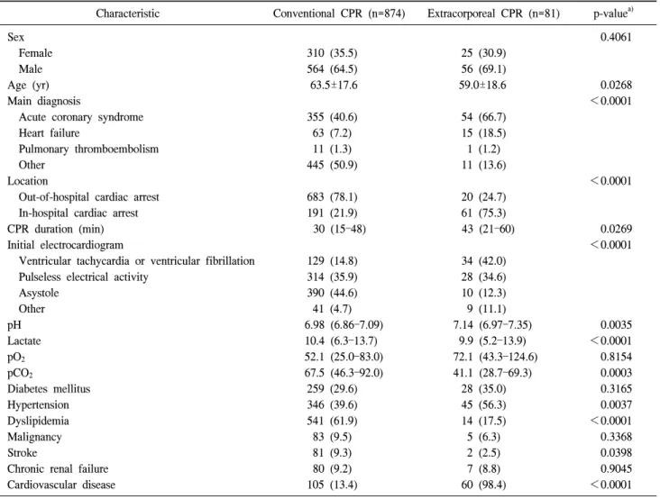
Table 1. Baseline characteristics
Characteristic Conventional CPR (n=874) Extracorporeal CPR (n=81) p-value
a)Sex 0.4061
Female 310 (35.5) 25 (30.9)
Male 564 (64.5) 56 (69.1)
Age (yr) 63.5±17.6 59.0±18.6 0.0268
Main diagnosis <0.0001
Acute coronary syndrome 355 (40.6) 54 (66.7)
Heart failure 63 (7.2) 15 (18.5)
Pulmonary thromboembolism 11 (1.3) 1 (1.2)
Other 445 (50.9) 11 (13.6)
Location <0.0001
Out-of-hospital cardiac arrest 683 (78.1) 20 (24.7)
In-hospital cardiac arrest 191 (21.9) 61 (75.3)
CPR duration (min) 30 (15–48) 43 (21–60) 0.0269
Initial electrocardiogram <0.0001
Ventricular tachycardia or ventricular fibrillation 129 (14.8) 34 (42.0)
Pulseless electrical activity 314 (35.9) 28 (34.6)
Asystole 390 (44.6) 10 (12.3)
Other 41 (4.7) 9 (11.1)
pH 6.98 (6.86–7.09) 7.14 (6.97–7.35) 0.0035
Lactate 10.4 (6.3–13.7) 9.9 (5.2–13.9) <0.0001
pO
252.1 (25.0–83.0) 72.1 (43.3–124.6) 0.8154
pCO
267.5 (46.3–92.0) 41.1 (28.7–69.3) 0.0003
Diabetes mellitus 259 (29.6) 28 (35.0) 0.3165
Hypertension 346 (39.6) 45 (56.3) 0.0037
Dyslipidemia 541 (61.9) 14 (17.5) <0.0001
Malignancy 83 (9.5) 5 (6.3) 0.3368
Stroke 81 (9.3) 2 (2.5) 0.0398
Chronic renal failure 80 (9.2) 7 (8.8) 0.9045
Cardiovascular disease 105 (13.4) 60 (98.4) <0.0001
Values are presented as number of patients (%), mean±standard deviation, or median (interquartile range), unless otherwise indicated.
CPR, cardiopulmonary resuscitation.
a)
Calculated using chi-square test, Student t-test, or Wilcoxon’s rank-sum test, as appropriate.
Because of the low survival rate after prolonged CPR, more aggressive methods have been suggested to increase the suc- cess rate. Hence, several mechanical devices and techniques that may extend the accepted duration of CPR and eventually increase the survival rate have been developed.
Since 2008, when Chen et al. [6] reported that extra- corporeal CPR (ECPR) significantly increases the survival rate in selective patients as compared to conventional CPR (CCPR), the application of ECPR has increased dramatically worldwide. Recently, advances in extracorporeal membrane oxygenation (ECMO) technologies and devices and the im- provements in biocompatible percutaneous cannulas have made
ECMO a more powerful resuscitation tool. Several studies have reported the advantages of ECPR such as hemodynamic stabilization, increased frequency of the return of spontaneous circulation (ROSC), and improved survival with a good neu- rologic outcome when compared to CCPR [7-11]. Further, re- cently, many studies have demonstrated that ECPR leads to more favorable outcomes in in-hospital CA (IHCA) patients than in out-of-hospital CA (OHCA) patients [12,13]. Moreover, primarily, an early decision and insertion of ECMO with ap- propriate indication is essential for improving the prognosis of patients with prolonged CPR [12-14].
In this study, we have attempted to evaluate the impact of
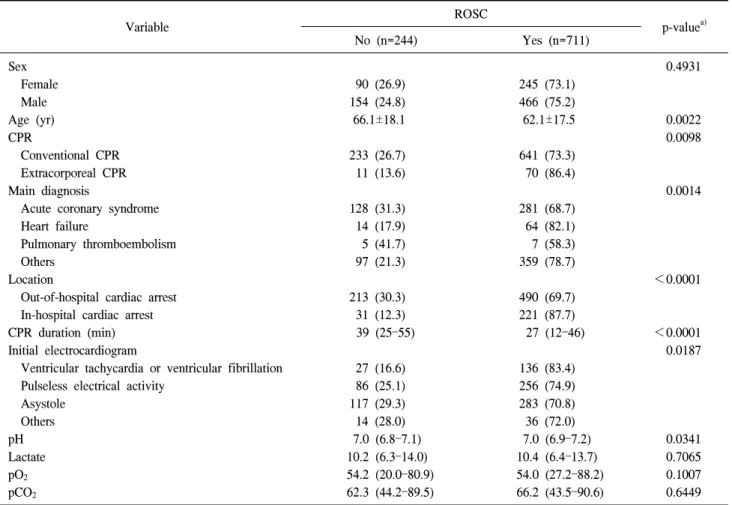
Table 2. ROSC analysis
Variable ROSC
p-value
a)No (n=244) Yes (n=711)
Sex 0.4931
Female 90 (26.9) 245 (73.1)
Male 154 (24.8) 466 (75.2)
Age (yr) 66.1±18.1 62.1±17.5 0.0022
CPR 0.0098
Conventional CPR 233 (26.7) 641 (73.3)
Extracorporeal CPR 11 (13.6) 70 (86.4)
Main diagnosis 0.0014
Acute coronary syndrome 128 (31.3) 281 (68.7)
Heart failure 14 (17.9) 64 (82.1)
Pulmonary thromboembolism 5 (41.7) 7 (58.3)
Others 97 (21.3) 359 (78.7)
Location <0.0001
Out-of-hospital cardiac arrest 213 (30.3) 490 (69.7)
In-hospital cardiac arrest 31 (12.3) 221 (87.7)
CPR duration (min) 39 (25–55) 27 (12–46) <0.0001
Initial electrocardiogram 0.0187
Ventricular tachycardia or ventricular fibrillation 27 (16.6) 136 (83.4)
Pulseless electrical activity 86 (25.1) 256 (74.9)
Asystole 117 (29.3) 283 (70.8)
Others 14 (28.0) 36 (72.0)
pH 7.0 (6.8–7.1) 7.0 (6.9–7.2) 0.0341
Lactate 10.2 (6.3–14.0) 10.4 (6.4–13.7) 0.7065
pO
254.2 (20.0–80.9) 54.0 (27.2–88.2) 0.1007
pCO
262.3 (44.2–89.5) 66.2 (43.5–90.6) 0.6449
Values are presented as number of patients (%), mean±standard deviation, or median (interquartile range), unless otherwise indicated.
ROSC, return of spontaneous circulation; CPR, cardiopulmonary resuscitation.
a)
Calculated using chi-square test, Fisher’s exact test, Student t-test, or Wilcoxon’s rank-sum test, as appropriate.
ECPR on the survival of patients who experienced prolonged CA and compared it with that of CCPR.
METHODS 1) Study population
This study enrolled patients who received CPR at our in- stitution between March 2009 and April 2014. The total num- ber of patients, including both in-hospital CPR patients and out-of-hospital CPR patients, was 955. Since the start of the ECPR program at our hospital in March 2010, there have been a total of 81 patients treated with ECPR; these patients were included in the ECPR group. The other 874 patients, who were treated with CCPR, were included in the CCPR
group. All data were retrospectively collected from the medi- cal records and CPR input forms of the emergency depar- tment.
2) Indications for extracorporeal cardiopulmonary resuscitation
The ECMO team was activated, and the team decided
whether or not to initiate ECPR in all patients who did not
show the ROSC after 10 minutes of advanced cardiac life
support or when the repetitive arrest events occurred without
ROSC for more than 20 minutes. The contraindications of
ECPR were patient conditions such as terminal malignancy,
irreversible brain damage, multi-organ failure, and certain cir-
cumstances when a patient’s family did not want additional
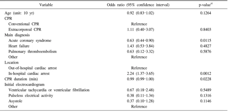
Table 3. Results of multiple logistic regression analysis of return of spontaneous circulation
Variable Odds ratio (95% confidence interval) p-value
a)Age (unit: 10 yr) 0.92 (0.83–1.02) 0.1264
CPR
Conventional CPR Reference
Extracorporeal CPR 1.11 (0.40–3.07) 0.8403
Main diagnosis
Acute coronary syndrome 0.63 (0.44–0.90) 0.0115
Heart failure 1.43 (0.53–3.84) 0.4827
Pulmonary thromboembolism 0.63 (0.12–3.32) 0.5876
Other Reference
Location
Out-of-hospital cardiac arrest Reference
In-hospital cardiac arrest 2.24 (1.37–3.65) 0.0012
CPR duration (min) 0.99 (0.99–1.00) 0.0228
Initial electrocardiogram
Ventricular tachycardia or ventricular fibrillation 0.67 (0.18–2.48) 0.5489
Pulseless electrical activity 0.38 (0.11–1.34) 0.1316
Asystole 0.37 (0.10–1.28) 0.1146
Other Reference
CPR, cardiopulmonary resuscitation.
a)
Calculated using chi-square test, Fisher’s exact test, Student t-test, or Wilcoxon’s rank-sum test, as appropriate.
treatment with the ECMO. Further, ECPR was not performed in OHCA cases of unwitnessed CA, missed previous perform- ance of bystander CPR, or in patients aged over 80 years. If the initial electrocardiogram (ECG) rhythm was at a stand- still, we did not perform ECPR.
3) Data collection
Clinical data such as the patient’s age, sex, and other basic characteristics, and the location of CA, CPR, and defib- rillation were collected. The peak level of lactate before the CPR, the results of an arterial blood gas analysis, and the ini- tial ECG rhythm were also collected and analyzed. The pri- mary end point in this study was all causes of death in hospital. The secondary end point was the ROSC rate.
4) Statistical analysis
All data were entered into an Excel spreadsheet (Microsoft, Bellevue, WA, USA). Data were analyzed using the SAS statistical program ver. 9.4 (SAS Institute Inc., Cary, NC, USA) to compare the clinical outcomes of each treatment modality. The univariate data analysis included t-tests for continuous variables and Fisher’s exact test for discrete
variables. A multivariate analysis with logistic regression was also performed. Data were reported as the mean±standard er- ror of the mean. A value of p<0.05 was considered statisti- cally significant.
RESULTS
There was no difference in sex between the two groups.
The patients were younger in the ECPR group than in the CCPR group (59.0±18.6 years vs. 63.5±17.6 years, p=0.027).
The diagnosis of acute coronary syndrome was significantly more frequent in the ECPR group (66.7% vs. 40.6%, p<
0.0001). Ventricular fibrillation and tachycardia, and pulseless electrical activity were the majority of the initial ECG find- ings in the ECPR group (42.0% and 34.6%, p<0.0001).
Asystole was noted only in 12.3% of the initial ECGs in the ECPR group. There were more cases of OHCA in the CCPR group than in the ECPR group (78.1% vs. 24.7%, p<0.0001).
Further, the CPR duration was significantly longer during the ECPR (43 minutes vs. 30 minutes, p=0.027). The peak lactate level was higher in the CCPR group (10.4 vs. 9.9, p<0.0001).
While there were more patients with hypertension and a past
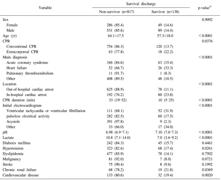
Table 4. Survival discharge rate
Variable Survival discharge
p-value
a)Non-survivor (n=817) Survivor (n=138)
Sex 0.9092
Female 286 (85.4) 49 (14.6)
Male 531 (85.6) 89 (14.4)
Age (yr) 64.1±17.5 57.3±18.0 <0.0001
CPR 0.0376
Conventional CPR 754 (86.3) 120 (13.7)
Extracorporeal CPR 63 (77.8) 18 (22.2)
Main diagnosis <0.0001
Acute coronary syndrome 346 (84.6) 63 (15.4)
Heart failure 52 (66.7) 26 (33.3)
Pulmonary thromboembolism 11 (91.7) 1 (8.3)
Other 408 (89.5) 48 (10.5)
Location <0.0001
Out-of-hospital cardiac arrest 625 (88.9) 78 (11.1)
In-hospital cardiac arrest 192 (76.2) 60 (23.8)
CPR duration (min) 33 (19–52) 10 (5–25) <0.0001
Initial electrocardiogram <0.0001
Ventricular tachycardia or ventricular fibrillation 111 (68.1) 52 (31.9)
pulseless electrical activity 282 (82.5) 60 (17.5)
Asystole 391 (97.8) 9 (2.3)
Other 33 (66.0) 17 (34.0)
pH 6.98 (6.9–7.1) 7.10 (7.0–7.3) <0.0001
Lactate 10.8 (7.1–14.0) 7.0 (3.6–9.2) <0.0001
Diabetes mellitus 242 (84.3) 45 (15.7) 0.4461
Hypertension 323 (82.6) 68 (17.4) 0.0261
Dyslipidemia 477 (85.9) 78 (14.1) 0.7502
Malignancy 81 (92.0) 7 (8.0) 0.0721
Stroke 75 (90.4) 8 (9.6) 0.1992
Chronic renal failure 68 (78.2) 19 (21.8) 0.0369
Cardiovascular disease 133 (80.6) 32 (19.4) 0.0020
Values are presented as number of patients (%), mean±standard deviation, or median (interquartile range), unless otherwise indicated.
CPR, cardiopulmonary resuscitation.
a)
Calculated using chi-square test, Student t-test, or Wilcoxon’s rank-sum test, as appropriate.
history of cardiovascular disease in the ECPR group (p<
0.05), the number of cases with a past history of stroke was higher in the CCPR group (9.3% vs. 2.5%, p<0.04) (Table 1).
Looking at the univariate analysis of ROSC results, factors such as age, CCPR, main diagnosis, CPR location, CPR dura- tion, initial ECG, and pH had a statistically significant rela- tionship with the ROSC rate (Table 2). However, in the mul- tivariate logistic regression analysis, the main diagnosis of acute coronary syndrome (ACS) and the CPR location of OHCA patients were the only significant negative risk factors
for ROSC. The ROSC rate of ACS patients was 0.63 times lower than that of the others (p=0.0115). IHCA in the CPR location was 2.24 times better than OHCA (p=0.0012). For every 1-minute increase in the CPR duration, the ROSC rate fell by 1% (p=0.0228) (Table 3).
In the univariate analysis, relatively young age, CPR loca-
tion, CPR duration, initial ECG, pH, peak lactate level, arte-
rial blood gas analysis, the history of hypertension, chronic
renal failure, and cardiovascular disease were the statistically
significant factors that showed a relationship with the survival
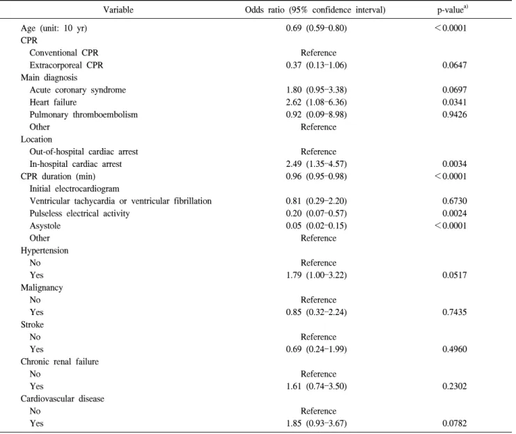
Table 5. Results of multiple logistic regression analysis of survival discharge
Variable Odds ratio (95% confidence interval) p-value
a)Age (unit: 10 yr) 0.69 (0.59–0.80) <0.0001
CPR
Conventional CPR Reference
Extracorporeal CPR 0.37 (0.13–1.06) 0.0647
Main diagnosis
Acute coronary syndrome 1.80 (0.95–3.38) 0.0697
Heart failure 2.62 (1.08–6.36) 0.0341
Pulmonary thromboembolism 0.92 (0.09–8.98) 0.9426
Other Reference
Location
Out-of-hospital cardiac arrest Reference
In-hospital cardiac arrest 2.49 (1.35–4.57) 0.0034
CPR duration (min) 0.96 (0.95–0.98) <0.0001
Initial electrocardiogram
Ventricular tachycardia or ventricular fibrillation 0.81 (0.29–2.20) 0.6730
Pulseless electrical activity 0.20 (0.07–0.57) 0.0024
Asystole 0.05 (0.02–0.15) <0.0001
Other Reference
Hypertension
No Reference
Yes 1.79 (1.00–3.22) 0.0517
Malignancy
No Reference
Yes 0.85 (0.32–2.24) 0.7435
Stroke
No Reference
Yes 0.69 (0.24–1.99) 0.4960
Chronic renal failure
No Reference
Yes 1.61 (0.74–3.50) 0.2302
Cardiovascular disease
No Reference
Yes 1.85 (0.93–3.67) 0.0782
CPR, cardiopulmonary resuscitation.
a)