• 검색 결과가 없습니다.

β 1 mRNA Expression in Skin Wound of Rat Electrical Stimulation Induces the Collagen Deposition and TGF

N/A
N/A
Protected

Academic year: 2021

Share "β 1 mRNA Expression in Skin Wound of Rat Electrical Stimulation Induces the Collagen Deposition and TGF"

Copied!
6
0
0

로드 중.... (전체 텍스트 보기)

전체 글

(1)

Jae-Hyoung Lee, PT, PhD; Chan-Eui Park, PT, MD

1

; Rae-Joon Park, PT, PhD

2

Department of Physical Therapy, Wonkwang Health Science University;

1

Department of Physical Therapy, College of Physical Therapy, Southwestern University;

2

Department of Physical Therapy, College of Rehabilitation Sciences, Daegu University

Purpose: The purpose of this study was to investigate the effect of electrical stimulation (ES) on the wound closure rate, collagen deposition, and TGF-β1 mRNA expression in skin wound of rat.

Methods: Twenty male Sprague-Dawley rats (222~271 g) were randomly divided into ES (n=10) and control group (n=10).

The ES group received a cathodal stimulation with 50 V at 100 pps for 30 minutes for 7 days, while the control group was not given electrical stimulation. The wound closure rate, collagen density and TGF-β1 mRNA ratio were measured.

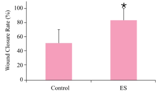
Results: The mean wound closure rates in the ES and control groups were 83.79±16.35% and 51.57±17.76%, respectively (p<0.001). The collagen density in the ES and control groups were 46.67±10.68% and 25.03±13.09%, respectively (p<0.001).

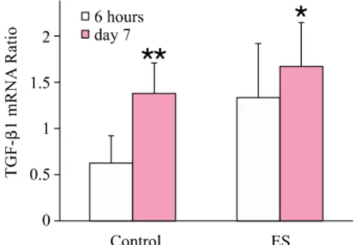
The TGF-β1 mRNA ratio in the ES and control groups were 1.35±0.60 and 0.63±0.30, respectively at 6 hours post-wound (p<0.01) and 1.69±0.47 and 1.32±0.28, respectively, at 7 days post-wound (p<0.05).

Conclusions: ES accelerated the wound closure rate of skin incision wounds and was accompanied by an increase in collagen deposition in the regenerating dermis. In addition, ES increased TGF-β1 mRNA expression during wound healing process. These findings suggest that ES may activate TGF-β1 expression, and may increase synthesis activities of fibroblasts in regenerating skin wounds in rats.

Keywords: Electrical stimulation, Wound healing, Collagen density, TGF-β1 mRNA, Rat Received: February 5, 2010

Revised: March 20, 2010 Accepted: March 31, 2010

Corresponding author: Jae-Hyoung Lee, [email protected]

TGF-β1 mRNA Expression in Skin Wound of Rat

The Journal Korean Society of Physical Therapy

I. Introduction

Many investigators have been used high voltage pulsed current (HVPC) stimulation to treat wounds in animals and human subjects. Brown et al

1

have reported that HVPC stimulation enhanced dermal wound healing rate in rabbits. Kloth and Feedar

2

have reported that HVPC stimulation accelerates the healing rate of stage IV decubitus ulcers, and Fitzgerald and Newsome

3

have reported that HVPC stimulation enhanced the healing rate infected thoracic spine wound. Burdge et al

4

have reported that HVPC stimulation promotes chronic diabetic wounds of the lower extremity. In 1994, a experts panel of the Agency for Health Care Policy and Research (AHCPR) re- commended that electrical stimulation for stage III and IV

pressure ulcers that do not respond to conventional therapy. In 2002, Centers for Medicare and Medicaid Services (CMS) decided coverage of ES for chronic wounds.

The wound healing process is a complex series of events. The healing process is initiated after injury by the release of various cytokines and growth factors. Many studies have demonstrated a beneficial effect of transforming growth factor (TGF)-β, platelet-derived growth factors (PDGF), fibroblast growth factor (FGFs) and granulocyte-macrophage colony stimulating factor (GM-CSF) on the healing process in animal and human models.

5,6

TGF-β1 has multiple functions during wound healing.

TGF-β1 stimulates infiltration of inflammatory cells, granula-

tion tissue formation, matrix formation and remodeling during

wound healing.

7

The proliferative phase starts with the migration

(2)

and proliferation of keratinocytes and fibroblasts in the edge of the wound. Fibroblasts play an important role during prolife- rative phase. Fibroblasts migrate into the wound area, where they proliferate and release extracellular matrix (ECM) in the granulation tissue, leading to collagen deposition and wound contraction. Both proliferation and migration of fibroblasts are stimulated by TGF-β1.

8

The effect of electrical stimulation on TGF-β1 in the heal- ing process remain largely unconfirmed. Many studies have reported that electrical stimulation can promote healing of soft tissue cause an increase of fibroblasts activity and collagen synthesis. However, these reports were based on in vitro experi- ment with cultured cells. The interaction between collagen deposition and TGF-β1 mRNA expression by ES during wound healing process still also not demonstrated. In this study, consequently, we used the rat full-thickness incision wound model and HVPC stimulation.

The purpose of this study was to investigate the effect of the ES on the wound closure rate, and collagen deposition in regenerating dermis and TGF-β1 mRNA expression in skin wound of rat.

II. Materials and Methods

1. Animals

Twenty male Sprague-Dawley rats weighing 170∼210 g were bio-clean cages housed in a temperature-controlled room with a 12 hour light-dark cycle. Food and water were allowed ad libitum. Experiments performed under normal room light and temperature. Three days before the operation, the hair were shaved the back and the abdominal region. Each rat received a 10 mm full-thickness incision wound on the back under general anesthesia by inhalation of halothane, maintained at a con- centration of 2∼3%. The wound was clean with a sterile gauze pad, and the rat was carefully observed until it had recovered fully from the anesthesia. The incision was not ligated and covered nor bandaged. Rats were then randomly divided into ES group (n=10) or control group (n=10).

2. Electrical Stimulation

The rats were placed in a clear plastic restrainer and rested comfortably. The incision area and abdominal area of rat skin

were covered with sterile saline-soaked gauze pads (2 × 2 cm), and carbon-silicone rubber electrodes (2 × 2 cm) were placed over each moist gauze, then secured by a restrainer. Positive and negative electric line cords connected the electrode on the abdo- men and the back, respectively. The ES rats were received electrical stimulation with a current intensity of 50 V at 100 pps for a duration of 30 minutes using a Pulsed High-Volt Stimu- lator (Intelect

HV2, Chattanooga Group Inc., TN, USA). The wave form of the HVPC stimulator consists of monophasic, twin-peak pulses that have a fixed pulse duration of 65 μs. For control group, rats were received a sham treatment without power supplied.

3. Wound closure rate measurement

The rats were anesthetized by halothane inhalation. The wound length was measured using a caliper at immediately after incision and at day 7 post-wound, and evaluated as percentage of wound closure using the equation;

wound closure rate (%)

= [(wound length at day 1-wound length at day 7)/wound length at day 1] × 100

The wound closure rate was calculated as a percentage of the reduction from the original incision length.

4. Measurement of collagen density and quantification

The rats from each group were euthanized and a 10 × 10 mm of

skin biopsies were harvested at day 7 post-wound. The harvested

tissue was fixed in 10% phosphate buffered formalin and

embedded in paraffin. Sections of 5 μm thickness were prepared

using a microtome, then deparaffinized, hydrated, cleared using

an automatic tissue processor (Citadel 1000, Shandon, Life

Sciences International Ltd., Rumcorn, England), and stained

with hematoxylin and eosin (H&E), and Masson trichrome

(MT). Sections were observed using a computerized image

analysis system (Image-Pro

Plus, Media Cybernetics, Inc.,

MD, USA). Images were recorded with a colour CCD camera

(IK-642K Toshiba CCD color camera, Toshiba Co., Tokyo,

Japan), attached to a light microscope (Olympus BX 50,

Olympus Optical Co. Ltd., Tokyo, Japan), and processing and

analysis using Image-Pro

Plus (ver 3.01). The density of

(3)

Table 1. Effects of electrical stimulation on wound healing rate, collagen density, TGF-β1 mRNA ratio at 6 hours and at day 7 in control and ES group

Control (n=10) ES (n=10) MD (95% CI) U p

Wound closure rate (%) 51.57±17.76 83.79±16.35 32.22 (16.18-48.26) 7 0.000

Collagen density (%) 25.03±13.09 46.67±10.68 21.64 (10.42-32.86) 7 0.000

TGF-β1 ratio at 6 hours 0.63±0.30 1.35±0.60 0.72 (0.27-1.17) 12 0.003

TGF-β1 ratio at day 7 1.32±0.28 1.69±0.47 0.37 (0.01-0.73) 23 0.043

Data are presented as mean±SD

ES: Electrical stimulation, MD: Mean difference, CI: Confidence interval

Figure 1. Rate of wound closure in control and ES groups.

The wound closure rate of ES group was increased signifi- cantly than control group (*p<0.001). The bar represents the standard deviation of values from 10 rats.

collagen were measured in 5 serial fields from regenerated dermis at 200× magnification. The mean density were calculated.

5. Reverse transcription-PCR analysis of TGF-β1 mRNA

A 10 × 10 mm of skin biopsies were harvested at 6 hours and day 7 post-wound. Total RNA from biopsies were prepared by the RNAzol™ B (Tel-Test Inc., TX, USA) following the in- structions provided by the supplier, and stored at -80℃. 1 μg of isolated RNA was reverse transcribed to first strand cDNA with 200 U/μl of Superscript II reverse transcriptase using 5 U/μl of Taq DNA polymerase in the presence of 10 U/μl RNase in- hibitor (Invirtogen Life Technologies, CA, USA). The cDNA synthesis reactions were performed at 42℃ for 60 min and the products were stored at -20℃. A 50 μl polymerase chain reaction (PCR) mixture containing 5 μl of the reverse transcription (RT) reaction was carried out PCR using specific primer pairs. The primers for TGF-β1 were chosen from coding region, sense 5′

-TGGGATTGTAACTGTGAACTG-3′ and antisense 5′-CAC GATCAT GTTGGACAACTGCTCC-3′, generating a 254-bp fragment, and β-actin were also chosen from coding region, sense 5′-GTGGGCCGCCCTAGGCA CCA-3′ and antisense 5′-CGGTTGGCCTTAGGGTTCAG-3′, generating a 245-bp fragment. The reaction mixtures were denatured at 94℃ for 45s, annealed at 55℃ for 45s and extended at 72℃ for 90s for 40 and 37 cycles, respectively for TGF-β1 and β-actin. The size of amplification products was estimated in 2% agarose gel with molecular weight markers and the amount was determined using a Gel Doc system and Quantity One 1-D analysis software (Bio-Rad Laboratories Inc., Richmond, CA, USA) on the negative image of ethidium bromide staining. For the relative level of TGF-β1 mRNA, an OD ratio (TGF-β1/β-actin) of 1.00 was designated to be 1.00 arbitrary unit.

6. Data Analysis

For a comparison of the percent of wound closure rate, collagen density and TGF-β1 mRNA ratio between the ES and control groups, Mann-Whitney U-testwas used. The statistical interpre- tation was based on a 0.05 significance test level. SPSS WIN (ver 10.0) software was used for the analyses.

III. Results

1. Wound closure rate

At day 7 post-wound, the wounds treated with ES were suppler and the surface was smoother than those control. In sham treated control group, the mean rate of wound closure was 51.57±17.76%. In ES group had mean wound closure rate was 83.79±16.35%. Mann-Whitney U-test showed a significantly higher the rate of wound closure in the ES group than control group (U=7, p<0.001)(Table 1, Figure 1). The ES increased wound closure rate by 32.22% than control group.

2. Collagen Density

The collagen fiber stained bluish color in the regenerated dermis

(4)

A B

Figure 2. The bluish-stained collagen fibers can be seen in the incised wound in the control (A) and the ES (B). The ES group displayed stronger staining (bluish) for collagen in regenerated dermis. Masson-trichrome stain. 200x. Scale bar: 5 μm.

Figure 3. The collagen density in regenerated dermis in control and ES groups. The collagen density of ES group was increased significantly than control group (*p<0.001).

The bar represents the standard deviation of values from 10 rats.

Figure 4. ES increases the expression of TGF-β1 at trans- criptional level. RT-PCR products were analyzed by 2%

agarose gel electrophoresis. β-actin was used as a reference gene. The resultant DNA bands of TGF-β1 was identified at the 254 bp position. Band intensity reflected the mRNA expression level.

1: Control 6 hours, 2: ES 6 hours, 3: Control day 7, 4:

ES day 7.

Figure 5. The TGF-β1 ratio in control and ES wound. The TGF-β1 ratio of ES group at 6 hours and 20.12% at day 7 post-wound were increased significantly than control group determined by RT-PCR followed by densitometry (**p<0.01, *0.05). The bar represents the standard de- viation of values from 10 rats.

by Masson trichrome stain (Figure 2). The collagen density in the control and ES group were 25.03±13.09% and 46.67±

10.68%, respectively. There was a significant increase of colla- gen density in the ES wound compared to the control group (U=7, p<0.001)(Table 1, Figure 3). The ES increased collagen density by 21.64% than control wound.

3. TGF‑β1 mRNA Ratio

The expression of TGF-β1 mRNA was upregulated by ES at 6 hours and at day 7 post-wound (Figure 4). The relative level of TGF-β1 mRNA (normalized to β-actin) was 0.63±0.30 and 1.35±0.60 arbitrary unit, respectively, in control and ES wound.

The TGF-β1 mRNA ratio at day 7 post-wound was 1.32±0.28 and 1.69±0.47, respectively, in control and ES wound. There was a significant increase of TGF-β1 mRNA ratio in the ES wound compared to the control group at 6 hours post-wound (U=12, p<0.01), and at day 7 post-wound (U=23, p<0.05) (Table 1). The ES had significantly increased the TGF-β1

mRNA ratio throughout the treatment (Figure 5). ES increased TGF-β1 ratio by 55.56% at 6 hours and 20.12% at day 7 post- wound than control wound determined by RT-PCR followed by densitometry.

IV. Discussion

The fibroblasts play a key role in skin wound healing process.

The activated fibroblasts synthesize and release ECM compo-

nents including collagen, fibronectin, hyaluronic acid, thrombo-

spondin, tenascin, and others.

9

The structural molecules of

newly formed ECM, termed the provisional matrix, contribute

to the formation of granulation tissue by providing a scaffold for

cell migration. The provisional ECM is gradually replaced with a

collagenous matrix. Once an abundant collagen matrix has been

deposited in the wound, the fibroblasts stop producing collagen,

and the fibroblast-rich granulation tissue is replaced by a

(5)

relatively acellular scar.

10

TGF-β1 is involved in all stages of wound healing. It facilitates the recruitment of inflammatory cells and augments macrophage mediated tissue debridement. It also deactivates superoxide production from macrophages.

TGF-β1 helps initiate granulation tissue formation by increasing the expression of genes associated with ECM formation in- cluding fibronectin, the fibronectin receptor, and collagen and protease inhibitors.

11,12

By stimulating recruitment of fibro- blasts to the wound site, the combined result isincreased collagen deposition and scar formation.

13,14

This study demonstrated that ES promotes the healing rate of skin wound with increases collagen density with HVPC 100 pps at intensity of 50 V for 30 minutes. ES was increased the collagen density in situ in rat skin wound model as high as 21.64%. In addition, ES increases the expression of TGF-β1 mRNA up to 56%, 20% at 6 hours and at day 7 post-wound.

This result suggested that ES could increases the collagen deposition in regenerative dermis and could enhances TGF-β1 activity from throughout the ES duration.

The parameters of ES used in this study are based on Bourguinon and Bourguinon’s experiment.15 They have reported that the HVPC increases maximally in synthesis of protein and DNA in fibroblast at 50 and 75 V, 100 pps, at negative pole. In our previous study, ES was demonstrated to stimulate fibroblasts migration and proliferation up to 24%, 10%, respectively, from tendon biopsy.

16

In another our study, ES increases fibroblasts density and collagen density up to 40%, 17%, respectively, from the skin wound with HVPC 100 pps at intensity of 50 V.

17

It is well known that various forms of ES influence fibro- blasts migration, proliferation, and structural ECM proteins synthesis. Thus, ES may contribute to fibroblasts activation in the process of wound healing.

18

The cellular mechanisms for the activation of fibroblasts by electrical stimulation have been postulated. At the cellular level, the electrical signals could alter the electrical properties of cell membranes. The electric signals could activate the voltage gated ion channel at the cell mem- brane. Besides, electric signals act an ionophoretic force on charged proteins and lipids in the cell membrane which exert to redistribution of membrane components. Therefore, the electrical stimulation induces cellular responses including lateral redistri- bution of membrane proteins such as ion channels and re- ceptors, and changes in intracellular calcium ion concentration.

Intracellular Ca

++

regulates many biological processes including signal transduction cascades, cytoskeletal reorganization, cell orientation, migration, differentiation and proliferation.

19,20

Such change could consequently modify intracellular signals for TGF-β1 gene expression. The migration and proliferation of fibroblasts are regulate by TGF-β1.

8

ES increase TGF-β1 gene expression, which enhance structural ECM proteins synthesis in fibroblasts resulting in a promotion in wound healing.

21,22

The results in this study are based on an in vivo model. This study is the first to document that ES enhances secretion of TGF-β1 at the transcriptional level in skin wound.

V. Conclusions

The results of this study suggested that ES which are similar to the current of injury promotes the healing of skin wound due to stimulates the cellular activity of fibroblast, and it may increases collagen deposition in skin wound sites. The molecular mecha- nism that accounts for the ES effect on may be modulated th- rough a TGF-β1 related pathway.

Author Contributions

Research design: Lee JH, Park CE, Park RJ Acquisition of data: Lee JH, Park CE, Park RJ

Analysis and interpretation of data: Lee JH, Park CE, Park RJ Drafting of the manuscript: Lee JH, Park CE, Park RJ Research supervision: Lee JH

Acknowledgements

1. This work was supported partly by the Academic Research Fund of Wonkwang Health Science University (2008).

2. This study was presented on the 4th World Congress of International Society of Physical and Rehabilitation Medicine (ISPRM), Seoul, 10-14 June, 2007.

References

1. Brown M, McDonnell MK, Menton DN. Electrical stimu- lation effects on cutaneous wound healing in rabbits. A follow-up study. Phys Ther. 1988;68(6):955-60.

2. Kloth LC, Feedar JA. Acceleration of wound healing with

high voltage, monophasic, pulsed current. Phys Ther. 1988;

(6)

68(4):503-8.

3. Fitzgerald GK, Newsome D. Treatment of a large infected thoracic spine wound using high voltage pulsed monophasic current. Phys Ther. 1993;73(6):355-60.

4. Burdge JJ, Hartman JF, Wright ML. A retrospective study of high-voltage, pulsed current as an adjunctive therapy in limb salvage for chronic diabetic wounds of the lower extremity.

Ostomy Wound Manage. 2009;55(8):30-8.

5. Harding KG, Morris HL, Patel GK. Science, medicine and the future: healing chronic wounds. BMJ. 2002;324(7330):

160-3.

6. Klass BR, Grobbelaar AO, Rolfe KJ. Transforming growth factor beta1 signalling, wound healing and repair: a multi- functional cytokine with clinical implications for wound repair, a delicate balance. Postgrad Med J. 2009;85(999):9-14.

7. Riedel K, Riedel F, Goessler UR et al. Tgf-beta antisense therapy increases angiogenic potential in human keratino- cytes in vitro. Arch Med Res. 2007;38(1):45-51.

8. Barrientos S, Stojadinovic O, Golinko MS et al. Growth factors and cytokines in wound healing. Wound Repair Regen.

2008;16(5):585-601.

9. Schultz GS, Wysocki A. Interactions between extracellular matrix and growth factors in wound healing. Wound Repair Regen. 2009;17(2):153-62.

10. Werner S, Grose R. Regulation of wound healing by growth factors and cytokines. Physiol Rev 2003;83(3):835-70.

11. Mani H, Sidhu GS, Kumari R et al. Curcumin differentially regulates TGF-beta1, its receptors and nitric oxide synthase during impaired wound healing. Biofactors. 2002;16(1-2):

29-43.

12. Rolfe KJ, Richardson J, Vigor C et al. A role for TGF- beta1-induced cellular responses during wound healing of the non-scarring early human fetus? J Invest Dermatol. 2007;

127(11):2656-67.

13. Martinez-Ferrer M, Afshar-Sherif AR, Uwamariya C et al.

Dermal transforming growth factor-beta responsiveness me- diates wound contraction and epithelial closure. Am J Pathol.

2010;176(1):98-107.

14. Heldin CH, Eriksson U, Ostman A. New members of the platelet-derived growth factor family of mitogens. Arch Bio- chem Biophys. 2002;398(2):284-90.

15. Bourguignon GJ, Bourguignon LY. Electric stimulation of protein and DNA synthesis in human fibroblasts. FASEB J.

1987;1(5):398-402.

16. Lee JH, Jekal SJ, Park RJ. Enhance of migration and pro- liferation of cells from tendon biopsies by high voltage pulsed current stimulation. J Kor Soci Phys Ther. 2002;14(2):53-8.

17. Lee JH, Song IY, Kim JG. Acceleration of wound healing and collagen deposition in rat skin by high voltage pulsed current stimulation. J Kor Soci Phys Ther. 2003;15(4):1-12.

18. Jennings J, Chen D, Feldman D. Transcriptional response of dermal fibroblasts in direct current electric fields. Bioelectro- magnetics. 2008;29(5):394-405.

19. Bourguignon GJ, Jy W, Bourguignon LY. Electric stimulation of human fibroblasts causes an increase in Ca

2+

influx and the exposure of additional insulin receptors. J Cell Physiol.

1989;140(2):379-85.

20. Kim YV, Conover DL, Lotz WG et al. Electric field-induced changes in agonist-stimulated calcium fluxes of human HL- 60 leukemia cells. Bioelectromagnetics. 1998;19(6):366-76.

21. Aaron RK, Boyan BD, Ciombor DM et al. Stimulation of growth factor synthesis by electric and electromagnetic fields.

Clin Orthop Relat Res. 2004;(419):30-7.

22. Zhao M. Electrical fields in wound healing-An overriding signal that directs cell migration. Semin Cell Dev Biol. 2009;

20(6):674-82.

수치

Figure 1. Rate of wound closure in control and ES groups.
Figure 4. ES increases the expression of TGF-β1 at trans- trans-criptional level. RT-PCR products were analyzed by 2%

참조

관련 문서

Purpose: The study was to investigate the organizational culture of middle and small sized hospital workers on organizational effectiveness and controlling effect of

The purpose of this study is to investigate the effect of the “problem solving and programming” area of ​​information subject on perception and

co-treatment with hispidulin and TGF-β up-regulated the protein of expression E-cadherin and occludin against TGF-β-induced in MCF-7 and HCC38 cells.. The

Objective: The purpose of this study was to investigate the effect of clinical practice satisfaction on professional self-concept among dental hygiene students.. Methods:

The purpose of this research was to identify the mediating effect of unconditional self-acceptance and the moderated mediating effect of emotional

The purpose of this study was to investigate the impact of social capital and positive psychology capital on the will of start-up according to experience

The purpose and necessity of this study was to investigate the effect of the 8-week GX complex exercise program on the stress index, blood lipid and APG

The purpose of this study is to investigate the effect of peer-mentoring on the achievement and learning attitudes of middle school students toward