KJTCVS
The Korean Journal of Thoracic and Cardiovascular SurgeryClinical Research Usefulness of 3-Dimensional Body Surface Scanning in the
Evaluation of Patients with Pectus Carinatum
Seung Hwan Song, M.D., Chong Hoon Kim, M.D., Duk Hwan Moon, M.D., Sungsoo Lee, M.D., Ph.D.
Department of Thoracic Surgery, Gangnam Severance Hospital, Yonsei University College of Medicine, Seoul, Korea
ARTICLE INFO Received May 11, 2020 Revised June 24, 2020 Accepted July 1, 2020 Corresponding author Sungsoo Lee
Tel 82-2-2019-3381 Fax 82-2-3461-8282 E-mail [email protected] ORCID
https://orcid.org/0000-0001-8998-9510
Background: Radiographic modalities have been commonly used to evaluate pectus carinatum (PC), and compressive orthotic bracing is the most widely accepted treatment method. The aim of this study was to determine the efficacy of 3-dimensional (3D) body surface scanning as an alternative modality for the evaluation of PC.
Methods: The medical records of 63 patients with PC who were treated with compres- sive orthotic bracing therapy between July 2017 and February 2019 were retrospectively analyzed. Using both 2-view chest radiography (posteroanterior and lateral view) and 3D body scanning, the height of maximal protrusion of the chest wall was measured both be- fore and after 2 weeks of bracing therapy. The difference between the pre- and post-treat- ment measurements was calculated for both modalities, and these differences were com- pared and analyzed.
Results: Based on the comparison between the pre- and post-treatment radiographs, bracing therapy produced favorable outcomes in all patients (p<0.001). The measure- ments obtained via 3D scanning were strongly correlated with those obtained via chest radiography (r=0.60).
Conclusion: Based on the findings of this study, 3D body surface scanning appears to be an effective, radiation-free, and simple method for the post-treatment follow-up evalua- tion of PC, and thus can be considered an alternative to radiography.
Keywords: Pectus carinatum, Three-dimensional, Three-dimensional body scan, Body surface scan, Braces
Copyright©The Korean Society for Thoracic and Cardiovascular Surgery. 2020. All right reserved.
This is an Open Access article distributed under the terms of the Creative Commons Attribution Non-Commercial License (http://creativecommons.org/licenses/
Introduction
Pectus carinatum (PC), also known as pigeon chest, is a relatively common chest wall deformity that is character- ized by anterior protrusion of the anterior chest wall. The prevalence of this condition has been reported to be ap- proximately 0.7% [1,2]. As with pectus excavatum, the cause may be an imbalance of growth between the ribs and the costal cartilage of the chest wall [3,4]. Most patients with PC are asymptomatic; instead, the chief complaint is the cosmetic appearance of the chest wall associated with PC. Traditionally, surgery was the mainstay of treatment, and surgical treatment was indicated based on the degree of deformity. However, in recent years, compressive orthot- ic bracing has been widely used as a non-invasive method with favorable results [5,6].
Most commonly, chest X-rays and computed tomogra- phy (CT) are used to evaluate patients with PC [7,8]. Pre- and post-treatment images are obtained using these radio- graphic modalities to determine the efficacy of treatment.
These modalities inevitably expose patients to multiple doses of harmful radiation. Recent studies have demon- strated that a 2-view chest X-ray (posteroanterior and later- al view) could replace CT in the evaluation of chest wall deformities, as the former entails less exposure to radiation [9]. At Gangnam Severance Hospital, the 2-view chest X- ray is used for the evaluation and follow-up of patients with PC. Although this technique does administer a lower radiation dose than CT, demand still exists for a new mo- dality that eliminates any unnecessary radiation exposure.
Recently, at our institution, clinicians have begun to use 3-dimensional (3D) body surface scanning to evaluate PC
https://doi.org/10.5090/kjtcs.20.042 pISSN: 2233-601X eISSN: 2093-6516
Korean J Thorac Cardiovasc Surg. 2020;53(5):301-305
KJTCVS
https://doi.org/10.5090/kjtcs.20.042treated with a compressive orthotic brace. Using 3D body surface scanning, we can obtain images that show post- treatment results on an objective scale. In this study, mea- surements obtained using 3D body surface scanning were compared with those obtained using chest X-ray scans to evaluate whether 3D body surface scanning can be an ef- fective and radiation-free alternative to evaluate the im- provement of PC after compressive orthotic brace therapy.
Methods
Patients
We retrospectively analyzed the medical records of 63 patients with PC who were treated with compressive or- thotic bracing between July 2017 and February 2019 at our institution. Both 3D body surface images and 2-view chest X-rays with posteroanterior and lateral views were ob- tained. The initial chest images were obtained at the first hospital visit, and the post-treatment images were obtained at the first follow-up visit, 2 weeks later. For both pre- and post-treatment scans, the height of maximal protrusion of the chest wall was measured on the 3D and chest X-ray im- ages. On the lateral chest X-ray images, this height was measured as the distance between the most prominent point of the sternum and the anterior edge of the vertebral body (Fig. 1). The results were compared and analyzed.
This study was approved by the institutional review board of Gangnam Severance Hospital, Yonsei University College
of Medicine (IRB approval no., 3-2017-0353).
Three-dimensional body scanner
The Pectus Metric Tool 3D body scanner was used to scan the body surfaces of patients. To obtain 3D surface images, Microsoft Kinect (Microsoft Corp., Redmond, WA, USA), which uses a pattern of projected infrared dots to generate 3D images, was incorporated to capture the depth and color. The device was mounted on a curved movable platform with a 120° arc of rotation from one side to the other (Fig. 2) and connected to a personal computer.
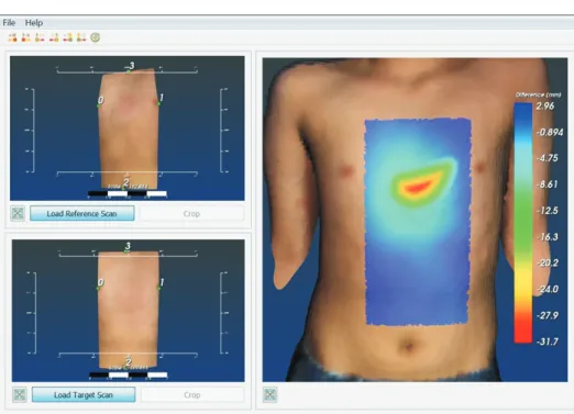
The software, which was newly developed for this purpose, could receive point cloud data using structured light scan- ning (Fig. 3). The real-time scanning software Recon- structMe (Profactor GmbH, Steyr-Gleink, Austria) was run on the computer, which was connected to Microsoft Ki- nect. Pre- and post-correction images were measured for comparison, and changes in the chest wall deformity were visualized using the developed software. With the patient in the supine position, the scan took 5 to 10 seconds to capture the necessary information. Once the initial scan- ning procedure was completed, the data file was saved in the standard polygon file (PLY) format for use in the soft- ware. After loading the pre- and post-correction images, the software automatically compared the point-to-point differences and generated a color map to visualize them.
The color map scale indicated, in millimeters, the differ- ence between the 2 images, which represented the im- provement of the chest wall deformity.
L
D
Fig. 1. Distance measurement on chest radiography (lateral view).
The point of the most anterior projection of the sternum was iden- Fig. 2. The Pectus Metric Tool 3-dimensional body scanner using
Seung Hwan Song, et al. 3-Dimensional Body Scan for Pectus Carinatum
KJTCVS
Protocol for pectus carinatum bracing
The application of the brace was based on the findings of the clinical examination. Through close collaboration with a certified orthoptist, we designed a custom-fitted com- pression orthotic brace tailored to each patient. Patients were instructed to wear the brace over a layer of clothing to protect the skin for 20 hours per day during the compres- sion period (2 weeks) and 10 hours per day during the maintenance period (6 months). The first follow-up visit was scheduled at the end of the compression period to evaluate patient compliance and the fit of the brace; there- after, patients were followed up with every 3 months.
Statistical analysis
Continuous variables are expressed as means and stan- dard deviations. Categorical variables are expressed as per- centages. For the 2 modalities, the paired t-test was used for continuous variables, and Pearson correlation analysis was used to assess the correlation between the 2 variables.
All statistical analyses were conducted using PASW SPSS for Windows ver. 18.0 (SPSS Inc., Chicago, IL, USA). All p-values <0.05 were considered to indicate statistical sig- nificance.
Results
A total of 63 patients with PC treated with compressive orthotic bracing were enrolled in the present study. All pa- tients underwent both chest radiography and 3D body sur- face scanning at the initial hospital visit (pre-treatment) and at the 2-week post-treatment follow-up. The baseline characteristics are presented in Table 1. The mean age was 10.52±4.21 years, and 56 of the patients were male (88.88%).
The mean chest wall protrusion height was 105.85±20.49 mm at the initial hospital visit and 89.84±20.00 mm at the 2-week follow-up visit (the post-treatment measurement).
Based on the comparison between the pre- and post-treat- ment radiographs, compressive orthotic bracing therapy produced favorable outcomes in all patients (p<0.001).
The height difference (from pre- to post-treatment) of the point of maximal protrusion of the chest wall was mea- sured using both 2-view chest X-rays and 3D body surface
Fig. 3. Software used to compare 2 scans of the chest wall: landmark- based alignment and registration (left) and difference color map in mm (right).
Table 1. Characteristics and measurements of patients obtained using lateral chest X-ray images
Characteristic Value p-value
Age (yr) 10.52±4.21 -
Male 56 (88.88) -
Chest wall protrusion (initial, mm) 105.85±20.49 0.001 Chest wall protrusion
(2 weeks after initial visit, mm)
89.84±20.00 Values are presented as mean±standard deviation or number (%).
KJTCVS
https://doi.org/10.5090/kjtcs.20.042scanning (Table 2). The mean difference was 19.44±17.46 mm on the chest radiographs and 16.00±9.28 mm on the 3D images. As shown in Fig. 4, the measurements obtained via 3D scanning were strongly correlated with those ob- tained via chest radiography (r=0.60).
Discussion
PC is an anterior chest wall deformity diagnosed at birth or in early childhood that has recently been treated via non-invasive, compressive orthostatic brace therapy with favorable outcomes. To date, the most conventional and widely used methods for evaluating the outcome of treat- ment and/or the severity of PC have mostly been radio- graphic modalities, such as chest X-rays and CT. However, given the nature of the disease (which is usually diagnosed at birth or in early childhood), those receiving treatment are generally young, and the issue of radiation exposure is crucial. Parents and guardians, and in some cases the pa- tients themselves, are generally opposed to receiving these modes of evaluation. Hence, the demand for a new modali- ty of evaluating the treatment outcomes and/or degree of PC with minimal or no radiation has been steadily increas- ing. Several studies have been conducted to attempt to reduce radiation exposure when evaluating PC severity and treat- ment outcomes. Khanna et al. [9] reported a statistically significant correlation between the Haller index as estimat- ed with radiographs and the same index as estimated with CT. We found a similar trend in our data. Of the 63 pa- tients, CT was performed in 7 patients at the initial hospi- tal visit and post-treatment follow-up. The pre- to post- treatment difference was statistically similar between CT and chest X-ray scanning (p=0.152).
Some recent studies have indicated the possibility of al- ternatives to radiographic tools for the evaluation of pa- tients with chest wall deformities [10-13]. Port et al. [12] re- ported the measurement and evaluation of the treatment outcome using white light scanning. Using a portable de-
vice, they obtained pre- and post-intervention images. The results they provided indicate that white light scanning may be a promising alternative to measure the severity of PC [12]. Wong et al. [13] used a 3D body scanner to evalu- ate the efficacy of compressive orthotic bracing. They show- ed that serial 3D body scan imaging can be used to mea- sure and monitor changes over time [13]. However, their study had some limitations that cannot be overlooked; no- tably, their procedure required a technician to acquire and analyze the images and modify the equations to calculate the treatment efficacy. This may have led to subjective re- sults.
In our study, the Pectus Metric Tool, which is a portable 3D body surface scanning device used to visualize the shape and contour of the thorax and to accurately assess chest wall deformity, was used to obtain 3D chest surface images in conjunction with Microsoft Kinect, an infrared laser projector. The software compared the pre- and post- treatment chest surface images to assess the change in the chest shape over the course of therapy. The system is sim- ple to use, and technicians, who could be a source of sub- jectivity, are not required. In this study, we showed that 3D chest surface scanning can be used to successfully visualize and quantify the changes over the course of therapy with- out radiation exposure.
This study had several limitations. First, it was a prelimi- nary study based on a relatively small cohort from a single institution. Although this study demonstrated the feasibili- ty of the Pectus Metric Tool system in evaluating patients with PC, only a few institutions are currently using this system. To better evaluate the efficacy of this system, a larger multi-institutional study may be necessary. Second, Table 2. Pre- to post-treatment difference in the height of maximal
protrusion of the chest wall as measured using 3D images and lateral chest X-ray images
Variable Value p-value
3D scan difference (mm) 19.44±17.46 0.057 Chest X-ray difference (mm) 16.00±9.28 - Values are presented as mean±standard deviation.
3D, 3-dimensional.
Fig. 4. Correlations of measurements obtained from 3D scan im- ages and lateral chest X-ray images of patients with pectus carina- tum (N=63). 3D, 3-dimensional.
50
40
30
20
10
0
10
100
ChestX-raydifference(mm)
3D scan difference (mm)
0 20 40 60 80
20
r=0.60
Seung Hwan Song, et al. 3-Dimensional Body Scan for Pectus Carinatum
KJTCVS
the 3D body surface images acquired in this study were only compared with chest X-ray images. Hence, for a more comprehensive evaluation, 3D body surface scanning should be compared with other modalities.
Nonetheless, 3D body surface scanning, at least prelimi- narily, appears to be an effective, radiation-free method for post-treatment follow-up evaluation of PC.
Conflict of interest
No potential conflict of interest relevant to this article was reported.
ORCID
Seung Hwan Song: https://orcid.org/0000-0002-3910-5585 Chong Hoon Kim: https://orcid.org/0000-0001-7833-3882 Duk Hwan Moon: https://orcid.org/0000-0003-1388-2471 Sungsoo Lee: https://orcid.org/0000-0001-8998-9510
References
1. Desmarais TJ, Keller MS. Pectus carinatum. Curr Opin Pediatr 2013;
25:375-81.
2. Westphal FL, Lima LC, Lima Neto JC, Chaves AR, Santos Junior VL, Ferreira BL. Prevalence of pectus carinatum and pectus excava- tum in students in the city of Manaus, Brazil. J Bras Pneumol 2009;
35:221-6.
3. Park CH, Kim TH, Haam SJ, Lee S. Does overgrowth of costal carti- lage cause pectus carinatum?: a three-dimensional computed tomog- raphy evaluation of rib length and costal cartilage length in patients with asymmetric pectus carinatum. Interact Cardiovasc Thorac Surg
2013;17:757-63.
4. Park CH, Kim TH, Haam SJ, Jeon I, Lee S. The etiology of pectus carinatum involves overgrowth of costal cartilage and undergrowth of ribs. J Pediatr Surg 2014;49:1252-8.
5. Emil S, Laberge JM, Sigalet D, Baird R. Pectus carinatum treatment in Canada: current practices. J Pediatr Surg 2012;47:862-6.
6. Jung J, Chung SH, Cho JK, Park SJ, Choi H, Lee S. Brace compres- sion for treatment of pectus carinatum. Korean J Thorac Cardiovasc Surg 2012;45:396-400.
7. Stephenson JT, Du Bois J. Compressive orthotic bracing in the treat- ment of pectus carinatum: the use of radiographic markers to predict success. J Pediatr Surg 2008;43:1776-80.
8. Haller JA Jr, Kramer SS, Lietman SA. Use of CT scans in selection of patients for pectus excavatum surgery: a preliminary report. J Pe- diatr Surg 1987;22:904-6.
9. Khanna G, Jaju A, Don S, Keys T, Hildebolt CF. Comparison of Haller index values calculated with chest radiographs versus CT for pectus excavatum evaluation. Pediatr Radiol 2010;40:1763-7.
10. Taylor JS, Madhavan S, Szafer D, et al. Three-dimensional optical imaging for pectus excavatum assessment. Ann Thorac Surg 2019;
108:1065-71.
11. Port E, Hebal F, Hunter CJ, Abdullah F, Malas B, Reynolds M. Mea- suring the impact of surgical intervention on pediatric pectus excava- tum using white light scanning. J Pediatr Surg 2019;54:2261-7.
12. Port E, Hebal F, Hunter CJ, Malas B, Reynolds M. Measuring the impact of brace intervention on pediatric pectus carinatum using white light scanning. J Pediatr Surg 2018;53:2491-4.
13. Wong KE, Gorton GE 3rd, Tashjian DB, Tirabassi MV, Moriarty KP.
Evaluation of the treatment of pectus carinatum with compressive orthotic bracing using three dimensional body scans. J Pediatr Surg 2014;49:924-7.