CONTENTS
Ⅰ. INTRODUCTION
Ⅱ. MATERIALS AND METHODS
Ⅲ. RESULTS
Ⅳ. DISCUSSION
Ⅴ. CONCLUSION REFERENCES KOREAN ABSTRACT
Ⅰ. INTRODUCTION
Temporomandibular disorders(TMD) is a collec- tive term embracing a number of clinical problems that involve the masticatory musculature, the temporomandibular joint(TMJ) and associated structures of both. TMD has been identified as a major cause of non-dental pain in the orofacial region and is considered to be a subclassification of musculo- skeletal disorders
1). Treatment options of TMD include patient education and self-care, cognitive intervention, pharmacotherapy, physical therapy, orthopedic appliances, occlusal therapy, and surgery. Physical therapy commonly includes thermotherapy, coolant application, ultra-sound therapy, transcutaneous electrical nerve stimulation (TENS), laser therapy and acupuncture.
Acupuncture is a component of the health care
system of China that can be traced back for at least 2,500 years. The general theory of acupuncture is based on the premise that there is pattern of energy flow(Qi) through the body that is essential for health. Imbalances of this flow are believed to be responsible for disease. Acupuncture may correct imbalances of flow at identifiable points close to skin
2). Acupuncture therapy was intended by ancient Korean oriental medical doctor to correct blockages or excess in the flow of the vital life force and to correct disharmonies of imbalances in the vital life force and elements on which physiologic function depended
3).
In several experimental clinical studies, an increase in the pain threshold during acupuncture stimulation by Andersson, et al.
4), Chapmarr, et al.
5)and a decrease in pain sensitivity sufficient to allow major surgical events by Kaade, et al.
6)have been reported. The analgesic action is probably multi-factorial, comprising segmental mechanism at the entry region in the central nervous system
7), supraspinal descending effects, including the release of endogenous opioids and other neuromodulatory substances
8,9), and psychological mechanisms
10). Kim
11)reported that EAST on S5 and Cv24 points showed an analgesic effect on mandibular canine. Han
12)evaluated the effects of EAST applied on Li4 point of the sensory and pain
A Clinical Effect of Electro-Acupuncture Stimulation Therapy on TMD Patients
Tae-Sik Park, D.D.S., M.S.D., June-Sang Park, D.D.S., M.S.D., Ph.D., Myung-Yun Ko, D.D.S., M.S.D., Ph.D.
Department of Oral Medicine, College of Dentistry, Pusan National University
threshold of electrically stimulated tooth pulp. In his study, EAST had an effect on mandibular anterior teeth area distant from the site of stimulation and he suggested that the reason was due to endorphin release which increased the pain threshold of whole body.
The purpose of this study was to evaluate the clinical effect of electro-acupuncture stimulation therapy(EAST) in TMD patients using pressure pain threshold and clinical indexes.
Ⅱ. MATERIALS AND METHODS 1. Subjects
Thirty eight patients, of which 5 were males and 33 were females, participated in this study. The mean age was 18.8 years, ranging from 13 to 54 years. All subjects were examined clinically and a comprehensive history was taken. Inclusion criteria were signs and symptoms of TMD - pain, noise and limitation of motion. Exclusion criteria were pregnancy, patients with a complex psycho-social situation, patients with too many uncontrolled factors, patients with illness or conditions which might influence TMD symptoms, and patients not interested in participating in this study.
2. Apparatus
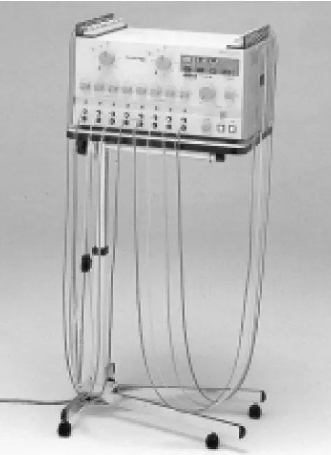
EAST was performed with Pulse Generator (PG)-8
Ⓡ(ITO Co. Tokyo, Japan) at the meridians.
PG-8
Ⓡproduced a biphasic wave current(120㎲
pulse width) of 3 Hz for 2 seconds, 15 Hz for 2 seconds and the current was slowly increased to cause a strong, but not painful, tingling sensation at the electrode attached area(Fig. 1).
The electronic algometer type Ⅰ(Somedic production, Stockholm, Sweden) used in this study consists of a gun shaped application handle with a round rubber tip, a main body that has a digital display panel, calibration knob and control knob of application rate slope, and a patient- operated switch.
The pressure pain threshold(PPT) was measured in
Fig. 1. Pulse Generator(PG)-8Ⓡ
Kpa by algometer. The algometer handle was applied perpendicularly to all the meridian and application speed was maintained at 30 Kpa/sec.
3. Procedure
The subjects were randomly assigned to two groups: the treated patient group(n=28) and the non-treated patient group(n=10). EAST was applied on the meridian according to signs and symptoms and PPT was measured in each meridian in the treated patient group(Fig. 2). Each treatment lasted twenty minutes, and was given three times in a week, totally six times during two weeks.
Meridian
1) B10(CHŎN JU, 天柱) 2) G2(CHŎNG HOI, 聽會) 3) G21(GYŎN JŎNG, 肩井) 4) S2(HA GWAN, 下關) 5) S3(HYŎP CHA, 頰車) 6) S8(DAE YOUNG, 大迎)
7) Li4(HAP GOK, 合谷) : origin between 1st and
2nd meta-carpal bone
Fig. 2. Meridian of head and neck
8) Si19(CHŎNG GUNG, 聽宮) 9) T17(YE POONG, 翳風)
The treated patient group was evaluated before first treatment and after final treatment. Pain intensity was evaluated with numerical analogue scale(NAS). The treated patients were asked to rate their pain using numerical scale of 0 to 10. The 0 on the scale was estimated to be "no pain" and 10 to be "pain as bad as can be". Noise frequency and limitation of motion(LOM) evaluated as same degrees.
Maximum comfortable opening (MCO) and active range of motion(AROM) were also measured by
"mm" degree.
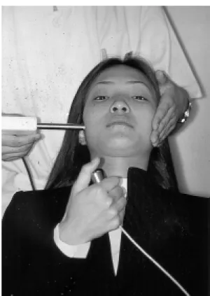
Before experiment, subjects were instructed to push button on patient-operated switch as soon as they recognized pain. When pain was felt, subject pushed button on patient- operated switch, digital display stopped for about 5 seconds, and red light turned on so operator could record the value.
During the test, subjects did not see the values of measurement. PPT was measured in each point.
The clinical indexes and PPT were taken before first EAST and after final EAST(Fig. 3).
The non-treated patient group without any
Fig. 3. Clinical application of Algometer
treatment was evaluated by same indexes before first experiment and two weeks later after first experiment, but the PPT value was measured only on S2 point, which was applied to all subjects.
4. Statistical Methods
The correlation in the paired t test was used to evaluate the significance of differences between mean values of before and after experiment. The same test was done in the non-treated patient group. ANOVA was used to test the difference of mean values according to chronicity in the treated patient group.
Ⅲ. RESULTS
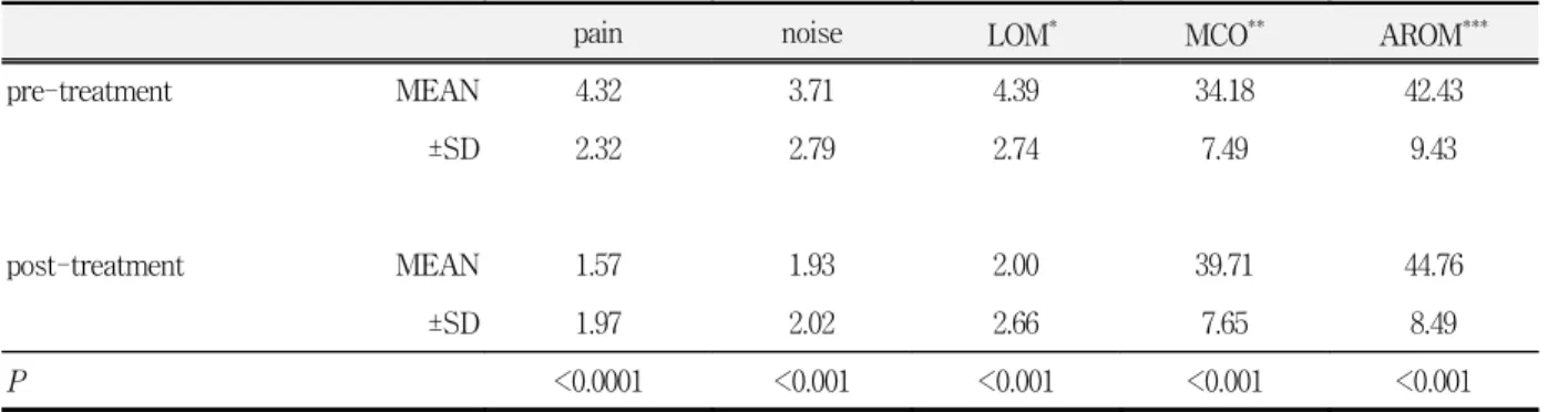
The clinical indexes in treated patient group
between pre- treatment and post-treatment are
shown in table 1. All indexes were very
significantly improved at post-treatment compared
with pre-treatment(P<0.001). Above all, there was best effect in pain control(P<0.0001).
The PPT values of the treated patient group between pre-treatment and post-treatment are shown in table 2. The PPT values were significantly increased in points of G2, G21, Li4, S2, S3, Si19, and T17(P<0.05). The PPT values of all points were increased except point of B10.
The differences of clinical indexes between before first experiment and after final experiment in the non-treated patient group are shown in table 3.
There were no significant differences.
In the non-treated patient group, there was no difference in PPT between before first experiment and after two weeks later on S2 points. Table 4 shows the results.
Table 2. Difference of pressure pain threshold according to meridian between pre-treatment and post- treatment in the treated patient group (Kpa, N=28)
B10 G2 G21 Li4 S2 S3 S8 Si19 T17
pre-treatment MEAN 85.26 43.84 79.81 104.06 78.19 41.73 53.76 58.33 42.79
±SD 12.64 8.42 20.68 15.63 18.07 11.35 13.60 13.39 13.26
post-treatment MEAN 85.11 49.93 94.93 130.31 83.11 56.53 62.60 63.52 47.77
±SD 9.60 7.73 21.36 31.16 17.29 4.95 12.56 14.55 12.96
P 0.487 0.033 <0.001 <0.001 0.012 0.045 0.018 0.005 0.002
Table 3. Clinical indexes before first experiment and after final experiment in the non-treated patient group (N=10)
pain noise LOM MCO AROM
Ⅰ MEAN 4.9 2.3 4.6 29.6 40.2
±SD 2.26 2.41 2.87 7.71 9.52
Ⅱ MEAN 4.6 2.7 4.6 31.7 39.5
±SD 2.37 2.53 2.65 7.56 9.18
P 0.270 0.051 0.500 0.363 0.168
Ⅰ : before first experiment
Ⅱ : two weeks later after first experiment Table 1. Clinical indexes between pre-treatment and post-treatment in the treated patient group(N=28)
pain noise LOM* MCO** AROM***
pre-treatment MEAN 4.32 3.71 4.39 34.18 42.43
±SD 2.32 2.79 2.74 7.49 9.43
post-treatment MEAN 1.57 1.93 2.00 39.71 44.76
±SD 1.97 2.02 2.66 7.65 8.49
P <0.0001 <0.001 <0.001 <0.001 <0.001
* : limitation of motion
** : maximum comfortable opening
*** : active range of motion
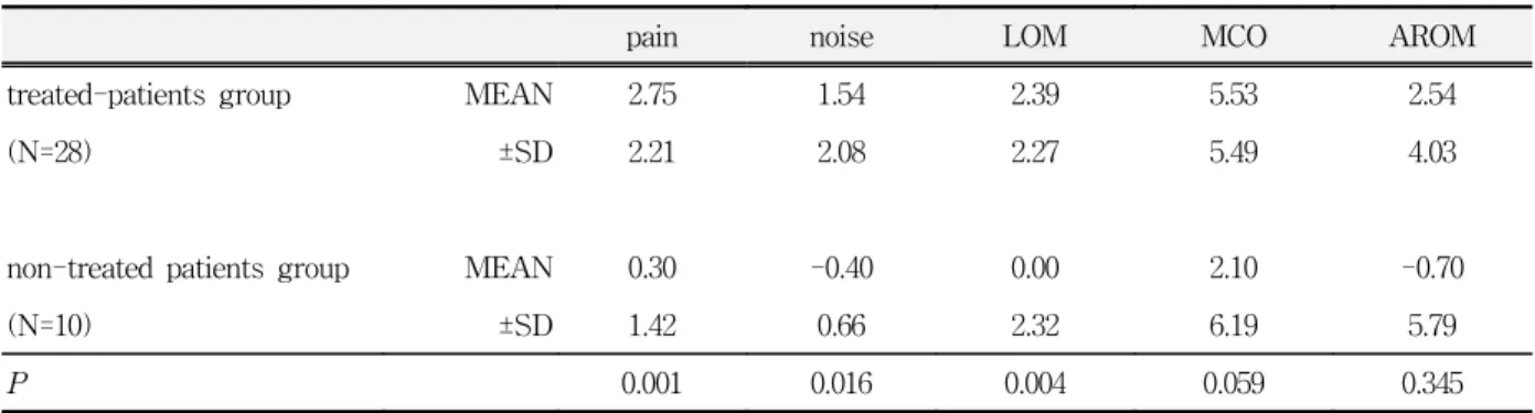
The changes in Table 5, 6, and 7 meant amount of subtract post-experiment from pre-experiment.
Negative numbers of subjective indexes - pain intensity, noise frequency, and limitation of motion - meant that symptoms were worsened. In MCO and AROM, positive numbers meant that the range of motion was increased and negative numbers meant that the range of motion was decreased.
The changes of clinical indexes are shown in table 5. There was significant difference between the treated patient group and the non-treated patient group in subjective indexes - pain intensity, noise frequency and limitation of motion(P<0.05).
Table 4. Difference of pressure pain threshold of S2 before and two weeks later after experiment in the non-treated patient group(Kpa, N=10)
PPT
Ⅰ MEAN 71.17
±SD 20.36
Ⅱ MEAN 69.39
±SD 16.70
P 0.305
Ⅰ : before first experiment
Ⅱ : two weeks later after first experiment
Table 5. Differences of change of clinical indexes before and two weeks later after experiment between the treated patient group and the non-treated patient group
pain noise LOM MCO AROM
treated-patients group MEAN 2.75 1.54 2.39 5.53 2.54
(N=28) ±SD 2.21 2.08 2.27 5.49 4.03
non-treated patients group MEAN 0.30 -0.40 0.00 2.10 -0.70
(N=10) ±SD 1.42 0.66 2.32 6.19 5.79
P 0.001 0.016 0.004 0.059 0.345
Table 6. Differences of change of pressure pain threshold between the treated patient group and the non-treated patient group in S2(Kpa)
PPT
treated patients MEAN 4.95
(N=28) ±SD 16.09
non-treated patients MEAN -1.79
(N=10) ±SD 15.07
P 0.055
Table 7. Differences of change of clinical indexes according to chronicity in the treated patient group(N=28)
pain noise LOM MCO AROM
acute MEAN 3.40 1.00 1.40 4.50 1.30
(N=10) ±SD 1.91 1.41 1.28 3.61 4.14
chronic MEAN 2.39 2.22 2.94 6.21 3.22
(N=18) ±SD 2.29 2.25 2.51 6.04 3.79
P 0.131 0.073 0.045 0.219 0.121
The PPT values on S2 point applied to all subjects between the treated patient group and the non-treated patient group were not significantly different(P>0.05). But there was a tendency to increase the PPT values of the treated patient group in contrast with the non-treated patient group. In the non-treated patient group, the PPT values became decreased(Table 6).
Table 7 shows that the clinical index according to chronicity in the treated patient group was significantly improved in limitation of motion (P<0.05).
Ⅳ. DISCUSSION
Since the presentation of the gate control theory, numerous reports have been published concerning various methods for the treatment of pain based on activation of afferent nerve fibers by electrical stimulation. EAST is the one case based on gate control therapy. There are considerable evidences that analgesic effects of acupuncture are mediated by an endogenous opiate-like mechanism, although this idea also remains somewhat controversial
7,9,14-25)
.
Andersson, et al.
4)reported that stimulation by the surface electrodes gave an onset and a decline of the effect upon pain threshold which were very similar to those obtained via needles. The surface electrodes were used in this study. The advantages of surface electrodes are that they are not invasive so the patient acceptance is better.
In this study, EAST statistically reduced the signs and symptoms after two weeks treatment compared with before treatment in the treated patient group. Subjective symptoms and objective signs were significantly improved. Johansson A., et al.
13)suggested that the clinical variables examined in actual acupuncture group showed statistically significant decreases. Carlsson
26)also reported reduced pain intensity following physiotherapy or acupuncture.
In this study, the PPT values of patient group showed significant increase in points of G2, G21,
Li4, S2, S3, Si19, and T17(P<0.05). Chung and Kim
3)suggested that the sensory and pain thresholds of mandibular posterior teeth were significantly increased during after EAST at both Li4 point, and concluded that EAST on Li4 point would be helpful in pain control of mandibular posterior teeth.
There were no changes of clinical indexes and PPT value between before experiment and after two weeks in the non-treated patient group. This result suggested that the effect of EAST was superior to spontaneous healing in short term treatment for TMD patients.
In the treated patient group, there were some different results according to chronicity. There were significant changes of LOM in chronic patients compared with acute patients.
The possible therapeutic effect has been explained by the gate control theory and endorphin system, and similar mechanisms are thought to act in TENS. In clinical trials, this ancient method has positive effects on longstanding chronic pain from the masticatory system of various origin
23)and positive effects have also been reported in patients resistant to earlier treatment
24).
In this study, several evaluation methods were used in order to give a good picture of the treatment effect. The results of the short-term evaluation show that the group receiving acupuncture responded favorably in all the indexes.
Both the subjective and clinically registered symptoms and signs improved significantly in the treated patient group, but were unchanged in the non-treated patient group. Clinically, particularly the pain intensity decreased.
Because a number of factors such as the size of algometer contact area, the rate of application, size of sample may influence the results obtained.
Further studies using large sample, standardized methods are needed.
This study confirmed clinical effect as well as
subjective effect, so EAST is one of useful method
to physical therapy for the patients with
temporomandibular disorders, especially for pain
control, though the mechanism of action is not fully understood.
Ⅴ. CONCLUSION
To evaluate the effect of electro-acupuncture stimulation therapy for TMD patients, we classified TMD patients into 2 groups, one was 28 treated patients group with EAST, the other group was 10 non-treated patients group without any therapy.
And then, we evaluated the result of therapy with following findings ; pressure pain threshold(PPT);
clinical index as a clinical findings; degree of pain intensity, frequency of noise, and limitation of motion as a subjective symptoms; MCO and AROM as an objective signs at before experiment and two weeks later after experiment.
The obtained results were as follows:
1. The clinical indexes of the treated patient group were significantly changed at post-treatment compared with pre-treatment (P<0.001).
2. The PPT values of meridian of the treated patient group were significantly increased in G2, G21, Li4, S2, S3, S8, Si19, and T17 at post-treatment (P<0.05).
3. The clinical indexes and the PPT values of the non-treated patient group were not changed at two weeks later compared with first experiment.
4. The clinical indexes of the treated patient group were more significantly changed than those of the non-treated patient group(P<0.05).
REFERENCES
1. Okeson J.P. : Bell's Orofacial Pains, 5th ed., Chicago, 1995, Quintessence, pp.123-133.
2. Acupuncture, NIH Consensus Development Panel on Acupuncture. JAMA, 280 : 1518-1524, 1998.
3. Chung, A.R. and Kim, K.S. : The effects of electro- acupuncture stimulation therapy on the pain threshold of mandibular posterior teeth using LI4(HAP GOK) points. J KAOM, 20 : 105-115, 1995.
4. Andersson S.A., Ericsson T., Holmgren E., and
Lindqvist G. : Electroacupuncture effect on pain thresholds measured with electrical stimulation of teeth. Brain Res, 63 : 393-396, 1973.
5. Chapmarr C.R., Chen A.C., and Bonica J.J. : Effects of intrasegmental electrical acupuncture on dental pain:
evaluation by threshold estimation and sensory decision theory. Pain, 3; 213-217, 1977.
6. Kaada B., Hoel E., Leseth K., Nygaard-Østby B., and Stovner J. : Acupuncture analgesia in the People's Republic of China. Tidsskr Nor Laegeforen, 94 : 417-442, 1974.
7. Melzack R. and Wall P.D. : Pain mechanism: a new theory. Science, 150 : 971-979, 1965.
8. Han J.S. and Terenius L. : Neurochemical basis of acupuncture analgesia. Am Rev Pharmacol Toxicol, 22 : 193-220, 1982.
9. Mayer D.J., Price D.D., and Rafii A. : Antagonism of acupuncture analgesia in man by the narcotic antagonist naloxane. Brain Res, 121 : 368-372, 1987.
10. Fields H.L. : Pain. New York, 1987, McGraw-Hill Book Co., pp309-315.
11. Kim, K.B. : The effects on the pain thresholds of mandibular anterior teeth of electro-acupuncture stimulation therapy on S5(Dae Yeoung) and CV24(Seung Jang) points. DanKook University Thesis, 1995.
12. Han, D.J. : The effects on the pain thresholds of mandibular anterior teeth of electro-acupuncture stimulation therapy on LI4(Hap Gok) point. DanKook University Thesis, 1995.
13. Johansson A., Wennegerg B., Wagersten C., and Haraldson T. : Acupuncture in treatment of facial muscular pain. Acta Odontol Scand, 49 : 153-158, 1991.
14. Terenius I., and Wahlstorm A.: Search for and endogenous ligand for the opiate receptor. Acta Physiol Scand, 94 : 74-81, 1975.
15. Chapman C.R., Colpitts Y.M., Benedetii C., and Gehring J.D. : Letter to Editor. Pain, 11 : 277, 1981.
16. Mayer D.J. and Price D.D. : Letter to Editor. Pain, 11 : 273, 1981.
17. Watkins L.R. and Mayer D.J. : Organization of endogenous opiate and nonopiate pain control systems. Science, 216 : 1185-1192, 1982.
18. Willer J.C., Roby A., Boulu P., and Boureau F.:
Comparative effects of electroacupuncture and transcutaneous nerve stimulation on the human reflex. Pain, 14 : 267-278, 1982.
19. Sjolund B., Terenius L., and Eriksson M. : Increased cerebrospinal fluid levels of endorphin after electro-acupuncture. Acta Physiol Scand, 100 : 382-384, 1977.
20. Cheng R.S. and Pomeranz B. : Electroacupuncture Analgesia is mediated by stereospecific opiate receptors and is reversed by antagonists of type Ⅰ receptors. Life Sci, 26 : 631-638, 1979.
21. McLenan H. : Some pharmacological observations on the analgesia induced by acupuncture in rabbits. Pain, 3 : 229-238, 1977.
22. Lovacky S., Lodin Z., Tauber O., et al. : Acupuncture treatment and its effect on low back pain: correlation with beta-endorphin immunoactivity. Am J Acupuncture, 15 : 245-251, 1987.
23. Johansson A., Wagersten C., Wenneberg B, Haraldson T., and Carlsson G.E. : Akupunkturbehandling vid kronisk smärta I ansikte och huvud.
Tandläkartidingen, 79 : 140-144, 1987.
24. Hiep N. and Stallard R. : Acupuncture - a valuable adjunct in the treatment of myofascial pain. J Dent Res, 53 : 203-211, 1974.
25. List T. and Helkimo M. : Acupuncture in the treatment of patients with chronic facial pain and mandibular dysfunction. Swed Dent J, 11 : 83-92, 1987.
26. Carlsson J. : The tension headache syndrome: Effects of acupuncture and physiotherapy. Thesis. University of Göteborg, 1990.
국문초록
측두하악장애환자에 대한 저주파 전자침 자극요법의 임상효과