서 론
뇌의 외상은 종합적인 손상으로서 이로 인해 뇌는 직 접적인 기계적 손상, 실질내출혈 및 거미막밑출혈
혈액뇌장 (intraparenchymal and subarachnoid hemorrhage),
벽(blood-brain barrier)파괴 흥분독성, (excitotoxicity) 그, 리고 허혈 (ischemia)등의 광범위한 손상을 입게 된다.
1)
지금까지 뇌외상을 유발시키는 실험적 모델로는 액체 타진손상법 (fluid percussion injury method)과 대뇌겉질 충돌손상법(cortical impact injury method)이 사용되었고, 이와 같은 실험 모델을 이용하여 외상 후 일어나는 신경세 포사망 축삭손실 미세아교세포, , (microglia)의 활성화 큰포,Vol. 25. No. 1, pp. 10 19, 2010∼
흰쥐 뇌의 냉동손상 후 항원제공세포와 신경교원섬유산단백의 발현 양상
강미영 ․ 김강련
1
대구산업정보대학 뷰티스타일리스트과
고신대학교 의과대학 해부학교실1
Mi-Young Kang ․ Kang-Ryune Kim
1
Department of Beauty Stylist, Daegu Polytechnic College University
1Department of Anatomy, Kosin University College of Medicine, Busan, Korea
――― Abstract ――――――――――――――――――――――――――――――――――――――――
Background : Traumatic brain injury is a multifaceted injury that involves direct mechanical damage, intraparenchymal and subarachnoid hemorrhage, breakdown of the blood-brain barrier, excitotoxicity, and ischemia. Even though much investigations were performed, acceptable mechanical informations were rare. The aim of this study was to reveal the migration of antigen presenting cells associated with inflammation and the expression pattern of GFAP associated with gliotic scars in cerebral cortex of rats after cryoinjury.
Material & Method : A total of 18 male Sprague-Dawley rats weighing 300 g, 2 months old, were used throughout the experiments. To injure the brain, rats were anesthetized for surgery with 3.5% chloral hydrate(1 ㎖/100 g, intraperitoneally); the frontal bones were exposed by elevating the skin; and craniectomies were performed adjacent to the central suture, midway between lambda and bregma. A cryoinjury was then created by applying a cold probe (3-mm-diameter steel rod chilled in liquid nitrogen) to the left frontal cortex (ipsilateral cortex) for 1 minute. Rats were sacrificed at 6 hours, 1, 4, 7 and 14 days postsurgery (n=3, per time point), and three rats were sacrificed as normal controls. Serial brain cryosections were made by cryostat. For immunohistochemistry, brain tissue sections were allowed to react with mouse anti-rat MHC class II antibody(1:500), mouse anti-rat ED2 antibody(1:500), and mouse anti-rat GFAP antibody(1:200).
Results : MHC class II positive dendritic cells were absent in normal cerebral cortex parenchyme, but were more 16 times number in injured rats at 7 days after cryoinjury. ED2 positive macrophages were rare in normal cerebral cortex parenchyme, and were more 14 times number in injured rats at 4 days after cryoinjury. GFAP positive astrocytes were not observed in normal rats, and increased suddenly at 4 days rats after cryoinjury. GFAP positive neurons were not expressed in normal rats, and in cryoinjured rats.
Conclusion : In conclusion, the above results suggest that the increases of the number of the two kinds of the antigen-presenting cells affect cell-mediated immune responses and phagocytosis, and GFAP positive cells attends in the formation of gliotic scars.
―――――――――――――――――――――――――――――――――――――――――――――――――
Key words : Antigen presenting cell, Dendritic cell, Macrophage, GFAP, Cryoinjury, Rat brain
교신저자 김 강 련:
주소 : 602-702 부산광역시 서구 암남동34번지 고신대학교 의과대학 해부학교실 TEL : 051-990-6411 FAX : 051-990-3081 E-mail : [email protected]
식세포의 여과(filtration),그리고 별아교세포(astrocyte)의 반응에 대한 정보를 얻을 수 있었다.
2-5)
그러나 위와 같은 방법들은 객관성과 일관성이 부족한 결점을 가지고 있어 이를 극복하기 위해 최근에는 냉동 손상법(cryoinjury method)이 일부 연구자들에 의해 시 도되고 있다 냉동손상법은 액체질소로 냉동시킨 쇠막대. 를 이용하여 직접 대뇌겉질(cerebral cortex)에 국소적인 손상과 염증(inflammation)을 유발시키기 때문에 일관성 과 객관성을 확보할 수 있는 장점이 있다.
6)
뇌가 손상된 후에 염증반응이 나타나고 회복되는 과정 에서 항원제공세포 (antigen presenting cell)인 수지상세 포(dendritic cell, DC)와 큰포식세포(macrophage)가 출 현하는 것으로 알려져 있다.
7,8)
그러나 이 세포들은 정 상적인 뇌막(meninges), 맥락얼기(choroid plexus), 그리 고 뇌 척수액(cerebrospinal fluid)에는 존재하나 뇌실질 에는 없는 것으로 알려져 있기 때문 (brain parenchyma)에 외부에서 손상부위로 이동할 것으로 추정되고 있 다.
9,10)
수지상세포와 큰포식세포는 같은 단핵백혈구계열에 속하는 세포이지만 시험관에 부착하는 성질 핵 및 세포, 질돌기의 형태 항원발현세포로서의 기능 용해소체, , 의 활성도 그리고 탐식능력 등에서 서로 다르 (lysosome)
다.
11)
그럼에도 불구하고 많은 학자들은 두 세포가 면역 반응에 필요한 잠재적 면역자극세포 또는 보조세포인 항원제공세포라는 점에서 같은 부류의 세포군으로 취급 하고 있다.12,13)
항원제공세포는 항원을 세포 내에서 처리한 다음, 소포체 (endoplasmic reticulum)에서 합성된 major 분자에 결합 histocompatibility complex (MHC) class II
시켜 T세포에 제공함으로써 T세포의 활성화에 관여하 는 것으로 알려져 있다.
14)
대부분의 학자들은 큰포식세포가 면역반응의 주된 항 원제공세포라고 생각해 왔지만 Steinman 등
15)
이 생쥐의 비장에서 최초로 수지상세포를 분리한 이후 최근 여러 학자들은 수지상세포가 다른 항원제공세포에 비해 세T 포 의존성 면역반응에 있어서 가장 효과적인 항원제공세 포로 간주하고 있다.16,17)
국제세망내피학회 용어위원회 (Reticuloendothelial Society 는 수지상세포를 출현부 Committee on the Nomenclature)
위 및 세포막특성에 따라 다음과 같은 종류의 세포군으3 로 분류하였다.
18)
첫째 부류는 피부에 분포하는 랑게르 한스세포(Langerhans cell)와 뇌를 제외한 대부분의 장기에 존재하는 수지상세포를 간질수지상세포 (interstitial 라고 하였고 둘째는 혈액 내에 있는 혈액 수지상세
DC) ,
포 (blood DC)와 림프 내에 존재하는 장막세포와 같은 수지상세포를 순환수지상세포(circulating DC)라고 하였 으며 셋째는 세포지역 및 흉선수질에 분포하는 깍지수, T 지상세포 (interdigitating DC)와 종자중심 (germinal
내에 위치한 소포수지상세포 등 림
center) (follicular DC) 프기관 내에 존재하는 수지상세포를 림프성수지상세포
라고 분류하였다 따라서 수지상세포는 신 (lymphoid DC) .
체내 전 부위에 걸쳐 광범위하게 분포하면서 세포에 항T 원을 제공하기 위해 특수하게 분화되어 있는 세포군이라 고 할 수 있다.
이러한 수지상세포들은 항원을 처리한 후 혈액이나 림 프액을 통해 림프기관으로 이주해 가서 세포에 항원을T 제공하기 때문에 존재하는 부위에 관계없이 기능적으로 유사하다고 할 수 있다 신체 내에서 수지상세포들은 비. 록 분명한 계통을 이루고 있지만 각 장기 미세환경 조건,
발생단계
(microenvironmental condition), (developmental
및 기능적 상태 에 따라 다양한
stage) (functional status)
표현형을 구성하며 복잡한 체계를 이루고 있다.
19,20)
수지상세포에 대한 지금까지의 연구는 대부분이 in vitro에 국한되어 왔고, in vivo 연구는 빈약한 형편이다.그리고 림프성 수지상세포에 대한 연구는 비교적 활발한 편이나 랑게르한스세포를 제외한 다른 간질 수지상세포 에 대한 연구는 부진한 실정이다.
21,22)
큰포식세포는 형태학적 및 기능적으로 이질적인 세포 군이다 큰포식세포는 세포의존성 면역 염증반응 조직. , , 재생 맥관 형성 종양파괴 및 미생물파괴에 관여한다, , . 또한 큰포식세포는 알파 종양괴사인자 (tumor necrosis
나 산화질산 과 같은 면역조절
factor alpha) (nitric oxide)
물질 (immunomodulator)을 분비한다고 알려져 있다 그. 러므로 큰포식세포는 면역반응에서 수지상세포의 기능 을 하향조절(down regulation)시킬 것으로 추측하고 있
다.
23-27)
그러므로 어떤 기관의 조직 내에 분포하는 큰포식세포와 수지상세포의 비율은 큰포식세포가 수지상 세포의 항원제공기능에 영향을 미칠 것으로 예상된다.
근래에 항원제공세포 중에서 수지상세포와 큰포 식세포에 존재하는 특이항원에 대한 단일클론항체 (monoclonal antibody)가 개발되었으며 이 중에서, MHC
항체는 수지상세포의 세포표면에 존재하는
class II MHC
항원을 감지하는 특성을 가지며
class II Ia
28)
, ED2항체는 조직 내에 상주하고 있는 큰포식세포 표면에 있는 특이한 항원에 결합하는 성질을 가지고 있어 조직 큰포식세 포를 구분 동정할 수 있게 되었다.
29,30)
뇌가 손상되면 신경아교흉터(glial scar)가 생겨 신경 세포 축삭 재생이 방해를 받게 되는데 이 때 중간세사단 백들이 신경아교흉터를 형성시키는 주된 역할을 한다고 알려져 있다.
31)
발생중이거나 성숙한 중추신경계통의 특수세포에는 또는 와 같은 중간 세사가 발견된 vimentin, nestin GFAP
다.
32,33)
이 중 GFAP 양성반응 다능성세포 (multipotent는 손상 자극을 받게 되면 그들의 표현형
cell) (phenotype)
이 별아교세포(astrocyte)로 변형된다.
34,35)
그러므로 뇌 가 손상 되고 회복되는 과정에서GFAP에 대한 발현 양 상을 추적하면 회복정도를 추정할 수 있을 것으로 생각 된다.중간세사는 일반적으로 세포내 단백질의 약1%를 차 지하지만 각질형성세포나 신경세포에서는 약85%를 차 지한다.
36)
중간세사는 직경이10nm되는 섬유로서 유전 자의DNA염기서열과 아미노산 구성에 따라 개의 종류6 로 분류된다.37)
제 형부터 제 형 그리고 제 형은 세Ⅰ Ⅳ Ⅵ 포질내 중간 세사이고 제 형은Ⅴ nuclear lamin으로서 핵 의 중간세사이다 사람에서는. 50가지 이상의 중간세사 유전자가 존재하며 신체 대부분의 세포에 중간세사가 존 재하지만 세포마다 다른 양상으로 발현된다.38)
세포질 내 중간세사는 핵 주위에서부터 세포막 쪽으로 방사상으 로 배열된 망상구조를 이루는데 세포의 종류에 따라서, 서로 다른 종류와 형태의 망상구조를 갖는다.39)
그러므로 본 연구자는 생후 개월 된 흰쥐 뇌에 냉동2 손상을 입힌 후 대뇌겉질의 파괴 시기와 회복 시기에 따 라 항원제공세포들인MHC class II양성반응 수지상세포 와ED2양성 반응 큰포식세포의 이동과정과 중간세사인 의 발현 양상을 면역세포화학적 방법을 이용하여 GFAP
밝히고자 이 연구를 시도하게 되었다.
연구대상과 방법 실험동물 1.
실험동물은 항온 항습이 유지되고12시간마다 밤낮이 교체되며 사료와 물을 자유로이 섭취할 수 있는 동일한 조건하에서 사육한 생후 개월 평균2 ( 300 g)된 웅성 흰쥐
를 사용하였다 (Sprague-Dawley) .
실험군은 냉동손상 후 경과시간에 따라 시간군6 , 1일
군 일군 일군, 4 , 7 , 14일군으로 분류하였고 정상군을 실험, 군에 대한 대조군으로 사용하였다 흰쥐는 각 실험군마. 다 마리씩 모두3 18마리를 사용하였다.
냉동손상방법 2.
흰쥐를3.5% chloral hydrate(1㎖/100 g)로 복강 주사하 여 마취시킨 후 두피를 절개하여 머리뼈를 노출시킨 다 음 정수리점(bregma)과 시옷점(lambda)사이에서micro 세신정밀 대구 한국 를 이 motor handpiece (Forte 300, , , ) 용하여 머리뼈절제 (craniectomy)를 시행하여 왼쪽 이마 엽(frontal lobe)을 노출시킨 뒤 액체질소로 미리 얼려 두 었던 직경3 mm철막대로 분간 이마엽 표면을 냉동 손1 상시켰다.
뇌절편 제작방법 3.
를 복강내주사하여 동 3.5% chloral hydrate(1 ㎖/100 g)
물을 마취시킨 상태에서 머리를 분리시킨 뒤 최단시간 내에 뇌를 적출하였다 뇌를. rat brain matrix (RBM)에 넣 고 면도날로 냉동손상시킨 이마엽 부위의 중앙을 통과하 는 관상절단 뇌절편을 만들었다.
조직표본제작 4.
뇌절편을 탄산가스 (CO
2
gas)로 동결고정한 후 동결절 편기 (cryocut, Reichert-Jung, Heidelberg, Germany)를 사 용하여 약6 ㎛두께로 조직절편을 제작하고 이들 조직, 절편을poly-l-lysine coated slide에 부착시켰다 조직절편. 을40℃의 조직 신전기에서40분정도 충분히 말린 다음에서 로 분간 고정하였고 이를
4℃ 4% paraformaldehyde 5 , 다시 조직 신전기에서 2 시간이상 충분히 말렸다.
면역조직화학적 염색 5.
조직절편을 0.02M인산염 완충액(phosphate buffered 에서 약 분 동안 회 세척한 후 조 saline; PBS, pH. 7.4) 10 2
직 내 과산화효소의 활성을 차단하기 위해0.3% 과산화 수소 (H
2
O2
)가 포함된 메틸알코올을 첨가시킨 용액에서 분간 처리하고 로 분간 회 세척한 다음 정20 , PBS 10 3 5%
상 말 혈청 (normal horse serum)에30분간 유치하였다. 그 후 조직절편들을 세척하지 않고 가볍게 털어 낸 다음 일차항체인 mouse anti-rat MHC class II antibody (OX6, 희석비율 1:500, Serotec, Oxford, UK)와 mouse anti-rat
희석비율 를 에서
ED2 antibody ( 1:500, Serotec) 4℃ 14-16 시간 동안 각각 적용시켰다.
일차항체를 적용시킨 뒤PBS로 10분씩 회 세척하였3 고 이어서 이차항체인, biotinylated horse anti-mouse IgG 를 실온에서 시간 동 (Vector Lab, Burlingame, CA, USA) 1 안 적용시켰다 그 후 조직절편들을. PBS로10분씩 회 수3 세한 다음 30분전에 미리 만들어 실온에 방치해 놓은 용액 ABC (avidin-biotin horseradish peroxidase complex)
을 실온에서 시간동안 적용시켰다 이어서
(Vector Lab) 1 .
이들 조직절편을 PBS로 10분씩 회 세척한 후3 , 0.05M 에
Tris-HCl buffer (Tris buffer, pH7.6) 0.05% 3,3' - diaminobenzine tetrahydrochloride; DAB, Sigma Co, St 와 과산화수소가 혼합된 용액으 Louis, MO, USA) 0.01%
로 처리하여 약10분간 실온에서 발색시켰다 조직 절편. 의 발색상태를 현미경하에서 확인한 후 Tris buffer, PBS 및 증류수로 각각 10분간씩 차례로 세척한 다음, Harris 으로 대조염색을 하고 통상적인 과정을 거 hematoxylin ,
쳐Permount (Polysciences, Warrington, PA, USA)로 봉입 하였다.
뇌조직을 공초점레이저현미경으로 관찰하기 위하여 일 차항체인mouse anti-rat GFAP antibody (희석비율1:200,
를 적용한 다음 이차항체인
Serotec) horse anti-mouse FITC (fluorescein isothiocyanate, Boehringer Mannheim, 를 실온에서 시간동안 조직절편에 적용시 Germany) 1
켰으며, PBS로 10분씩 3회 세척한 뒤 Acquimount 로 봉입하였다 (Polysciences, Warrington, PA, USA) .
면역염색의 정확도를 검증하기 위해 염색과정에서 일 차항체나ABC용액 적용과정을 생략시켜 본 결과 이 과, 정을 거친 표본에서는 어떤 면역반응성도 나타나지 않았 기 때문에 면역염색의 정확성이 입증되었다.
검경 및 계수 6.
뇌조직 표본을 광학현미경 (Axiophot, Zeiss, Hallbergmoos, 과 공초점레이저현미경
Germany) (CLSM510, Zeiss)을 이용 하여 검경하였으며 관찰부위로는 왼쪽 이마엽의 관상절단 면의 대뇌겉질(cerebral cortex) 이었다.
항원제공세포의 계수는 개의 실험군으로 구분하고6 한 개체에서10개의 표본을 무작위로 선택하여 각 실험 군 당50개의 표본을 계수하였다 각 실험군의 수적 변화. 의 통계적 유의성을 확인 하기 위해서 일원분산분석
을 이용하여 검증하였다 (one-way ANOVA) .
결 과
흰쥐 대뇌겉질에 분포하는 항원제공세포들이 국소적 인 냉동손상을 받았을 때 이 세포들이 손상부위로 이동 하는 양상과 중간세사인GFAP의 발현 여부를 알아보기 위해 생후 개월 된 흰쥐의 뇌를 대상으로 냉동 연속조2 직절편을 제작하여 면역조직화학적 염색을 시행한 뒤 양 성 면역 반응성을 보인 세포를 관찰하고 계수하여 얻은 결과는 다음과 같다.
수지상세포와 큰포식세포의 세포수의 변화 1.
정상군을 대조군으로 하여 왼쪽 이마엽의 대뇌겉질 뇌 실질에 분포하는 세포수를 계수하였다.
양성반응 수지상세포는 정상군에서는 관 MHC class II
찰되지 않았고 냉동손상 후 시간째에는 단위면적당6 ( )㎟ 평균1.0 ,개 손상 후 일째에는1 1.4 ,개 손상 후 일째에는4 개로 증가하다가 손상 후 일째에는 개로 급격히
7.5 7 16.3
증가하였으며 손상 후, 14일째에는10.1개로 세포수가 유 의성 있게 감소하였다(p<0.05, Table 1).
양성반응 큰포식세포는 정상군에서는 평균 개
ED2 1.2
가 관찰되었고 냉동손상 후 시간째에는, 6 3.6 ,개 손상 후 일째에는 개로 조금 증가하다가 손상 후 일째에는
1 4.4 4
개로 급격한 증가를 보였으며 손상 후 일째에는
14.2 7
개로 급격하게 감소하였고 손상 후 일째에는 개
5.2 , 14 3.4
로 세포수가 유의하게 감소하였다(p<0.05, Table 1).
Cell types Time after cryoinjury
Normal 6 hours 1 day 4 day 7 day 14 day Dendritic cells 0.0±0.0* 1.0±0.2* 1.4±0.5* 7.5±0.4* 16.3±0.3*10.1±0.6* Macrophages 1.2±0.2* 3.6±0.4* 4.4±0.7* 14.2±0.5* 5.2±0.4* 3.4±0.3* The number represents mean±standard deviation.
Cells were counted per unit area( ) on 50 slides in each rat groups.㎟
* : p<0.05 on one-way ANOVA.
Table 1. The number of MHC class II positive dendritic cells and ED2 positive macrophages in the ipsilateral frontal cortex parenchyme of normal and cryoinjured rat brains
조직화학적 변화 2.
정상군 1)
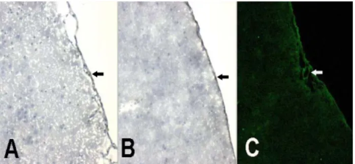
정상군의 이마엽 대뇌겉질의 실질에서는 MHC class II 양성반응 수지상세포는 관찰되지 않았 으나(Fig. 1A),
양성반응 큰포식세포는 혈관벽에서 관찰되었다
ED2 (Fig.
그리고 연질막에서는 수지상세포와 큰포식세포가 1B).
관찰되었다(Fig. 1A, 1B). GFAP는 혈관벽과 연질막에 매우 약한 양성반응을 나타내었다(Fig. 1C).
Fig. 1. Serial sections of cerebral cortex of normal rat brain.
(A) A (OX6) antibody for dendritic cells. MHC class II positive cells were absent in parenchyme, but several dendritic cells were located on pia mater (black arrow). (B) ED2 positive macrophages were visible on the wall of a blood vessel (white arrow) and on pia mater (black arrow). (C) A very weak immunoactivity of GFAP was detected on the wall of blood vessels (white arrow). A: OX6, DAB; B: ED2, DAB;
C: GFAP, FITC, Confocal microscope. x 100.
시간군 2) 6
냉동손상을 준 후 시간째에 대뇌겉질의 손상부위에6 서 연질막밑공간 (subpial space)이 넓어 졌으며 출혈과 약간의 괴사가 있었다. 소수의MHC class II 양성반응 수지상세포들이 연질밑막(subpial mater)에서 관찰되었 으나 손상된 뇌실질에서는 발견되지 않았다(Fig. 2A).
양성 반응 큰포식세포들이 손상된 뇌실질에서 발 ED2
견되었다(Fig. 2B). 뇌실질에서 발견된 큰포식세포는 혈관옆에 위치하고 있었고 둥근핵을 갖고 있었다(Fig.
양성반응 중간세사들이 연질막과 연질막 2B). GFAP
밑막 그리고 손상된 뇌실질에서 관찰되었다, (Fig. 2C).
일군 3) 1
냉동손상을 준 후 일째에 대뇌겉질의 손상부위에서1 광범위한 괴사가 나타났다. 약간의MHC class II 양성 반응 수지상세포들이 손상된 뇌실질에서 관찰되었다 매우 큰 수지상세포가 손상 받지 않은 뇌 (Fig. 3A).
실질에 나타났다(Fig. 3A). 소수의 ED2 양성반응 큰 포식세포들이 손상된 뇌실질에서 관찰되었다(Fig. 3B).
다수의 GFAP 양성반응 세포들이 손상된 뇌실질에서 관찰되었다(Fig. 3C).
Fig. 3. Serial sections of cerebral cortex lesions at 1 day after cryoinjury. There was a extensive necrosis. (A) Several MHC class II positive dendritic cells were observed in injured parenchyme (black arrows). A large dendritic cell was appeared in uninjured parenchyme (white arrow). (B) A small number of ED2 positive macrophages were visible in injured parenchyme (white arrows). (C) Many GFAP positive cells were observed in injured areas (white arrows). A: OX6, DAB;
B: ED2, DAB; C: GFAP, FITC, Confocal microscope. x 100.
일군 4) 4
냉동손상을 준 후 일째에 대뇌겉질의 손상부위에서4 괴사와 신경아교흉터가 관찰되었다. MHC class II 양 성반응 수지상세포들이 손상된 뇌실질 부위의 중심부 와 주변부에서 관찰되었다(Fig. 4A). ED2 양성반응 큰포식세포들이 손상된 뇌실질 부위에서 관찰되었다 강하게 염색된 양성반응 세포들이 (Fig. 4B). GFAP
손상된 뇌실질에서 많이 관찰되었다(Fig. 4C).
Fig. 2. Serial sections of cerebral cortex lesions at 6 hours after cryoinjury. There were extension of subpial space, hemorrhage and necrosis. (A) Several MHC class II positive dendritic cells were observed in subpial mater (black arrows) and other positive cells were not in injured parenchyme. (B) ED2 positive macrophages were visible in injured parenchyme (black arrows). A macrophage with round nuclei was located adjacent a blood vessel (white arrow). (C) GFAP positive intermediate filaments were observed in pia mater and subpial mater and in injured parenchyme (white arrow). A: OX6, DAB; B: ED2, DAB; C: GFAP, FITC, Confocal microscope. x 100.
Fig. 4. Serial sections of cerebral cortex lesions at 4 days after cryoinjury. There were necrosis and gliotic scar. (A) Several MHC class II positive dendritic cells were observed in core area (white arrows) and peripheral area(black arrows) of injured parencyme. (B) ED2 positive macrophages were observed in injured parenchyme (white arrows). (C) Many GFAP strongly immunoreactive cells were observed in injured area (white arrow). A: OX6, DAB; B: ED2, DAB; C: GFAP, FITC, Confocal microscope. x 100.
일군 5) 7
냉동손상을 준 후 일째에 대뇌겉질의 손상부위에서7 괴사와 신경아교흉터가 관찰되었다. 다수의 MHC 양성반응 수지상세포들이 손상된 뇌실질 부위 class II
에서 발견되었다(Fig. 5A). 다수의 수지상세포들이 군 집을 이루고 있었다(Fig. 5A). 소수의 ED2 양성반응 큰포식세포들이 손상된 뇌실질 부위에서 관찰되었다 약하게 염색된 양성반응 세포들이 (Fig. 5B). GFAP
손상된 뇌실질에서 관찰되었다(Fig. 5C).
Fig. 5. Serial sections of cerebral cortex lesions at 7 days after cryoinjury. There were necrosis and glial scar. (A) Many MHC class II positive dendritic cells were observed in injured parencyme(black arrows). There was a cluster of dendritic cells in injured area (white arrow). (B) Several ED2 positive macrophages were observed in injured parenchyme (white arrows). (C) Pale GFAP positive cells were observed in injured parenchyme (white arrow). A: OX6, DAB; B: ED2, DAB; C: GFAP, FITC, Confocal microscope. x 100.
일군 6) 14
냉동손상을 준 후 14일째에 대뇌겉질의 손상부위에 서 괴사와 신경아교흉터가 관찰되었다. 소수의 MHC 양성반응 수지상세포들이 손상된 뇌실질 부위 class II
에서 관찰되었다(Fig. 6A). 소수의 ED2 양성반응 큰 포식세포들이 손상된 뇌실질 부위에서 관찰되었다(Fig.
약하게 반응된 양성반응 세포들이 손상된
6B). GFAP
뇌실질에서 관찰되었다(Fig. 6C).
Fig. 6. Serial sections of cerebral cortex lesions at 14 days after cryoinjury. There were necrosis and glial scar. (A) Several MHC class II positive dendritic cells were observed in injured parencyme(white arrows). (B) Several ED2 positive macrophages were observed in injured parenchyme (white arrows). (C) Small GFAP positive cells were observed in injured parenchyme (black arrows). A: OX6, DAB; B: ED2, DAB; C: GFAP, FITC, Confocal microscope. x 100.
고 찰
경증뇌외상 (mild traumatic brain injury)은 뇌손상의 흔한 형태로서 야외 여가생활이 많은 현대사회에서 증 가되는 추세에 있다.
40,41)
경증뇌외상은 순간의식불명 기억상실 인식과 감정의, , 불균형 등을 수반하며 환자는 수주일 내에 정상으로 회 복되는 경우도 있지만 일부 환자들은 수개월 내지 수년 간 학습능력과 기억장애를 호소하는 경우도 있다.
42-44)
지금까지 경증뇌외상으로 인한 생물학적 기전을 여러 동물모델을 이용하여 시도하였지만 연구결과는 아직 미 진한 상태이다.
45)
본 연구에서 시도된 냉동손상법은 인 간의 경증뇌외상과 완전히 같을 수는 없지만 뇌에 너무 심한 손상을 입히지 않는 점과 국소적이고 반복적인 손 상을 줄 수 있다는 점에서 이 방면의 연구에 채택되어도 좋을 것으로 생각되어진다.수지상세포와 큰포식세포와 같은 항원제공세포는 단
백항원을 섭취하여 분쇄한 다음 적절한 입체구조를 갖 도록 처리하여 소포체에서 합성된MHC class II 분자에 비공유결합으로 부착시키고 이, peptide-MHC 복합체를 세포 표면에 발현시키는 역할을 한다.
46)
이러한 방법으 로 처리된 항원은 림프구의 세포표면에 존재하는 세T T 포 수용체(T cell receptor)에 의해 인식될 수 있다. T세 포 중에서도MHC class II분자를 인식할 수 있는 세포T 는 조력 세포T (helper T cell)로서 이 세포의 특이 수용, 체와 복합체를 이루게 된다 이러한 과정은 세포 활성. T 화에 필수적이다.47-49)
정상적인 뇌에서 수지상세포와 큰포식세포는 뇌막이 나 맥락얼기에 주로 분포되어 있다고 알려져 있다.
9)
수 지상세포는 뇌실이나 지주막밑공간 (subarachnoid space) 에서도 관찰되는 반면 큰포식세포는 경질막이나 지주막, 의 결합조직 맥락얼기의 버팀질, (stroma),그리고 연질밑 막(subpial mater)에서 발견되어진다.50)
뇌실질에 있는 토박이큰포식세포(resident macrophage)는 미세아교세포로 존재하며 주로 혈관주위에 위치한다
(microglia) .
51)
본연구자는 정상군에서 이런 사실을 재확인할 수 있었다 (Fig. 1).
이와 같이 정상뇌에도 항원제공세포들이 존재하나 다 른 조직에 비해 매우 적은 편이다. MHC class II 양성반 응 수지상세포는 동물의 종에 따라 다르고 같은 기관일 지라도 부위에 따라 다르다 성숙한 흰쥐 마우스 사람. , , 의 홍채와 모양체에 분포하는 수지상세포는 흰쥐 홍채의 경우 1 mm
2
당 평균400개가 존재하며 홍채의 기저부 중, 간부 가장자리에 따라 차이가 난다,52)
고 하며 성숙한 흰, 쥐의 기관지 상피에 분포하는MHC class II 양성반응 수 지상세포는 큰 기관지의 경우 1 mm2
당 평균600-800 ,개 작은 기관지의 경우 1 mm2
당 평균75개 정도였고 배쪽부 위보다는 등쪽부위에서 그 수가 많다고 한다.53)
또한ED2 양성반응 큰포식세포는 림프절 수질 골수,
54,55)
, 폐포사이중격 눈의 홍채 및 모양체,56,57)
등 결합조직에 널리 분포한다고 알려져 있다. ED2양성반응 큰포 식세포는 성숙한 흰쥐의 홍채에서 1 mm
2
당 평균 600 -개정도 분포하여 양성반응 수지상세포
800 MHC class II 보다 약 1.4배 정도 많다고 한다.
56,57)
본 연구에서 정상군의 뇌실질에서는MHC class II양성 반응 수지상세포를 관찰하기 어려웠으나, ED2양성반응 큰포식세포는 1 mm
2
당 평균 1.2개 정도 분포하는 것을 확인하였다(Table 1). 발견된 큰포식세포들이 혈관벽을 따라 분포하기 때문에 미세아교세포로 여겨진다.그러나 본 연구에서 냉동손상 후 항원제공세포들이 손 상된 부위에서 다수 관찰되었고 세포군에 따라 차이를 보였다. MHC class II양성반응 수지상세포는 손상 후7 일째에 평균16.3개였으며, ED2 양성반응 큰포식세포는 손상 후 일째에 평균4 14.3개였다(Table 1). 이와 같은 현상은 항원제공세포들이 외부에서 이동했다는 것을 의 미하는 것이다 많은 큰포식세포가 수지상세포보다 더. 일찍 손상부위에 도달할 수 있었던 것은 뇌실질내에 있 는 혈관을 통해 용이하게 이동하였고 냉동손상에 의한 초기의 출혈과 괴사로 인해 출현한 염증세포들을 포식했 을 것이며 연질막에서 이동한 큰포식세포도 있었을 것으 로 생각된다 정상군과는 달리 손상된 부위의 수지상세. 포는 큰포식세포보다 세포수가 더 증가되어 있었는데 그 이유는 수지상세포가 연질막 뿐만 아니라 지주막밑 공간의 뇌척수액에도 있었기 때문에 이동시간이 더 걸렸 지만 이동한 전체 세포수는 더 많아졌다고 여겨진다.
뇌가 손상받으면 다능전구세포 (multipotent progenitor 가 활성화되어 신경세포 별아교세포 희소돌기아교
cell) , ,
세포와 같이 뇌조직을 회복시키는 세포로 분화한다.
34,35)
그러므로 이 전구세포들의 증식과 분화를 조절하면 뇌의 재구성 형태를 자유롭게 바꿀 수 있을 것이다 발생중인. 뇌에서 세포가 이동하거나 성장할 때GFAP이나 nestin 같은 발생중간세사 (embryonic intermediate filament)가 관여하는 것처럼 손상된 뇌에서도 중간세사들이 뇌를 회 복시키기 위해 증가한다는 보고가 있다.58)
그러므로 양성반응 세포들은 대부분 별아교세포들이라고 GFAP할 수 있다.
이 연구에서 신경세포가 정상군과 실험군 모두에서
에 반응하지 않았지만 양성반응 세
GFAP (Fig. 1C), GFAP
포들이 손상 후 일째에 손상부위에서 가장 많이 나타났4 다(Fig. 4C). 이 세포들은 신경아교흉터(glial scar)를 형 성하는 별아교세포라고 할 수 있겠다 한편 혈관을 둘. , 러싸는 세포들이 GFAP에 강한 양성반응을 보였다 이. 세포들은 정상군에서도 발견되었으며(Fig. 1C), 손상 후 일째에 혈관에서 아주 많았다 이 세포들은 혈
4 (Fig 4C).
관을 재구성하는데 필요한 중간세사를 갖는 세포들로 보 여진다.
결 론
흰쥐 대뇌겉질에 분포하는 항원제공세포들이 국소적 인 냉동손상을 받았을 때 이 세포들이 손상부위로 이동 하는 양상과 중간세사인GFAP의 발현 여부를 알아보기 위해 생후 개월 된 흰쥐의 뇌를 대상으로 냉동 연속조2 직절편을 제작하여 면역조직화학적 염색을 시행한 뒤 양 성 면역 반응성을 보인 세포를 관찰하고 계수하였다.
양성반응 수지상세포는 정상적인 이마엽 대 MHC class II
뇌겉질의 뇌실질에서는 관찰되지 않았지만 냉동손상 후, 일째에 정상보다 배 이상 증가하였고 양성반응
7 16 , ED2
큰포식세포는 정상적인 이마엽 대뇌겉질의 뇌실질에에 서 소수 관찰되었으나 냉동손상 후 일째에는 정상보다4 배 이상 증가하였으며 그리고 양성반응 별아교
14 , GFAP
세포들은 정상군에서 관찰되지 않았으나 손상 후 일 때, 4 에 급격한 증가를 보였으며 신경세포는 정상군 뿐만 아, 니라 냉동손상을 준 실험군 모두에서GFAP양성반응을 보이지 않았다는 실험결과를 얻 었다.
위와 같은 실험결과로 미루어 보아 두 종류의 항원제 공세포는 세포의존성 면역반응과 포식작용을 하며, 양성반응세포들은 신경아교흉터형성에 관여한다 GFAP
고 생각되어진다.
참고문헌
1) McIntosh TK, Saatman KE, Raghupathi R, Graham DI, Smith DH, Lee VM, Trojanowski JQ : The Dorothy Russell Memorial Lecture. The molecular and cellular sequelae of experimental traumatic brain injury : pathogenetic mechanisms. Neuropathol Appl Neurobiol 24:251-267, 1998.
2) Sun D, Tani M, Newman TA, Krivacic K, Phillips M, Chernosky A, Gill P, Wei T, Griswold KJ, Ransohoff RM, Weller RO : Role of chemokines, neuronal projections, and the blood-brain barrier in the enhancement of cerebral EAE following focal brain damage. J Neuropathol Exp Neurol 59:1031-1043, 2000.
3) Aihara N, Hall JJ, Pitts LH, Fukuda K, Noble LJ : Altered immunoexpression of microglia and macrophages after mild head injury. J Neurotrauma 12:53-63, 1995.
4) Baldwin SA, Scheff SW : Intermediate filament change in astrocytes following mild cortical contusion. Glia 16:266-275, 1996.
5) Sato M, Chang E, Igarashi T, Noble LJ : Neuronal injury and loss after traumatic brain injury: time course and regional variability. Brain Res 917:45-54, 2001.
6) Kim MD, Min DS, Sim KB, Cho HJ, Shin T : Expression and potential role of phospholipase D1 in cryoinjured cerebral cortex of rats. Histol Histopathol 19:1015-1019, 2004.
7) Austyn JM, Wood KJ : Antigen-presenting cells. In principles of cellular and molecular immunology. Oxford, Oxford University Press, 1993, pp115-177.
8) Pashenkov M, Teleshova N, Link H : Inflammation in the central nervous system : the role for dendritic cells. Brain Pathol 13:23-33, 2003.
9) McMenamin PG, Wealthal RJ, Deverall M, Cooper SJ, Griffin B : Macrophages and dendritic cells in the rat meninges and choroid plexus : three-dimentional localisation by environmental scanning eletron microscopy and confocal microscopy. Cell Tissue Res 313:259-269, 2003.
10) Newman TA, Ian Galea I, van Rooijen N, Perry VH : Blood-derived dendritic cells in an acute brain injury. J Neuroimmunol 166:167-172, 2005.
11) Steinman RM, Nussenzweig MC : Dendritis cells : Features and functions. Immunol Rev 53:127-147, 1980.
12) Van Voorhis WC, Witmer MD, Steinman RM : The phenotypes of dendritic cells and macrophages. Federation Proc, 42:3114-3118, 1983.
13) Jekins MK, Desilva DR, Johnson JG : Costimulating factors and signals relevant for antigen presenting cell function. Adv Exp Med Biol, 329:87-92, 1993.
14) Jekins MK : The ups and downs of T cell costimulation.
Immunity, 1:443-446, 1995.
15) Steinman RM, Cohn ZA : Identification of novel cell type in peripheral lymphoid organs of mice. J Exp Med 137:1142-1162, 1973.
16) Bronstein BR, Murphy GF, Harrist TJ, Ruiter DJ, Mihm MC, Bhan AK : Location of HLA-A, B, C antigens in dendritic cells of normal human skin : An immunoelectron microscopic study. J Invest Dermatol 80:481-484, 1983.
17) Steinman RM : The dendritic cell system and its role in immunogenecity. Annu Rev Immunol 9:271-296, 1991.
18) Tew JG, Thorbecke J, Steinman RM : Dendritic cells in the immune response : Characteristics and recommended nomenclature : A report from the Reticuloendothelial Society Committee on the Nomenclature. J Reticuloendothel Soc 31:371-380, 1982.
19) Crowley M, Inaba K, Steinman RM : Dendritic cells are the principal cells in mouse spleen bearing immunogenic fragments of foreign proteins. J Exp Med 172:383-386, 1990.
20) Forrester JV, McMenamin PG, Holthouse I, Lumsden L, Liversidge J : Localization and characterization of major histocompatibility complex class Ⅱ-immunoreactive cells in the posterior segment of the eye : Implications for induction of autoimmune uveoretinitis. Invest Ophthalmol Vis Sci 35:64-77, 1994.
21) Hart DNJ, McKenzie JL : Interstital dendritic cells. Intern Rev Immunol 6:127-138, 1990.
22) Inaba K, Inaba M, Deguchi M, Hagi K, Yasumizu R, Ikehara S, Muramatsu S, Steinman RM : Granulocytes,
macrophages, and dendritic cells arise from common major histocompatibility complex class Ⅱ-negative progenitor in mouse bone marrow. Proc Natl Acad Sci 90:3038-3042, 1993.
23) Holt PG : Down-regulation of immune responses in the lower respiratory tract : The role of alveolar macrophages.
Clin Exp Immunol 63:261-270, 1986.
24) Holt PG, Schon-Hegrad MA, Oliver J : MHC class Ⅱ antigen-bearing dendritic cells in pulmonary tissues of the rat : Regulation of antigen presentation activity by endogenous macrophage populations. J Exp Med 167:262-274, 1987.
25) Solbach W, Moll H, Rollinghoff M : Lymphocytes play the music but the macrophage calls the tune. Immunol today 12:4-6, 1991.
26) Holt PG, Oliver J, Biltk N, McMenamin C, McMenamin PG, Kraal G, Theper T : Down regulation of the antigen presenting cell function(s) of pulmonary dendritic cells in vivo by resident alveolar macrophages. J Exp Med 177:397-407, 1993.
27) Strickland DH, Thepen T, Kees UR, Kraal G, Holt PG : Regulation of T-cell function in lung tissue by pulmonary alveolar macrophages. Immunology 80:266-277, 1993.
28) McMaster WR, Williams AF : Identification of Ia glycoproteins in rat thymus and purification from rat spleen. Eur J Immunol 9:426-433, 1979.
29) Dijkstra CD, Dopp EA, Joling P, Kraal G : The heterogenecity of mononuclear phagocytes in lymphoid organs : Distinct macrophage subpopulations in the rat recongnized by monoclonal antibodies ED1, ED2 and ED3.
Immunology 54:589-599, 1885.
30) Beelen RHJ, Eestermans IL, Dopp EA, Dijkstra CD : Monoclonal antibodies ED1, ED2, and ED3 against rat macrophages : Expression of recognized antigens in different stages of differentiation. Transplantation Proceeding 109:3166-3170, 1987.
31) Lin J, Cai W : Effect of vimentin on reactive gliosis : in vitro and in vivo analysis. J Neurotrauma 21:1671-82, 2004.
32) Eliasson C, Sahlgren C, Berthold CH, Stakeberg J, Celis JE, Betsholtz C, Eriksson JE, Pekny M : Intermediate filament protein partnership in astrocytes. J Biol Chem 274:23996-24006, 1999.
33) Wei LC, Shi M, Chen LW, Cao R, Zhang P, Chan YS : Nestin containing cells express glial fibrillary acidic protein in the proliferative regions of central nervous system of postnatal developing and adult mice. Dev Brain Res 139:9-17, 2002.
34) Calvo JL, Carbonell AL, Boya J : Co-expression of glial fibrillary acidic protein and vimentin in reactive astrocytes following brain injury in rats. Brain Res 566:333-336, 1991.
35) Calza L, Fernandez M, Giuliani A, Aloe L, Giardino L : Thyroid hormone activates oligodendrocyte precursors and increases a myelinforming protein and NGF content in the spinal cord during experimental allergic encephalomyelitis.
Proc Natl Acad Sci USA 99:3258-3263, 2002.
36) Fuchs E, Cleveland DW : A structural scaffolding of intermediate filaments in health and disease. Science 279:514-519, 1978.
37) Stewart M : Intermediate filaments structure and assembly.
Curr Opin Cell Biol 5:3-11, 1993.
38) Fuchs E, Weber K : Intermediate filaments : structure, dynamics, function, and disease. Annu Rev Biochem 63:345-382, 1994.
39) Goldman RD, Gold AE, Green KJ, Jones JC,Jones SM, Yang HY : Intermediate filament networks : Organization and possible functions of a diverse group of cytoskeletal elements. J Cell Sci Suppl 5:69-97, 1986.
40) Kraus J, Nourjah P : The epidemiology of mild head injury. In: Benton A (Ed.), Mild Head Injury. Oxford, Oxford Univ Press, 1989, pp8-22.
41) Zhang L, Yang KH, King AI : Biomechanics of neurotrauma. Neurol Res 23:144-156, 2001.
42) Blostein PA, Jones SJ, Buechler CM, Vandongen S : Cognitive screening in mild traumatic brain injuries:
analysis of the neurobehavioral cognitive status examination when utilized during initial trauma hospitalization. J Neurotrauma 14:171-177, 1997.
43) Arciniegas D, Adler L, Topkoff J, Cawthra E, Filley CM, Reite M : Attention and memory dysfunction after traumatic brain injury : cholinergic mechanisms, sensory gating, and a hypothesis for further investigation. Brain Inj 13:1-13, 1999.
44) Ryan LM, Warden DL : Post concussion syndrome. Int Rev Psychiatry 15:310-316, 2003.
45) Thompson HJ, Lifshitz J, Marklund N, Grady MS, Graham DI, Hovda DA, Mcintosh TK : Lateral fluid percussion brain injury : a 15-year review and evaluation.
J Neurotrauma 22:42-75, 2005.
46) Beckman IGR, Bradley J, Brooks D, Zola H : Delenation of serologically distinct monomorphic determinants of human MHC classⅡantigens : Evidence of heterogenecity in their topographical distribution. Mol Immunol 21:205-214, 1984.
47) Rochester CL, Goodell EM, Stoltenborg JK, Bowers WE : Dendritic cells from rat lung are potent accessory cells.
Am Rev Respir Dis 138:121-128, 1988.
48) Young JW, Steinman RM : Dendritic cells stimulate primary human cytolytic lymphocyte responses in the abscence of CD4+ helper T cells. J Exp Med 171:1315-1332, 1990.
49) Deng H, Fosdick L, Sercarz E : The involvement of antigen processing in determinant selection by class Ⅱ MHC and its relationship to immunodominance. APMIS 101:655-662, 1993.
50) Weller RO, Engelhardt B, Phillips MJ : Lymphocyte targeting of the central nervous system : a review of afferent and efferent CNS-immune pathways. Brain Pathol 6:275-288, 1996.
51) McMenamin PG : The distribution of dendritic cells and resident tissue macrophages in the dura mater, leptomeninges and choroid plexus of the normal rat brain as demonstrated by wholemount preparations. J Comp
Neurol 405:553-562, 1999.
52) McMenamin PG, Crewe J, Morrison S, Holt PG : Immunomorphologic studies of macrophages and MHC class II-positive dendritic cells in the iris and ciliary body of the rat, mouse, and human eye. Invest Ophthalmol Vis Sci 35:3234-3250, 1994.
53) Schon-Hegrad MA, Oliver J, McMenamin PG, Holt PG : Studies on the density, distribution, and surface phenotype of intraepithelial class Ⅱ major histocompatibility complex antigen(Ia)-bearing dendritic cells(DC) in the conducting airway. J Exp Med 173:1345-1356, 1991.
54) Dijkstra CD, Dopp EA, Joling P, Kraal G : The heterogenecity of mononuclear phagocytes in lymphoid organs : Distinct macrophage subpopulations in the rat recongnized by monoclonal antibodies ED1, ED2 and ED3.
Immunology 54:589-599, 1985.
55) Holt PG, Schon-Hegrad MA : Localization of T cells, macrophages and dendritic cells in rat respiratory tract tissue: implications for immune function studies.
Immunology 62:349-356, 1987.
56) McMenamin PG, Crewe J, Morrison S, Holt PG : Immunomorphologic studies of macrophages and MHC class -immunoreactive dendritic cells in the iris and ciliary
Ⅱ
body of the rat, mouse, and human eye. Invest Ophthalmol Vis Sci 35:3234-3250, 1994.
57) McMenamin PG : Optimal methods for preparation and immunostaining of iris, ciliary body, and choroidal wholemounts. Invest Ophthalmol Vis Sci 41:3043-3048, 2000.
58) Shin T, Lee Y, Sim KB : Embryonic intermediate filaments, nestin and vimentin, expression in the spinal cords of rats with experimental autoimmune encephalomyelitis. J Vet Sci 4:9-13, 2003.