ISSN 0378-6471 (Print)⋅ISSN 2092-9374 (Online)
http://dx.doi.org/10.3341/jkos.2015.56.10.1635
Case Report
눈꺼풀올림근 절제술 후 과교정된 선천 눈꺼풀처짐 환자에서 눈꺼풀 신전 운동
Eyelid Stretching Exercise Performed in Overcorrection after Levator Resection in Congenital Ptosis
문가영1⋅유인천1,2,3⋅안 민1,2,3
Ka Young Moon, MD1, In Cheon You, MD, PhD1,2,3, Min Ahn, MD, PhD1,2,3 전북대학교 의학전문대학원 안과학교실1, 전북대학교 임상의학연구소2,
전북대학교병원 의생명연구원3
Department of Ophthalmology, Chonbuk National University Medical School1, Jeonju, Korea Research Institute of Clinical Medicine, Chonbuk National University2, Jeonju, Korea Biomedical Research Institute, Chonbuk National University Hospital 3, Jeonju, Korea
Purpose: To report the clinical effect of an eyelid stretching exercise in 8 cases of overcorrected congenital ptosis after levator resection and present a literature review.
Case summary: Eyelid stretching exercise was performed by pushing the margin of the upper eyelid down. If margin reflex dis- tance 1 (MRD1) asymmetry was larger than 1.5 mm compared with the other eyelid after the stretching exercise for 2 weeks, the exercise was extended for 1 more week. This study included 8 eyes of 8 patients who performed eyelid stretching exercise for 2 weeks when overcorrected 1 week after undergoing levator resection. We compared preoperative MRD1 and postoperative MRD1s for 1 month. Overcorrection was corrected satisfactorily in all patients with bilateral symmetry with eyelid stretching ex- ercise for 2 weeks. Additional exercise did not affect MRD1.
Conclusions: In congenital ptosis overcorrected after levator resection, medical treatment including eyelid stretching exercise could be considered before undergoing a secondary operation. Eyelid stretching exercise can be effective when performed early before fibrous tissue bonds are too firm.
J Korean Ophthalmol Soc 2015;56(10):1635-1639
Key Words: Congenital ptosis, Eyelid stretching exercise, Levator resection
■Received: 2014. 12. 19. ■ Revised: 2015. 6. 1.
■Accepted: 2015. 7. 31.
■Address reprint requests to Min Ahn, MD, PhD
Department of Ophthalmology, Chonbuk National University Hospital, #20 Geonji-ro, Deokjin-gu, Jeonju-si, 54907, Korea Tel: 82-63-250-1965, Fax: 82-63-250-1960
E-mail: [email protected]
* This study was presented as a narration at the 109th Annual Meeting of the Korean Ophthalmological Society in 2013.
* This paper was supported by Fund of Biomedical Research Institute, Chonbuk National University Hospital.
ⓒ2015 The Korean Ophthalmological Society
This is an Open Access article distributed under the terms of the Creative Commons Attribution Non-Commercial License (http://creativecommons.org/licenses/by-nc/3.0/) which permits unrestricted non-commercial use, distribution, and reproduction in any medium, provided the original work is properly cited.
눈꺼풀올림근의 발달장애가 원인으로 알려진 선천눈꺼 풀처짐의 수술 방법은 눈꺼풀올림근 기능에 따라 결정해 볼 수 있다. 눈꺼풀올림근절제술은 눈꺼풀올림근 기능이 불량한 선천눈꺼풀처짐에서도 효과적인 치료방법으로 알 려져 있다.1 눈꺼풀올림근절제술 후 과교정이 발생할 경우 공막노출로 인해 미용적으로 좋지 않고, 토안, 노출각막염 등이 나타날 수 있다.2 현재 과교정 시 치료로 마사지를 해 볼 수 있으나 그 치료효과가 미미하므로 수술적 교정을 효 과적인 것으로 제시하고 있다.3 또한 눈꺼풀을 마사지하는 방법에 대한 구체적인 합의가 없는 상황이다. 이에 저자들
Figure 1. Photograph showing eyelid stretching exercise.
Table 1. Summary of cases
Sex Age
(years)
MRD1 (mm) 1st week
Asymmetry (mm) MRD1 changes
(mm) Reoperaion Result
1st week 3rd week 4th week
1 M 13 7.0 5.0 0.0 0.0 4.5 No Excellent
2 F 5 6.5 4.5 0.0 0.0 3.5 No Excellent
3 F 40 4.5 2.5 2.0 2.0 0.5 No Good
4 F 3 7.0 6.0 1.5 1.5 3.5 No Good
5 F 11 5.0 2.0 1.0 1.0 1.0 No Excellent
6 M 5 4.0 2.0 0.0 0.0 2.0 No Excellent
7 F 56 7.0 3.0 1.5 1.5 4.0 No Good
8 M 3 5.0 2.0 0.0 0.0 2.0 No Excellent
Average 17 5.8 3.4 0.8 0.8 2.6
MRD1 = margin reflex distance 1; M = male; F = female.
은 선천눈꺼풀처짐으로 눈꺼풀올림근절제술 후 과교정이 발생하였을 때 일차적으로 눈꺼풀신전운동을 시행하였으 며, 그 임상적인 효과를 경험하였기에 문헌고찰과 함께 그 증례들을 보고하고자 한다.
증례보고
대상 및 방법
2008년부터 2012년까지 본원 안과에서 선천 단안 근성 눈꺼풀처짐으로 최대눈꺼풀올림근절제술 시행 후 수술 1주 째에 과교정이 발생하여 눈꺼풀신전운동을 시행한 8명, 8안 을 대상으로 환자의 나이, 성별, 술 전 눈꺼풀올림근 기능, 술 전 눈꺼풀각막반사간거리(Marginal reflex distance 1, MRD1), 눈꺼풀올림근절제량 등에 대하여 의무기록을 분석하였다. 대 상 환자는 남자 3예, 여자 5예였으며, 연령은 3-56세로 평균 17세였고, 술 전 눈꺼풀올림근 기능은 8예 모두 4.0 mm 미만 으로 불량하였고, 술 전 MRD1은 평균 0.3 mm, 반대편 눈과 의 MRD1 차이, 즉 대칭성은 평균 -3.1 mm였다. 술 후 1주 째 과교정이 발생한 8예의 MRD1은 +4.0 mm~+7.0 mm로
평균 +5.8 mm, 양안 비대칭성은 +2.0 mm~+6.0 mm로 평균 +3.4 mm였다. 치료 결과의 판정은 양안 비대칭 정도에 따 라 +2.0 mm보다 큰 ‘불량’, +1.0 mm 이내일 경우 ‘우수’, 두 가지의 중간에 해당하는 경우에 ‘양호’로 정의하였다. 8예 중 5예가 ‘불량’, 나머지 3예는 모두 +2.0 mm로 ‘양호’에 해 당하였다.
눈꺼풀신전운동은 환자로 하여금 엄지와 검지로 수술 받 은 눈의 눈꺼풀테 바로 위 피부를 잡은 상태에서 위를 보게 한 다음 아래로 당겨 눈꺼풀을 신장시키고자 하였다(Fig. 1).
한 번 시행 시 당긴 후 약 1초씩 유지하게 하였고, 6회씩 하루 5번 시행하도록 교육하였다. 눈꺼풀신전운동은 2주간 시행하고 나서(술 후 3주째) 반대편 정상안과의 MRD1 차 이가 1.00 mm 미만인 경우 운동을 종료하였고, 1.50 mm 이상 남은 경우 눈꺼풀신전운동을 추가로 1주간 더 시행하 였다. 수술 전, 수술 후 1주, 수술 후 3주, 수술 후 4주째의 MRD1을 측정하여 절대치 변화 및 반대편 정상안과의 대 칭성을 비교하였다(Table 1).
결과
2주간 눈꺼풀신전운동 시행 후(수술 1주째와 비교해) MRD1 감소량은 평균 2.6 mm를 보였다. 수술 3주째 8예의 평균 MRD1은 +3.2 mm, 반대편 눈과의 MRD1 차이는 평균 +0.8 mm였다. 이때 양안대칭성이 +1.5 mm 이상을 보여서 운동을 1주간 추가로 지속시킨 3예 모두 술 후 3주째와 비 교해 술 후 4주째의 MRD1 호전은 보이지 않았다. 또한 운 동을 2주간만 시행 후 경과 관찰한 5예 모두 술 후 3주째와 4주째의 MRD1 차이는 보이지 않았다. 2주 이상의 눈꺼풀 신전운동 후, 최종적으로 ‘불량’ (양안 차이가 2.0 mm보다 큰 경우)한 경과를 보이는 증례는 없었고 양안 MRD1 차이 가 +1.0 mm 이내로 ‘우수’한 경우가 5예, ‘양호’ (+1.5 mm 또는 +2.0 mm)한 경우가 3예였다. 8예 모두에서 주관적인 만족을 보였다(Fig. 2, 3). 6개월간 경과관찰하였을 때 8예 모두에서 유의한 MRD1 변화를 보이는 경우는 없었다. 이
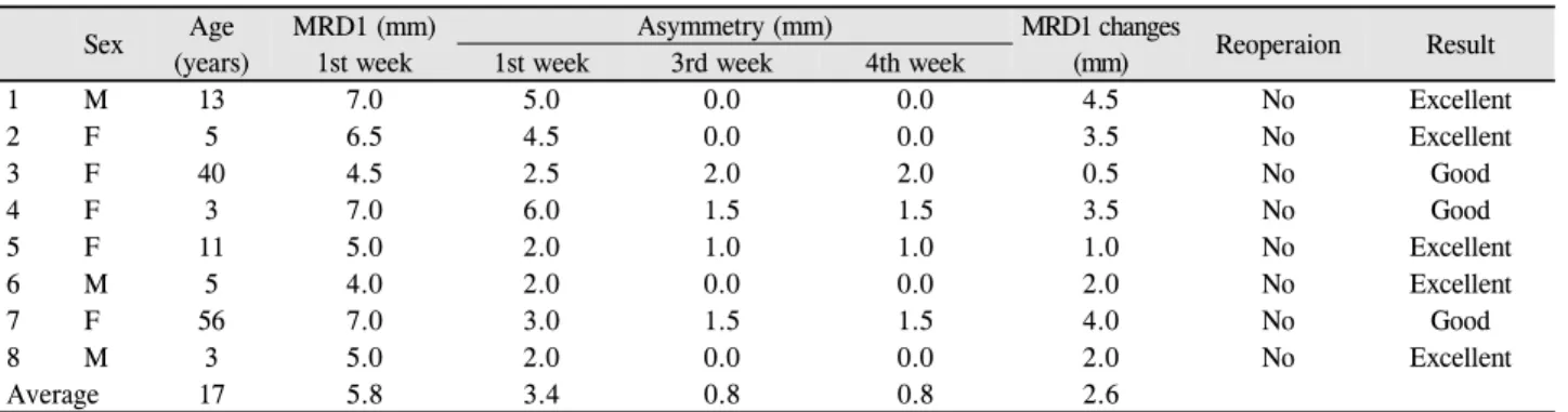
Figure 2. Photographs of patient 1 (1A-1C), 2 (2A-2C), 3 (3A-3C) and 4 (4A-4C). (1A, 2A, 3A, 4A) Before the operation of levator
resection. (1B, 2B, 3B, 4B) One week after the operation. (1C, 2C, 3C, 4C) Final postoperative appearance after eyelid stretching exercise for two or three weeks.후 3예는 내원하지 않아 경과관찰이 종료되었고 1년까지 경과관찰한 5예 모두에서 유의한 MRD1 변화가 관찰되지 않았다.
고 찰
눈꺼풀처짐의 분류는 저자마다 다양해서 Callahan and Beard2는 선천성과 후천성으로, Frueh4는 눈꺼풀올림근 이 상발육형, 신경성, 건막성, 기계성으로 분류한 바 있다. 그 중에 선천눈꺼풀처짐은 전체 눈꺼풀처짐의 80-90%로 가장
많은 비중을 차지한다.5 원인은 태생기 4개월에 정상적으로 분화되어야 하는 눈꺼풀올림근의 발달장애에 의한 것으로 알려져 있으며, 눈꺼풀올림근 기능이 불량한 경우가 많다.
가림약시, 부등시, 눈을 치켜뜨고 이마주름, 이상두위 등이 발생할 때 수술을 고려하게 되며 수술 방법은 눈꺼풀올림 근 기능이 가장 중요한 요소로 알려져 있다.2 기존에는 눈 꺼풀올림근기능이 3 mm 또는 5 mm 이하로 불량한 경우에 이마근걸기술이 주된 수술 방법으로 제시되어 왔지만 선택 의 정확한 기준은 없는 상태이며, 이전의 보고에 따르면 눈 꺼풀올림근 기능이 불량한 중증 선천눈꺼풀처짐에서도 눈
1A 1B 1C
2A 2B 2C
3A 3B 3C
4A 4B 4C
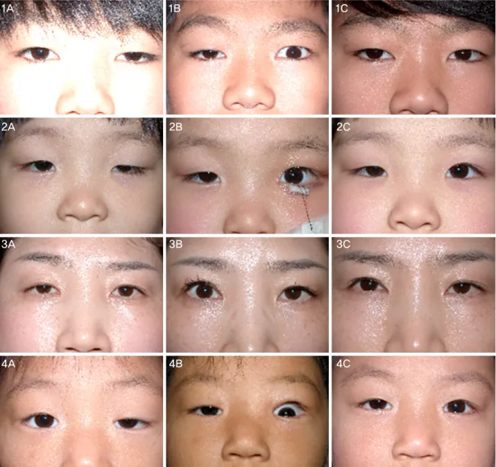
Figure 3. Photographs of patient 5 (1A-1C), 6 (2A-2C), 7 (3A-3C) and 8 (4A-4C). (1A, 2A, 3A, 4A) Before the operation of levator
resection. (1B, 2B, 3B, 4B) One week after the operation. (1C, 2C, 3C, 4C) Final postoperative appearance after eyelid stretching exercise for two or three weeks.꺼풀올림근절제술이 효과적인 치료방법이며, 눈꺼풀올림근 절제술을 시행하였을 때 눈꺼풀올림근 기능이 호전되어 좋 은 결과를 보였다고 한다.4,6,7 Byon and Choi1에 따르면 눈 꺼풀올림근 기능이 불량한 선천눈꺼풀처짐에서 눈꺼풀올 림근절제술은 이마근걸기술과 유사한 결과를 보이며, 재발 시에 추가수술이 가능하고 자가근막채취가 필요 없다는 장 점이 있다고 하였다.
눈꺼풀처짐 수술 후 과교정 시 공막의 과다노출로 인해 미용상 문제, 토안, 노출각막염 및 그로 인한 시력저하 등 이 나타날 수 있다. Fox8는 과교정 발생원인을 위눈꺼풀 수
술 부위의 반흔 형성과 유착 때문으로 설명하였다. 따라서 눈꺼풀마사지는 눈꺼풀올림근절제술 후 눈꺼풀 뒤당김을 일으킬 수 있는 섬유조직과 근육부착부위를 신전시킴으로 써 효과가 있을 것으로 사료된다. 시기는 상처치유과정 중 염증기(술 후 1-2일)가 지나 섬유모세포기(술 후 4-6일경) 에 시행하여 반흔화 시기로 진행되기 전에 하여야 할 것으 로 생각된다.9 Edmonson and Wulc10는 눈꺼풀을 아래 방향 으로 마사지하는 것이 과교정에 도움이 될 수 있다고 한 바 있지만, 마사지의 구체적인 방법, 횟수나 시기에 관한 연구 는 현재 없는 상태이다.
1A 1B 1C
2A 2B 2C
3A 3B 3C
4A 4B 4C
= 국문초록 =
눈꺼풀올림근 절제술 후 과교정된 선천 눈꺼풀처짐 환자에서 눈꺼풀 신전 운동
목적: 선천 눈꺼풀처짐 환자에서 눈꺼풀올림근절제술을 시행 후 과교정이 발생한 8명의 환자를 대상으로 눈꺼풀신전운동을 시행하여 치료효과를 얻어 문헌고찰과 함께 이를 보고하고자 한다.
증례요약: 눈꺼풀신전운동은 환자로 하여금 위를 보게 한 다음 수술 받은 눈의 눈꺼풀테 바로 위 피부를 아래로 당겨 눈꺼풀을 신장시 키고자 하는 것이다. 눈꺼풀신전운동을 2주간 시킨 후 반대편 정상안과의 차이가 1.5 mm 이상 비대칭성이 남은 경우 눈꺼풀신전운동 을 1주간 추가로 시행하였다. 선천 단안 근성 눈꺼풀처짐으로 눈꺼풀올림근절제술 후 1주째에 과교정이 발생한 환자 8명 8안에서 눈꺼풀신전운동을 시행한 후 수술 전, 수술 후 1주, 수술 후 3주, 수술 후 4주째의 margin reflex distance 1 (MRD1) 및 경과관찰을 시행하였다. 8예 모두에서 눈꺼풀올림근절제술 후 과교정이 비교적 성공적으로 교정되었으며, 양안의 대칭성이 회복되었고 주관적인 만족을 보였다. 추가로 1주간 눈꺼풀신전운동을 시킨 경우 MRD1의 차이는 없었다.
결론: 선천 눈꺼풀처짐 환자에서 눈꺼풀올림근절제술을 시행 후 과교정이 발생하였을 때 눈꺼풀올림근후전술 등의 수술적인 치료를 시행하기에 앞서 눈꺼풀신전운동을 포함한 비수술적 치료를 먼저 고려해 볼 수 있으며, 눈꺼풀올림근절제술 후 조직 섬유화가 단단하 게 이루어지기 전, 비교적 이른 시기에 눈꺼풀신전운동을 시행해야 하겠다.
<대한안과학회지 2015;56(10):1635-1639>
본 연구에서는 수술 후 1주째 과교정이 발생하였을 때 눈 꺼풀신전운동을 2주간 시행하였고 효과를 보인 경우 중단 하였으며 이후에 MRD1의 감소나 증가는 관찰되지 않았다.
효과가 부족할 시 1주간 추가로 더 시행한 경우 또한 MRD1 추가 변화는 보이지 않았다. 안검신전운동 후 8예 모두에서 MRD1의 감소를 보였고, 환자가 미용적으로 만족하였으며, 이후에 눈꺼풀올림근후전술 등의 수술적 치료를 시행한 경 우는 없었다.
선천눈꺼풀처짐으로 눈꺼풀올림근절제술 시행 후 과교 정 시에는 수술적인 치료를 시행하기에 앞서 눈꺼풀신전운 동을 포함한 비수술적 치료를 먼저 고려해 볼 수 있으며, 섬 유조직이 단단하게 진행되기 전 비교적 이른 시기에 눈꺼 풀신전운동을 시행하여야 하겠다. 술 후 초기에 눈꺼풀신전 운동효과가 미미할 경우에는 조기에 교정술을 고려하는 것 이 좋을 것으로 사료된다.
참고문헌
1) Byon IS, Choi HY. Outcomes of anterior levator resection and frontalis sling in congenital ptosis with poor levator function. J
Korean Ophthalmol Soc 2005;46:1605-10.
2) Callahan MA, Beard C. Beard’s ptosis, 4th ed. Birmingham:
Aesculapius Publishing Co., 1990;52-87.
3) Seo SW. Treatment for ptosis. In: Kim YD, Lee SY, Kim SJ, et al, eds. Ophthalmic Plastic and Reconstructive Surgery, 2nd ed.
Seoul: Naewaeh-haksool, 2009; v. 1. chap. 6.
4) Frueh BR. The mechanistic classification of ptosis. Ophthalmology 1980;87:1019-21.
5) Kim MH, Kim HS. Surgical comparative observation of congenital blepharoptosis. J Korean Ophthalmol Soc 1988;29:275-81.
6) Göncü T, Çakmak S, Akal A, Karaismailoğlu E. Improvement in levator function after anterior levator resection for the treatment of congenital ptosis. Ophthal Plast Reconstr Surg 2015;31:197-201.
7) Putnam JR, Nunery WR, Tanenbaum M, McCord CD. Blepharoptosis.
In: MacCord CD, Tanenbaum M, Nunery WR, eds. Oculoplastic Surgery, 3rd ed. New York: Lippincott Williams & Wilkins, 1995;
175-220.
8) Fox SA. Surgery of Ptosis (Blepharoptosis), 1st ed. New York:
Grune and Stratton, 1968;91-111.
9) Lee YJ. Principles of ophthalmic plastic and reconstructive surgery. In: Kim YD, Lee SY, Kim SJ, et al, eds. Ophthalmic Plastic and Reconstructive Surgery, 2nd ed. Seoul: Naewaehs, 2009; v. 1. chap. 2.
10) Edmonson BC, Wulc AE. Ptosis evaluation and management.
Otolaryngol Clin North Am 2005;38:921-46.