ISSN 0378-6471 (Print)⋅ISSN 2092-9374 (Online)
http://dx.doi.org/10.3341/jkos.2016.57.12.1897
Original Article
광역학치료와 베바시주맙을 이용한 특발 중심와부근 모세혈관확장증 I형의 치료
Combination Therapy with Photodynamic Therapy and Intravitreal Bevacizumab in Idiopathic Macular Telangiectasia Type I
김인근1⋅이지은2⋅박정민1
In Geun Kim, MD1, Ji Eun Lee, MD, PhD2, Jung Min Park, MD, PhD1
메리놀병원 안과1, 부산대학교 의학전문대학원 부산대학교병원 안과학교실2 Department of Ophthalmology, Maryknoll Medical Center1, Busan, Korea
Department of Ophthalmology, Pusan National University Hospital, Pusan National University School of Medicine2, Busan, Korea
Purpose: We investigated the clinical manifestations of combination therapy with photodynamic therapy (PDT) and intravitreal bevacizumab in idiopathic macular telangiectasia type I patients.
Methods: The present study included 7 patients who visited our clinic from May 2008 to February 2011 complaining of decreas- ing visual acuity and diagnosed as idiopathic macular telangiectasia type I including visible aneurysms at juxtafoveal area and telangiectatic vessels leakage based on fluorescein angiography. Additionally, all patients were treated with combination ther- apy with PDT and intravitreal bevacizumab.
Results: Visual acuity improved from 0.48 ± 0.14 (log MAR) to 0.18 ± 0.17 (log MAR) after the combination therapy, however, there was no change in intraocular pressure between before (17.9 ± 3.1) and after (16.8 ± 2.3) therapy. After combination ther- apy, fluorescein angiography showed decreased leakage of telangiectatic vessels and optical coherence tomography showed only minimal intraretinal edema. Central subfield macular thickness decreased from 301.9 ± 50.7 μm to 193.6 ± 58.8 μm after the combination therapy.
Conclusions: Combination therapy with intravitreal bevacizumab injection and PDT in patients with idiopathic macular te- langiectasia type Ⅰ can result in a rapid decrease of macular edema, which can lead to rapid visual recovery.
J Korean Ophthalmol Soc 2016;57(12):1897-1902
Keywords: Idiopathic macular telangiectasia type Ⅰ, Intravitreal bevacizumab, Photodynamic therapy
■Received: 2016. 8. 11. ■ Revised: 2016. 10. 4.
■Accepted: 2016. 11. 23.
■Address reprint requests to Jung Min Park, MD, PhD
Department of Ophthalmology, Maryknoll Medical Center, #121 Junggu-ro, Jung-gu, Busan 48972, Korea
Tel: 82-51-461-2469, Fax: 82-51-462-3534 E-mail: [email protected]
ⓒ2016 The Korean Ophthalmological Society
This is an Open Access article distributed under the terms of the Creative Commons Attribution Non-Commercial License (http://creativecommons.org/licenses/by-nc/3.0/) which permits unrestricted non-commercial use, distribution, and reproduction in any medium, provided the original work is properly cited.
특발성 중심와부근모세혈관확장증은 망막모세혈관의 확 장을 보이는 망막혈관장애로 중년 혹은 그 이상의 나이에 서 원인 모르게 발생하고 망막주변부 및 후극부에 모세혈
관의 불규칙한 확장 및 관류 장애가 나타나고 황반 부종이 나 경성 삼출물이 발생하거나 맥락막 신생혈관이 발생하는 경우 시력저하가 올 수 있는 질병이다. 이러한 모세혈관확 장증이 다른 원인 없이 중심와 주변부 모세혈관에만 나타 날 경우, 특발성 중심와부근모세혈관확장증(parafoveal te- langiectasis)이라고 한다.1 Gass and Blodi2는 Group I과 Group II로 나누었으며 특발성 중심와부근모세혈관확장증 type Ⅰ은 선천성 혹은 후천성으로 발병하며 주로 중년 남 자에게 발견되고 대부분 한쪽 눈에 발병하는 형태이며 Yannuzzi et al3은 Group I을 aneurysmal telangiectasia (type I)
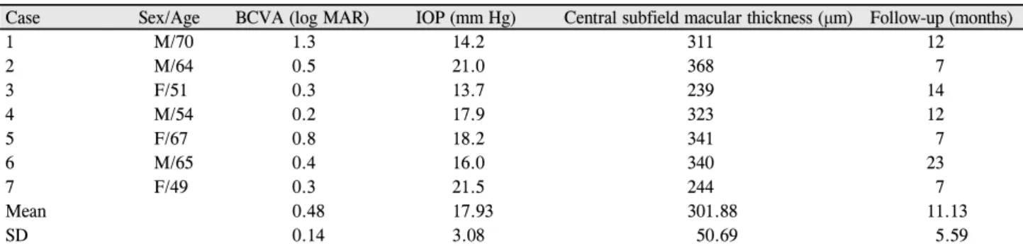
Table 1. Clinical characteristics of the patient at presentation
Case Sex/Age BCVA (log MAR) IOP (mm Hg) Central subfield macular thickness (μm) Follow-up (months)
1 M/70 1.3 14.2 311 12
2 M/64 0.5 21.0 368 7
3 F/51 0.3 13.7 239 14
4 M/54 0.2 17.9 323 12
5 F/67 0.8 18.2 341 7
6 M/65 0.4 16.0 340 23
7 F/49 0.3 21.5 244 7
Mean 0.48 17.93 301.88 11.13
SD 0.14 3.08 50.69 5.59
BCVA = best corrected visual acuity; log MAR = logarithm of minimal angle resolution; IOP = intraocular pressure; SD = standard deviation.
로, Group II A를 perifoveal telangiectasia (type II)로 분류하 였다.
시력 저하는 주로 황반 부종과 지질 삼출물로 인해 발생 하고 치료 없이 저절로 좋아지는 경우가 많아 치료 없이 경 과 관찰하는 경우도 있지만, 황반 부종 및 지질 삼출물에 의해 지속적인 시력 저하가 있을 경우 격자레이저 광응고 술, 유리체강내 트리암시놀론 주입술(intravitreal triamcinolone injection), 베바시주맙 유리체강내 주입술 등이 치료에 이용되 고 있다. 그러나 이 질병의 유병률이 낮아 대규모 연구가 힘 들어 효과적인 치료 방법이 아직 정립되지 않았다. 중심와 부근모세혈관확장증 type II에서는 베바시주맙에 의해 효과 적인 치료를 보인 보고가 있다.4,5 하지만 중심와부근모세혈 관확장증 type I에서 베바시주맙이 시력호전에 효과가 있다 는 보고6가 있으나 베바시주맙 투여 후 조직학적으로는 호 전을 보이지만 시력의 호전은 보이지 않는 등7 효과적인 치 료 방법에 대한 이견이 많다.
이에 저자들은 중심와부근모세혈관확장증 type I으로 진 단된 환자에서 좀 더 나은 효과적인 치료방법을 알아보고자 베바시주맙 유리체강내 트리암시놀론 주입술과 광역학치료 (photodynamic therapy, PDT) 병합요법의 단기적 임상결과 에 대해 알아보았다.
대상과 방법
시력 이상을 주소로 2008년 5월부터 2012년 2월까지 본 원에 내원하여 특발성 중심와부근모세혈관확장증 type Ⅰ 으로 진단 받고 삼출성 병변으로 유리체강내 베바시주맙 주사와 광역학요법 병합치료를 시행한 뒤 6개월 이상 경과 관찰이 가능했던 7명을 대상으로 의무기록 후향적 분석을 시행하였다. 병력, 시력검사, 안압검사, 세극등검사, 안저검 사, 형광안저혈관조영술(fluorescein angiography, FA), 빛 간섭단층촬영(optical coherence tomography [OCT], Stratus OCT, Carl Zeiss Inc., Dublin, CA, USA) 등을 조사하였다.
최종 시력은 마지막 방문 때 측정한 교정시력으로 하였으 며, 시력은 한천석 시력표로 측정하였고 logMAR로 변환하 여 분석하였다.
안저소견상 황반부 이측에 이상소견에 관찰되고 이상소 견에 해당하는 부위가 OCT상 망막부종이 확인되고 형광안 저촬영에서 모세혈관 확장, 미세혈관류가 관찰되고 후기에 확장된 모세혈관에서 형광누출을 보인 경우 중심와부근모 세혈관확장증 type I으로 진단하였다. 진단된 환자 중 시력 저하를 동반하고 OCT상 황반 중심 오목을 소실시키는 망막 부종이 보이고 형광안저촬영상 황반 주위에 모세혈관확장 이 발견되고 후기에 형광누출이 발견된 경우 유리체강내 베 바시주맙(Avastin, Genentech Inc., San Francisco, CA, USA) 주사와 광역학요법(PDT)을 병행한 복합치료를 시행하였다. 광 역학치료는 Treatment of Age-Related Macular Degeneration with Photodynamic Therapy Study의 치료 지침에 따라 시행 하였고, 레이저 조사 범위는 확장된 모세혈관과 형광누출 부 위를 포함하여 시행하였다. 광역학치료를 시행 후 7일째 유 리체강내 베바시주맙 0.05 mL (1.25 mg)를 주사하였다. 치 료 이후 1달 경과 관찰 후 OCT상 망막 부종의 호전이 보이 지 않으면 재치료를 시행하였다.
안압은 골드만 안압계를 통하여 측정하였고 세극등을 이 용한 전안부 검사, 안저 검사를 통해 합병증 유무를 조사하 였다. 형광안저촬영에서 형광 누출이 감소하는 것과 OCT 에서 황반 부종이 감소하는 것을 병변 호전으로 판단하였 다.
통계적인 분석은 SPSS® version 12.0 (SPSS Inc., Chicago, IL, USA)을 사용하였다. 시술 전후 값의 비교는 Wilcoxon 검정을 이용하였고 p값이 0.05 미만인 경우에 통계학적으로 유의하다고 판단하였다.
결 과
분석에 포함된 7명 중 남자는 4명, 여자는 3명이었고 연령
Table 2. Clinical outcomes after combined therapy for idiopathic macular telangiectasia type I
BCVA (log MAR) p-value CSMT (μm) p-value
Baseline 0.48 ± 0.14 301.9 ± 50.7
1 month 0.30 ± 0.20 0.06 227.9 ± 53.6 0.02
2 months 0.30 ± 0.21 0.06 211.1 ± 61.3 0.01
6 months 0.18 ± 0.17 0.02 193.6 ± 58.8 0.01
Values are presented as mean ± SD unless otherwise indicated.
BCVA = best corrected visual acuity; log MAR = logarithm of minimal angle resolution; CSMT= central subfield macular thickness.
Table 3. Change of best corrected visual acuity
Visual acuity (log MAR) Initial number (%) Final number (%)
VA ≤ 0.3 3 (42.8) 7 (100)
0.3 < VA ≤ 1.0 3 (42.8) 0 (0)
1.0 < VA 1 (14.4) 0 (0)
Total 7 (100) 7 (100)
Values are presented as n (%).
log MAR = logarithm of minimal angle resolution; VA = visual acuity.
Figure 1. Fluorescein angiography and opti-
cal coherence tomography before treatment.Fluorescein angiography of telangiectatic vessels at the early phase (A) and leakage at the late phase (B). (C) Optical coherence to- mography of intraretinal edema before com- bination treatment.
A B
C
분포는 49세에서 70세까지 평균 58.6 ± 8.7세였다(Table 1).
평균 관찰 기간은 11.1 ± 5.6개월이었고 초진 진단 후 치료 가 시작된 간격은 5.1 ± 5.6개월이었다. 전신질환으로서는 고혈압이 6명, 당뇨가 2명에서 있었다.
최초 내원 시 평균시력(logMAR)은 0.4 ± 0.21이었고 시 술 전 평균시력(logMAR)은 0.48 ± 0.14였다. 광역학치료 시 대상 병변 부위 중 가장 긴 길이(greatest linear dimension)의 평균은 1,764 ± 453 μm였다. 6개월 경과 관찰 중 3명에서
추가적인 1회의 주사 치료를 시행하였고 1명에서는 추가적 인 2회의 주사치료를 추가적으로 시행하였다.
시술 후 평균시력은 1개월 0.30 ± 0.20, 3개월 0.30 ± 0.21 그리고 6개월 0.18 ± 0.17로 유의하게 호전되었으며 (Table 2) 7안 중 6안에서 3줄 이상의 시력호전이 있었다 (Table 3).
형광안저촬영에서 시술 전에 있었던 모세혈관 확장 부위 형광 누출이 시술 후 감소되는 것을 7안 중 6안에서 관찰할
Figure 2. Fluorescein angiography and optical
coherence tomography after treatment.Fluorescein angiography of a regression of the telangiectatic vessels (A) and a decreased leak- age of telangiectatic vessels at the late phase af- ter combination therapy(B). (C) Optical coher- ence tomography of minimal intraretinal edema and restored foveal depression after treatment.
A B
C
수 있었다. OCT에서 central subfield macular thickness는 시술 전 301.9 ± 50.7 μm에서 1개월 227.9 ± 53.6 μm, 3개 월 211.1 ± 61.3 μm 그리고 6개월 193.6 ± 58.8 μm로 감소 하였다(p=0.01, Table 2). 시술 후 6개월의 관찰기간 동안 안압 상승, 안내염, 유리체 출혈 등 심각한 합병증은 발견 되지 않았다.
증례 1
2년 전부터 시작된 우안의 변형시를 주소로 내원한 65세 남자가 내원하였다. 과거력상 2년 동안 고혈압 치료 중이었 다. 내원 시 나안 시력은 우안 0.4 (logMAR)였고 세극등 검 사상 전안부에서는 특이소견이 관찰되지 않았으며 안저 검 사상 황반부에서 황반 부종이 관찰되었다. 좌안은 안저 검 사상 특이소견이 없었다. OCT에서 우안 황반의 망막 내 부 종이 확인되었고 형광안저촬영에서 우안 황반부의 확장된 모세혈관주위로 누출이 있어(Fig. 1). 이에 저자들은 특발성 중심와모세혈관확장증 type I으로 진단하였다. 모세혈관이 확장된 영역에 대해 광역학치료를 시행하고 1주일 후 유리 체강내 베바시주맙을 주사하였다. 6개월 후 시력은 0.15 (logMAR)로 호전되었으며 OCT와 형광안저촬영에서 황반 부의 모세혈관 확장과 형광누출 및 황반부종이 감소된 소견 을 보였다(Fig. 2).
고 찰
중심와부근 모세혈관의 미세동맥류, 낭성 확장 및 비관 류를 동반하는 질환을 중심와부근모세혈관확장증이라고 한다. 1993년 Gass and Blodi2는 이 질환을 세 군으로 분류 하였으며 I군은 편측성 여부에 따라 다시 I A군과 I B군으 로 나누었다. I A군은 일측성 선천성 중심와부근 모세혈관 확장증(visible and exudative idiopathic juxtafoveal retinal telangiectasis)으로 지칭하였는데 진단 시 평균 연령은 40세 로 주로 남자에서 편측성으로 생기며 지질 삼출물을 특징 으로 하며 코오츠병(Coats’ disease)의 경한 형태로 보이기 도 한다. 안저검사상 2유두직경넓이 이상으로 황반부 이측 으로 명백한 모세혈관 확장 및 다양한 크기의 미세혈관류 가 관찰되며 Group II A에서 보이는 직각소정맥, 망막표층 의 결절 침착 망막색소상피 증식, 색소침착 등은 보이지 않 는다. OCT상 황반부 중심에 낭포 혹은 비낭포성 황반부종 이 관찰되며 비정상적으로 큰 망막 내 혈관이 외핵층에서 관찰되는 것이 특징이다. Group I 중에서 2시각 이하의 작 은 모세혈관 확장을 보이는 경우 Group I B로 구분하였고 Yannuzzi et al3은 이 두 가지를 통틀어 간단히 aneurysmal telangiectasia (type I)로 분류하였다.
Group II A (occult and nonexudative idiopathic juxtafoveal retinal telangiectasis)는 모세혈관확장증의 가장 흔한 형태
이며 중심와부근 망막의 위축이 특징이다.2 Group Ⅲ는 occlusive idiopathic juxtafoveolar retinal telangiectasis라 하 며 아주 드문 형태이며 중심와주변의 모세혈관폐쇄 및 모세 혈관 확장과 함께 전신적인 중추신경계 이상과 연관되어 시 력소실이 일어난다.2
중심와부근모세혈관확장증 type I에서 시력 저하는 주로 황반 부종과 지질 삼출물로 인해 발생하고 치료 없이 저절 로 좋아지는 경우가 많아 치료 없이 경과 관찰하는 경우도 있지만, 황반 부종 및 지질 삼출물에 의해 지속적인 시력 저하가 있을 경우 격자레이저 광응고술이 사용되기도 한 다.8 경과 관찰한 기간의 차이로 인해 정확한 비교는 어려 우나 Gass and Blodi2도 중심와부근모세혈관확장증 type I 환자에게서 레이저 광응고 치료가 좋은 반응을 나타냄을 보고하였고, Park et al9도 유사한 결과를 보고하였다.
레이저 광응고술은 확장된 모세혈관 부위의 퇴행을 가져 올 수 있으나, 레이저 광응고술을 받지 않은 망막의 다른 부위에 다시 이상 혈관이 재발할 수 있으며, 레이저 광응고 술 자체로도 망막출혈, 망막박리 등의 합병증이 발생될 수 있고 병변이 명확하지 않거나 광범위할 경우 레이저치료의 범위를 결정하기 어렵고, 병변이 황반에서 가까울 때는 레 이저반흔에 의한 황반 손상의 위험성이 있어 사용하기 어 렵다는 한계점이 있어 직접 레이저 광응고술을 시행하기 어려운 환자들에서 다른 다양한 치료가 시도되었다. 레이 저 광응고 치료 외에 Alldredge and Garretson10은 양안 중 심와부근모세혈관확장증에서 단안에만 유리체강내 트리암 시놀론 주입술을 시도하여, 경과관찰을 시행한 반대 안에 비해 유의하게 좋은 결과를 얻었다고 보고하였다.
광역학 치료는 verteporfin을 이용하여 광독성 효과로 신 생혈관을 치료하는 방법으로 신생혈관이 발생하는 여러 질 환에 사용되고 있으나 중심장액맥락망막병증에서처럼 신 생혈관이 없는 경우에도 이러한 광역학 치료를 통해 황반 부종의 호전과 시력향상을 보일 수 있다.11-13 중심와부근모 세혈관확장증 type I에서 광역학 치료 후 시력호전과 함께 형광누출 감소 및 황반 출혈 및 삼출물의 감소를 보인 보고 도 있다.14
항혈관내피성장인자항체인 베바시주맙은 선택적으로 혈 관내피성장인자와 결합하여 활성을 막아 결과적으로 혈관 신생을 억제하는 역할을 한다고 알려져 있어 증식 당뇨망 막병증이나 맥락막 신생혈관 등의 치료에 많이 쓰이고 있 다.15 중심와부근모세혈관확장증 type II에서는 맥락막 신생 혈관이 합병된 경우 베바시주맙으로 효과적인 치료를 시행 한 보고가 있다.4,5 이에 반해 중심와부근모세혈관확장증 type I에서 베바시주맙의 역할은 아직 확실히 밝혀지지 않 았다. 중심와부근모세혈관확장증 type I에서 베바시주맙이
시력호전에 효과가 있다는 보고6도 있으며 베바시주맙 투 여 후 조직학적 호전이 있지만 시력호전은 보이지 않았다 는 보고도 있다.7 이는 모세혈관확장을 호전시키지는 못하 지만 비정상적인 혈관투과성을 안정화시켜 황반내 부종을 호전시킬 것으로 생각되고 있다.
베바시주맙의 vascular endothelial growth factor in- hibition 효과는 신생혈관의 발생시기에 그 예방 효과가 좋 으나 그 신생혈관이 자리를 잡으면 그 효과가 떨어지는 경 향을 보인다.16 물론 이러한 시기에 베바시주맙의 효과가 없다는 것이 밝혀지지는 않았지만 광역학치료와 같은 광응 고 치료를 먼저 시행한 후 베바시주맙을 주사하면 추가 주 사 횟수도 줄이고 좀 더 나은 결과를 보일 것으로 예상하였 고 실제로 idiopathic macular telangiectasia Type 2에서 광 역학치료 이후 ranibizumab을 사용하여 좋은 효과를 보인 보고도 있다.17
처음 치료로 광역학치료 시 대상혈관에 대한 focus를 쉽 게 잡을 수 있다는 장점이 있으나 레이저 범위가 커져 이에 대한 부작용 위험도 커지는 단점이 있어 좀 더 많은 연구가 필요할 것으로 사료된다. 저자들은 중심와부근모세혈관확 장증 type I에서 베바시주맙과 함께 광역학치료를 같이 시 행한 후 형광안저촬영상 형광 누출이 줄어들고 OCT상 황 반 부종이 감소되는 소견과 시술 전에 비해 시력이 호전되 는 것을 관찰할 수 있었다. 광역학치료를 통해 확장된 모세 혈관을 직접적으로 치료하여 확장되었던 혈관의 직경이 정 상화되고 혈관 외 누출이 줄어드는 효과가 있었으며 베바 시주맙의 효과로 광역학치료 후 일시적으로 증가한 혈관내 피증식인자를 억제하며 비정상적인 혈관투과성을 안정화 시켜 황반내 부종을 호전시킨 것으로 생각된다. 이러한 복 합치료를 통해 서로 간의 상승작용으로 안내 주사의 횟수 를 줄일 수 있고 좀 더 영구적인 효과를 가져올 것으로 생 각된다.
본 연구에서는 경과 관찰 기간이 충분하지 않고 다른 치 료방법과의 비교가 없었다는 한계점이 있다. 또한 장기적 경과 관찰 시 광역학치료로 인한 망막과 모세혈관의 손상 및 반흔 형성의 가능성18에 대한 관찰이 없다는 단점도 있 다. 따라서 광역학치료와 베바시주맙유리체주입술 병합 치 료 효과에 대한 좀 더 광범위하고 장기적인 연구가 필요할 것으로 사료되며 다른 치료 방법 효과와의 비교 조사도 필 요할 것으로 생각된다.
결론적으로 중심와부근모세혈관확장증 type I에서 황반 부종으로 인해 시력저하가 생길 때 광역학치료와 베바시주 맙유리체강내 주입술 병합 치료를 통해 단기간에서 효과적 인 병변 호전 및 시력 개선을 가져올 수 있었다.
= 국문초록 =
광역학치료와 베바시주맙을 이용한 특발 중심와부근 모세혈관확장증 I형의 치료
목적: 중심와부근모세혈관확장증 type I으로 진단된 환자에서 베바시주맙 유리체강내 주입술과 광역학치료 병합요법의 효과에 대해 알아보았다.
대상과 방법: 2008년 5월부터 2012년 2월까지 시력저하를 주소로 내원한 환자 중 안저소견상 황반부 이측에 이상소견이 관찰되고 형광안저혈관조영술에서 모세혈관 확장으로 인한 누출이 관찰되어, 중심와부근모세혈관확장증 type I으로 진단된 7명을 대상으로 유 리체강내 베바시주맙주사와 광역학 치료를 병행한 복합치료를 시행하였다.
결과: 시술 전 시력(logMAR)은 0.48 ± 0.14였고 시술 후 0.18 ± 0.17로 호전되었다. 안압은 시술 전 17.9 ± 3.1에서 16.8 ± 2.3으로 의미있는 차이를 보이지는 않았으며 시술 후 형광안저혈관조영술상에서 시술 전에 있었던 모세혈관 확장 부위 형광 누출이 감소되는 것을 관찰할 수 있었다. 빛간섭단층촬영에서 황반내 부종이 호전되어 central subfield macular thickness가 시술 전 301.9 ± 50.7 μm에 서 시술 후 193.6 ± 58.8 μm로 감소하였다.
결론: 중심와부근모세혈관확장증 type I에서 망막하모세혈관확장과 황반부종으로 인해 시력저하가 생길 때 광역학치료와 베바시주맙 유리체강내 주입술 병합 치료를 통해 단기간에 효과적인 병변 호전 및 시력 개선을 가져올 수 있었다.
<대한안과학회지 2016;57(12):1897-1902>
REFERENCES
1) Hutton WL, Snyder WB, Fuller D, Vaiser A. Focal parafoveal reti- nal telangiectasis. Arch Ophthalmol 1978;96:1362-7.
2) Gass JD, Blodi BA. Idiopathic juxtafoveolar retinal telangiectasis.
Update of classification and follow-up study. Ophthalmology 1993;100:1536-46.
3) Yannuzzi LA, Bardal AM, Freund KB, et al. Idiopathic macular telangiectasia. Arch Ophthalmol 2006;124:450-60.
4) Kovach JL, Rosenfeld PJ. Bevacizumab (avastin) therapy for idio- pathic macular telangiectasia type II. Retina 2009;29:27-32.
5) Charbel Issa P, Holz FG, Scholl HP. Findings influorescein angiog- raphy and optical coherence tomography after intravitreal bev- acizumab in type 2 idiopathic macular telangiectasia. Ophthalmology 2007;114:1736-42.
6) Gamulescu MA, Walter A, Sachs H, Helbig H. Bevacizumab in the treatment of idiopathic macular telangiectasia. Graefes Arch Clin Exp Ophthalmol 2008;246:1189-93.
7) Takayama K, Ooto S, Tamura H, et al. Intravitreal bevacizumab for type 1 idiopathic macular telangiectasia. Eye (Lond) 2010;24:
1492-7.
8) Chopdar A. Retinal telangiectasis in adults: fluorescein angio- graphic findings and treatment by argon laser. Br J Ophthalmol 1978;62:243-50.
9) Park DW, Schatz H, McDonald HR, Johnson RN. Grid laser photo- coagulation for macular edema in bilateral juxtafoveal telangiectasis.
Ophthalmology 1997;104:1838-46.
10) Alldredge CD, Garretson BR. Intravitreal triamcinolone for the
treatment of idiopathic juxtafoveal telangiectasis. Retina 2003;23:
113-6.
11) Battaglia Parodi M, Da Pozzo S, Ravalico G. Photodynamic ther- apy in chronic central serous chorioretinopathy. Retina 2003;23:235-7.
12) Khosla PK, Rana SS, Tewari HK, et al. Evaluation of visual func- tion following argon laser photocoagulation in central serous retinopathy. Ophthalmic Surg Lasers 1997;28:693-7.
13) Klais CM, Ober MD, Ciardella AP, et al. Retina, 4th ed. Vol. 2. Los Angeles: Mosby, 2006; 1153-9.
14) Kotoula MG, Chatzoulis DZ, Karabatsas CH, et al. Resolution of macular edema in idiopathic juxtafoveal telangiectasis using PDT.
Ophthalmic Surg Lasers Imaging 2009;40:65-7.
15) Kurup S, Lew J, Byrnes G, et al. Therapeutic efficacy of intravitreal bevacizumab on posterior uveitis complicated by neovascularization.
Acta Ophthalmol 2000;87:349-52.
16) Dhalla MS, Shah GK, Blinder KJ, et al. Combined photodynamic therapy with verteporfin and intravitreal bevacizumab for choroi- dal neovascularization in age-related macular degeneration. Retina 2006;26:988-93.
17) Rishi P, Shroff D, Rishi E. Combined photodynamic therapy and intravitreal ranibizumab as primary treatment for subretinal neo- vascular membrane (SRNVM) associated with type 2 idiopathic macular telangiectasia. Graefes Arch Clin Exp Ophthalmol 2008;
246:619-21.
18) Bashshur ZF, Schakal A, Hamam RN, et al. Intravitreal bev- acizumab vs verteporfin photodynamic therapy for neovascular age-related macular degeneration. Arch Ophthalmol 2007;125:
1357-61.