CASE REPORT
악성 위궤양과 감별이 어려웠던 Epstein-Barr Virus 감염이 동반된 양성 위궤양 증례 1예
곽진욱, 유지원, 서승오, 김재연, 오인수, 배지윤1
경찰병원 내과, 병리과1
Benign Gastric Ulcer with Epstein-Barr Virus Infection Mimicking Malignant Gastric Ulcer
Jin Wuk Gwak, Jiwon Yoo, Seong O Suh, Jaeyeon Kim, In Soo Oh and Ji Yoon Bae1 Departments of Internal Medicine and Pathology1, National Police Hospital, Seoul, Korea
Epstein-Barr virus (EBV) is the cause of infectious mononucleosis, which is characterized by fever, lymphadenopathy, and sore throat.
On the other hand, gastrointestinal symptoms of EBV infection like dyspepsia, abdominal pain are non-specific and rarely encoun- tered, which means it is difficult to diagnose gastric involvement of EBV infection without suspicion. The relation between gastric carci- noma and gastric lymphoma associated with EBV infection is well defined, but relations with other EBV-associated gastrointestinal diseases such as gastritis and peptic ulcer disease have rarely been reported. We report a case of benign gastric ulcer with EBV in- fection confirmed by endoscopic and histological findings. (Korean J Gastroenterol 2019;73:177-181)
Key Words: Epstein-Barr virus infections; Stomach ulcer; Peptic ulcer; In situ hybridization; Helicobacter pylori
Received June 7, 2018. Revised August 9, 2018. Accepted August 21, 2018.
CC This is an open access article distributed under the terms of the Creative Commons Attribution Non-Commercial License (http://creativecommons.org/licenses/
by-nc/4.0) which permits unrestricted non-commercial use, distribution, and reproduction in any medium, provided the original work is properly cited.
Copyright © 2019. Korean Society of Gastroenterology.
교신저자: 유지원, 05715, 서울시 송파구 송이로 123, 경찰병원 내과
Correspondence to: Jiwon Yoo, Department of Internal Medicine, National Police Hospital, 123 Songi-ro, Songpa-gu, Seoul 05715, Korea. Tel: +82-2-3400-1519, Fax: +82-2-3400-1680, E-mail: [email protected], ORCID: https://orcid.org/0000-0003-0337-158X
Financial support: None. Conflict of interest: None.
서 론
Epstein-Barr virus (EBV)는 일차 감염을 통하여 전염성 단핵구증을 일으키는 원인이 되는 바이러스로 열감, 피로, 인 두염 등의 증상을 흔하게 발생시킨다.1하지만 소화불량, 복통 등의 EBV 연관 위장관 증상은 비특이적이고 드물게 나타난 다. EBV의 위 침범은 드물게 알려져 있고 위의 악성 림프종2 및 위암종3과도 연관이 있는 것으로 보고되고 있다. 한편 EBV와 연관된 급성 위염에 대한 보고는 국내에 몇 차례 있었 으나,4,5 EBV와 연관되어 양성 위궤양이 발현된 예는 국내에 서는 현재까지 보고된 바가 없다. 저자들은 EBV와 연관되어 발생한 것으로 추정되는 양성 위궤양 환자 1예를 경험하였기 에 이를 보고하는 바이다.
증 례
특이 병력이 없는 36세 남자가 내원 2주 전부터 발생한 속 쓰림 및 소화불량을 주소로 내원하였다. 환자는 내원 한 달 전에 기침, 열감 및 오한 증상이 있었다.
말초혈액 검사에서 백혈구 4,900/mm3, 혈색소 14.2 g/dL, 혈소판 182,000/mm3였고, 혈청 C-반응성 단백 0.13 mg/dL, 혈청 생화학 검사에서 총 단백 7.1 g/dL, 알부민 4.6 g/dL, AST 44 IU/L, ALT 48 IU/L였다. 간염바이러스 표지자 검사 에서는 HBsAg 음성, anti-HBs 항체 양성, anti-HCV 항체 음성 소견을 보였다.
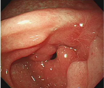
속쓰림과 소화불량 증상에 대하여 상부위장관 내시경을 시 행하였고, 위 전정부 소만에 삼출물과 응고된 혈액으로 덮여
Fig. 1. Initial endoscopic examination revealed a large ulcer with thick yellowish exudate and an irregular margin on the lesser curvature of antrum of the stomach.
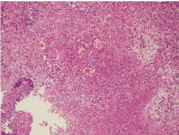
Fig. 2. Histopathological findings showing chronic active gastritis with necrotic detritus (H&E, ×100).
Fig. 3. Immunohistochemistry results of the gastric specimen were negative for pan cytokeratin [AE1/AE3] (A) and L26 (B) (original magnification, ×40).
있는 경계가 불분명한 궤양침윤형 병변이 관찰되어 진행성 위 암을 배제하기 위하여 다수의 조직생검을 시행하였다(Fig. 1).
내시경을 통하여 얻은 조직의 H&E염색에서는 만성 활동성 위염 소견을 보였고(Fig. 2) 면역조직화학염색에서 anti-pan cytokeratin (AE1/AE3) 항체 음성으로 판명되었다. 2주 후 상부위장관 내시경 추적 검사를 시행하였으며 궤양침윤형 병변 이 변화 없이 관찰되었고, 조직생검을 시행한 결과 면역조직화 학염색에서 anti-pan cytokeratin (AE1/AE3) 항체(Fig. 3A) 와 anti-CD20 항체(L26) (Fig. 3B)가 음성으로 판명되었다.
하지만 EBV 잠복 감염 세포에서 풍부하게 발현되어 조직에 서의 EBV 감염과 질병의 연관성을 알아보는데 유용한 Epstein-Barr virus-encoded RNA (EBER)를 표적으로 하는
EBER 제자리부합법(in situ hybridization)6,7 검사에서 양성 반응을 보여(Fig. 4) 최종적으로 EBV와 연관되어 발생한 궤 양으로 추정할 수 있었다. 또한 조직 검사 결과 헬리코박터균 양성 소견을 보여 3제 1차 제균 요법을 시행하였으며, 양성자 펌프억제제를 복용 후 한달 뒤 증상은 호전되었다. 3개월 뒤 상부위장관 내시경을 시행하였고 위 전정부 소만에 있었던 궤양은 백태가 감소하고 재생상피가 동반된 치유기의 궤양 형태를 보였고, 6개월 뒤 치유된 반흔의 형태로 관찰되었다 (Fig. 5).
위궤양 병변 조직에서 EBER 제자리부합법 양성으로 EBV 와의 연관성이 의심되어 6개월이 지났지만 EBV 항체 검사를 시행하였다. 이종친화성 항체(heterophil Ab) 검사에서는 음 성을 보였고, anti-EBV viral capsid antigen (VCA) IgM 항 체 음성(<10.0), anti-EBV VCA IgG 항체 양성(>750.0), an-
A B
Fig. 4. EBER in situ hybridization demonstrated positive signals (original magnification, ×200). EBER, Epstein-Barr virus-encoded RNA.
Fig. 5. Follow-up esophagogastroduodenoscopy at 6 months after ulcer treatment showed a healed, reddish scar had replaced the ulcer.
ti-EBV Epstein-Barr nuclear antigen (EBNA) IgM 항체 음 성(1 U/mL), anti-EBV EBNA IgG 항체 양성(568.0 U/mL) 소견을 보였다. 환자는 현재 건강한 상태로 증상 없이 소화기 내과 외래 추적 관찰 중이다.
고 찰
EBV는 혈청학적 역학 연구를 통하여 전 세계적으로 95%
이상의 성인에서 감염된 것으로 알려져 있다.8상대적으로 위 암 발병 위험이 높은 동아시아 국가에서 EBV 항체 양성률이 99% 이상이었다는 최근 보고가 있었고9 국내에서는 VCA IgG 항체가 위암 환자군의 94%, 대조군의 92%에서, EBNA IgG 항체가 위암 환자군의 79%, 대조군의 82%에서 혈청 양 성임을 보고한 바 있다.10 또한 EBV와 위암과의 연관성이 있 다고 알려진 이래로 위암 병변의 조직생검을 통한 EBV 검출 보고가 최근 늘어나고 있는데, 한 체계적 문헌고찰에 의하면 위암 조직의 EBER 제자리부합법 검사에서 5.0-17.9%에 이르 는 양성률이 보고된 바 있다.11
EBV 감염과 연관된 위장관 질환은 위염, 위궤양 등의 양성 질환에서부터 위의 림프종 및 위암종 등의 악성 종양까지 드 물지만 다양하게 보고된 바 있다. 그중에서도 악성 질환에 비 하여 위염, 위궤양 등의 양성 질환은 드물게 보고되고 있다.
EBV 감염에 의한 위염은 급성 및 만성 위염 모두 발생 가능 한데, 만성 위염은 보통 위축성 위염으로 나타나는 경우가 많 으며 장상피화생을 동반하기도 한다.12 국내에서 보고된 EBV 연관 위염의 경우 전염성 단핵구증과 동반되어 나타나 상부위 장관 내시경에서 위 체부에 다발성으로 산재된 미란의 형태로 관찰되며, 조직의 EBER 제자리부합법 검사 양성으로 EBV
연관 위염으로 최종 진단된 증례가 있었다.4또한 고열 및 복 통 증상을 호소하는 소아에서 상부위장관 내시경 검사상 위 체부와 전정부의 점막부종 소견을 보이며, 조직의 EBER 제자 리부합법 검사에서 양성을 보여 EBV 위염으로 진단한 증례 가 국내에서 보고된 바 있다.5
한편, EBV 감염과 소화성 궤양의 연관성에 대하여 아직까 지도 명확하게 밝혀진 바는 없다. 한 연구에서 십이지장 궤양 과 높은 혈청 anti-EBV IgG 항체 역가 및 위궤양과 높은 혈 청 anti-EBV IgA 항체 역가 간의 유의한 연관성이 있다는 연구 결과가 있었다.13 또한, 비궤양성 소화불량(Non-ulcer dyspepsia) 환자에 비하여 소화성 궤양 환자의 병변 조직에 서 EBV DNA 복제수(DNA copy number)가 유의하게 높았 다는 연구 결과가 보고된 바 있었으나,14 실제 EBV와 연관되 어 발현된 소화성 궤양의 증례는 드물게 보고되고 있다. 일본 에서 고령의 면역 저하 환자의 위 체부 다발성 궤양에서 생검 한 조직의 중합효소연쇄반응에서 EBV DNA가 검출되어 진 단된 이래15면역 저하 소아에서 위 조직의 EBER 제자리부합 법 양성 소견과 중합효소연쇄반응에서 EBV DNA 검출로 EBV 유발 위궤양으로 진단된 증례가 있었다.16 하지만 국내 에서는 EBV 연관 소화성 궤양에 대하여 아직 보고된 바가 없다.
본 증례에서는 처음에 시행한 상부위장관 내시경에서 악성 이 의심되는 궤양성 병변이 관찰되었으나 반복적인 조직생검에 서 만성 염증 소견을 보이고 anti-pan cytokeratin (AE1/AE3) 항체 염색에서 음성을 보여 위상피암종을 배제하였다. 또한 B세포 표지자인 anti-CD20 항체 염색 소견이 B세포 림프종과 맞지 않아 이를 배제할 수 있었다. EBV 감염과 위궤양 병변의
연관성을 밝히기 위하여 조직생검에 EBER 제자리부합법을 시행하였고, 양성 소견을 보여 EBV와 연관되었을 것으로 생각 되는 양성 위궤양으로 추정할 수 있었다. 최근 EBV 감염 세포 에서 분비되는 EBER이 toll-like receptor 3 신호 체계를 통하 여 I형 인터페론 및 염증성 사이토카인의 생성을 유도함으로써 염증 반응에 관여함을 보고하는 연구가 있었다.6 이는 병변 조직의 EBER 제자리부합법 양성 소견이 EBV의 단순 잠복 감염 상태만을 뜻하는 것이 아닌 염증 반응의 진행에 있어 어느 정도 역할을 수행한다는 것을 말해준다.
한편, 정상 위 조직의 중합효소연쇄반응에서 EBV DNA가 거의 검출되지 않았지만 위염 조직에서의 EBV DNA가 46%
에서 양성이었다는 이전 연구에 비추어 볼 때17 본 증례에서 EBV를 병변에 대한 직접적인 원인으로 지목하기에는 다소 어려운 점이 있다. 하지만 조직에서의 EBV 감염을 증명하는 데 있어서는 앞서 언급한 EBER 제자리부합법이 현재 표준 검사법으로 알려져 있고 이와 비교하였을 때 EBV DNA 중합 효소연쇄반응의 특이도는 더 낮은 것이 사실이다. 실제로 최 근 한 체계적 문헌고찰에서 총 177명의 소화성 궤양 환자의 병변 조직에서 시행한 EBV DNA 중합효소연쇄반응 양성률 은 50.2%를 보인 반면 총 106명의 소화성 궤양 환자의 병변 조직에서 시행한 EBER 제자리부합법 검사에서는 모두 음성 을 보였다.11
본 증례에서는 헬리코박터균의 중복 감염도 관찰되었다.
멕시코에서 시행된 복통이 있는 소아를 대상으로 한 연구에 따르면 헬리코박터균과 EBV의 중복 감염 시 각각의 단독 감 염에 비하여 중증 위염과의 연관성이 유의하게 높았다고 보고 하였다.18 이를 설명하는 기전으로 중복 감염 시 조직 손상을 일으키는 각각의 염증 반응이 단순히 더해져서 위염의 증증도 가 증가한다는 것과 헬리코박터균과 EBV 유전자 사이의 밀 접한 상호작용으로 위염이 심해진다는 가설을 제시하고 있다.
실제로 헬리코박터균에 의하여 생성이 유도되는 클로라민 (NH2CL)이 잠복기 감염 상태의 EBV를 용해기(lytic phase) 로 전환하여 염증 반응을 더욱 촉발한다는 연구결과가 있다.19 헬리코박터균과 EBV의 상호작용에 대하여 아직도 명확한 기 전이 알려져 있지는 않아 이에 대한 추가적 연구가 필요할 것으로 보인다. 본 증례의 경우 악성 병변을 의심할 정도의 중증 위궤양이 관찰되었고, 이는 헬리코박터균과 EBV의 중 복 감염에 의한 것으로 생각하였다.
양성 위궤양이 관찰될 때 EBER 제자리부합법을 항상 시행 하지 않기 때문에 위궤양에서 EBER 양성인 예가 그동안 없 었을 가능성도 있다. 하지만 이전의 문헌고찰에서 106명의 소 화성 궤양 환자의 병변 조직에서 시행한 EBER 제자리부합법 검사가 모두 음성인 것에 반하여 본 증례의 경우 EBER 제자 리부합법 양성이었던 것은 의미가 있다. 이에 저자들은 속쓰
림을 주소로 내원한 환자의 양성 위궤양에서 EBER 제자리부 합법 검사를 시행하여 EBV 감염이 동반됨을 확인한 1예를 경험하여 이를 보고하는 바이다.
REFERENCES
1. Hisamatsu A, Nagai T, Okawara H, et al. Gastritis associated with Epstein-Barr virus infection. Intern Med 2010;49:2101-2105.
2. Lee SS, Jang JJ, Cho KJ, Khang SK, Kim CW. Epstein-Barr virus-as- sociated primary gastrointestinal lymphoma in non-immunocom- promised patients in Korea. Histopathology 1997;30: 234-242.
3. Sidagis J, Ueno K, Tokunaga M, Ohyama M, Eizuru Y. Molecular epidemiology of Epstein-Barr virus (EBV) in EBV-related malignancies. Int J Cancer 1997;72:72-76.
4. Lee JH, Eum SW, Kim GY, et al. One case of infectious mono- nucleosis concurrent with acute erosive EBV gastritis. Korean J Gastrointest Endosc 2007;35:91-95.
5. Kim JM, Song CW, Song KS, Kim JY. Acute gastritis associated with Epstein-Barr virus infection in a child. Korean J Pediatr 2016;59(Suppl 1):S68-S71.
6. Iwakiri D. Epstein-Barr virus-encoded RNAs: key molecules in vi- ral pathogenesis. Cancers (Basel) 2014;6:1615-1630.
7. Gulley ML, Tang W. Laboratory assays for Epstein-Barr virus-re- lated disease. J Mol Diagn 2008;10:279-292.
8. Luzuriaga K, Sullivan JL. Infectious mononucleosis. N Engl J Med 2010;362:1993-2000.
9. Varga MG, Cai H, Waterboer T, et al. Epstein-Barr virus antibody titers are not associated with gastric cancer risk in East Asia. Dig Dis Sci 2018;63:2765-2772.
10. Kim Y, Shin A, Gwack J, et al. Epstein-Barr virus antibody level and gastric cancer risk in Korea: a nested case-control study. Br J Cancer 2009;101:526-529.
11. Chen XZ, Chen H, Castro FA, Hu JK, Brenner H. Epstein-Barr virus infection and gastric cancer: a systematic review. Medicine (Baltimore) 2015;94:e792.
12. Yanai H, Takada K, Shimizu N, Mizugaki Y, Tada M, Okita K.
Epstein-Barr virus infection in non-carcinomatous gastric epithelium. J Pathol 1997;183:293-298.
13. Cárdenas-Mondragón MG, Torres J, Flores-Luna L, Carreón-Talavera R, Camorlinga-Ponce M, Fuentes-Pananá EM. Epstein-Barr virus association with peptic ulcer disease. Anal Cell Pathol (Amst) 2015;2015:164840.
14. Shukla SK, Prasad KN, Tripathi A, et al. Epstein-Barr virus DNA load and its association with Helicobacter pylori infection in gas- troduodenal diseases. Braz J Infect Dis 2011;15:583-590.
15. Kusano Y, Terui Y, Ueda K, Hatake K. Epstein-Barr virus gastric ulcer associated with ruxolitinib. Ann Hematol 2016;95:1741-1742.
16. Hiejima E, Yasumi T, Kubota H, et al. Gastric ulcer and gastro- enteritis caused by Epstein-Barr virus during immunosuppressive therapy for a child with systemic juvenile idiopathic arthritis.
Rheumatology (Oxford) 2012;51:2107-2109.
17. Ryan JL, Shen YJ, Morgan DR, et al. Epstein-Barr virus infection is common in inflamed gastrointestinal mucosa. Dig Dis Sci 2012;57:1887-1898.
18. Cárdenas-Mondragón MG, Carreón-Talavera R, Camorlinga- Ponce M, Gomez-Delgado A, Torres J, Fuentes-Pananá EM.
Correction: Epstein Barr virus and Helicobacter pylori co-infection are positively associated with severe gastritis in pediatric patients. PLoS One 2013;8:e62850.
19. Minoura-Etoh J, Gotoh K, Sato R, et al. Helicobacter pylori- associated oxidant monochloramine induces reactivation of Epstein-Barr virus (EBV) in gastric epithelial cells latently infected with EBV. J Med Microbiol 2006;55(Pt 7):905-911.