증례요약: 59세 남자환자가 6년 전부터 시작된 시력저하 및 좌안 돌출을 주소로 내원하였다. 환자는 10여 년 전 림프종 의심하에 타 병원에서 항암 치료를 받았으나 이후 자의적으로 치료를 중단하였으며 이후 안와 주위 부종 완화 위해 임의로 스테로이드를 복용하여 왔다. 안와 자기공명영상에서 양측 시신경 및 외안근을 침범하는 미만성의 침윤성 병변이 관찰되었다. 좌측 눈물샘 후면과 외직근 외측 사이에서 절개생검을 시행하였고, 면역화학염색에서 IgG4 양성을 보이는 림프형질세포의 침윤 소견을 보여, IgG4 연관 경화성 질환 진단하에 고용량 스테로이드 치료를 시행하였다.
<대한안과학회지 2012;53(12):1879-1884>
■ 접 수 일: 2012년 4월 17일 ■ 심사통과일: 2012년 7월 12일
■ 게재허가일: 2012년 10월 29일
■ 책 임 저 자: 염 정 훈
경기도 고양시 일산서구 주화로 170 인제대학교 일산백병원 안과
Tel: 031-910-7240, Fax: 031-911-7241 E-mail: [email protected]
* 이 논문의 요지는 2011년 대한안과학회 제106회 학술대회에서 포스터로 발표되었음.
IgG4 연관 경화성 질환군은 주로 췌장, 턱밑샘, 눈물샘, 림프절 등을 침범하는 최근에 알려진 염증성 질환이다.
IgG4 연관 질환군은 2003년 Kamisawa et al1에 의해 처음 기술되었다. 이 질환은 특징적으로 혈청 내의 높은 IgG4 수 치를 보이며, 병리학적으로 IgG4 양성을 보이는 림프형질 세포의 침윤을 특징으로 하고 있다. 전 세계적으로 눈부속 기를 침범한 IgG4 연관 질환군의 증례는 다수 보고되고 있 으며,1-4국내에서도 보고된 예가 있다.5눈부속기를 침범하 는 경우 주로 눈물샘, 눈꺼풀, 결막 및 외안근을 포함하고 있으나 시신경을 침범하는 경우는 아직 국내에 발표된 바 없다. 또한, 다른 눈부속기를 침범하는 림프종과 같은 림프 증식성 질환이나 안와 거짓종양과 같은 질환과 감별질환이 필요하다. 이에 본 저자들은 장기간 림프종으로 진단받아 온 환자에서 시신경을 침범한 IgG4 연관 경화성 질환의 증 례를 문헌고찰과 함께 보고하는 바이다.
증례보고
59세 남자 환자가 6년 전부터 시작된 시력저하 및 좌안
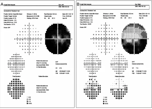
돌출을 주소로 내원하였다. 환자는 2001년 타 병원 건강검 진에서 혈액이상 소견을 보여 시행한 림프절 조직검사에서 림프종 진단하에 항암치료를 시행 받은 병력이 있었다. 환 자는 총 10차의 항암치료 중 치료에 따른 부작용으로 9차 까지만 시행 받은 후 추적관찰을 중단하였다고 한다. 이후 특별한 증상 없이 지내던 중 6년 전 시력저하와 좌안 부종 및 돌출이 발생하여 시행한 자기공명영상에서 림프종이 의 심되는 소견을 보였으나 양전자 단층촬영에서 이상소견이 보이지 않아 치료는 시행하지 않았다. 이후 환자는 증상 호 전을 위하여 임의적으로 스테로이드제 경구투여하며 지내 다가 수개월 전부터 시력저하 및 안구돌출이 심해져 본원 에 내원하였다. 환자는 좌안 눈꺼풀 부종과 함께 양측 턱밑 샘에서 무통성의 단단한 종창이 촉지되었다. 내원 당시 교 정시력은 우안 20/30, 좌안 20/200으로 측정되었고, 안압 은 양안 모두 정상범위 내였으며, Hertel 안구돌출계 검사 에서 우안 18 mm 좌안 20 mm로 측정되었다. 안구 운동 장 애 및 복시는 보이지 않았으며, 안구 운동 시 통증을 호소 하지는 않았다. Ishihara 색각검사표를 이용한 적녹색각검 사에서 우안은 정상이었으나 좌안은 중등도의 제2색각이상 소견을 보였으며, Humphrey 자동시야계를 이용한 시야검 사에서 우안은 하측 수평시야결손 소견을 보였으며, 좌안은 전반적인 시야협착 소견으로 보였다(Fig. 1).
환자는 안와 자기공명영상을 촬영하였으며 양측 해면정 맥동과 안와를 침범하는 침윤성의 병변이 관찰되고 있었고, 이 병변은 양측 시신경과 외안근을 포함하고 있었다. 또한 양측 상악동, 벌집굴, 우측 나비굴 내에 미만성의 조영증강 이 관찰되어 부비동염 소견을 보이고 있었으며, 이전 내시
A B
Figure 1. Humphrey visual field test reveals marked visual field defects in the left eye (A), and inferior
altitudinal visual field defects in the right eye (B).A B
Figure 2. Orbit MRI reveals diffuse well enhancing infiltrative lesions involving both optic nerves, extraocular
muscles, and cavernous sinus (A). Diffuse heterogeneous lesions involving the right maxillary sinus suggests sinusitis (B).경코곁굴수술을 받은 흔적이 관찰되었다(Fig. 2). 복부 초 음파를 포함한 전신 검사상 타 장기로의 병변의 침범 소견 은 보이지 않았다. 환자는 원인 감별 진단을 위하여 좌측 눈물샘 후면과 외직근 외측 사이의 안와 조직에서 절개 생 검을 시행하였다. 병리학적 조직 검사 결과 림프형질세포의 침윤과 간질섬유화가 관찰되었으며, 면역화학염색에서 IgG4 양성 형질세포를 확인할 수 있었다(Fig. 3). 또한 면역 화학염색에서 IgG4/IgG 양성 형질세포의 비율이 40% 이상 으로 관찰되었으며, 혈청단백전기영동검사에서 IgG 1,341
mg/dl, IgG4 267 mg/dl로 측정되어 IgG4의 혈청 수치가 정 상보다 높게 나타난 것을 확인할 수 있었다. 이는 최근에 발 표된 IgG4 연관 질환의 진단기준에 부합되는 소견으로 안와 를 침범한 IgG4 연관 경화성 질환으로 진단할 수 있었다.6 환자는 고용량 스테로이드 요법으로 methylprednisolone 1 g씩 3일간 정맥주사를 시행 받았으며 이후 외래 통하여 스 테로이드 경구 복용을 시작하였다.
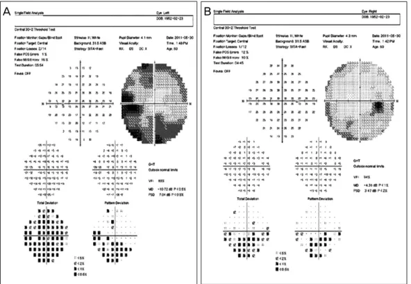
치료 2개월 후 환자의 교정 시력은 양안 20/30으로 호전 되었으며, 색각검사에서 양안 모두 정상 소견을 보였고, 시
Figure 3. (A) Biopsied specimen shows interstitial fibrosis and lymphoplasmacytic infiltrations with lymphoid
follicles (H&E, ×400). (B) Immunochemistry staining for IgG4 expression: IgG4-positive plasma cells have infiltrated the lesion (Anti-IgG4 Ab, ×400).A B
Figure 4. Two months after steroid therapy, there is significant improvement in the visual field defects of
both eyes (A, B).야검사 역시 호전된 소견을 보였다(Fig. 4). 안와 전산화단 층촬영에서도 양측 시신경과 외안근을 침범하던 침윤성 병 변의 감소를 확인할 수 있었으며, 이와 함께 부비동염 소견 의 호전도 관찰되었다(Fig. 5). 환자는 치료 6개월 후 특별 한 불편감 및 합병증 없이 외래를 통하여 경과관찰 중이다.
고 찰
IgG4 연관 경화성 질환군은 최근에 알려진 질환군으로서
자가면역이 원인인 것으로 생각하고 있으며, 한 개 이상의 외분비샘이나 림프절 외 장기로의 종양성 병변을 특징적으 로 하고 있고, 조직 내의 IgG4 양성의 형질세포와 높은 혈 청 내 IgG4 수치를 동반한 림프형질구성 침윤 및 경화에 의 한 것으로 생각되고 있다.1-3과거 IgG4 연관 경화성 눈물 샘염은 Mikulicz씨병으로 혼동되어 불려져 왔다. Mikulicz 씨병은 눈물샘과 침샘의 대칭적인 비대를 특징적으로 하는 질환으로써 1953년에 Morgan and Castleman7에 의해 처 음 명명되었으며, 병리학적으로 비슷한 소견으로 보이는 쇼
A B
Figure 5. Orbit CT images reveals interval decreased extents of infiltrative lesions involving both optic nerves
and extraocular muscles after steroid therapy (A, B).그렌 증후군의 아형으로 구분되었다. 최근 연구에 따르면 Mikulicz씨병은 쇼그렌 증후군과는 별개의 질병으로 밝혀 졌으며,8 특징적인 혈청 IgG4의 상승과 IgG4 염색 양성을 보이는 림프구형질세포의 침윤과 조직의 섬유화 소견을 보 여 자가면역성 췌장염, 후복막강 섬유화, 경화성 담관염, 그 리고, 만성 경화성 침샘염 등을 포함하는 IgG4 연관 경화성 질환의 같은 질병단위에 포함되었다.8-10
눈부속기는 특발성 염증성 질환이 발생할 수 있는 주요 부위 중 하나로서 이에는 안와 거짓종양이나 특발성 안와 경화증, 그리고 림프구증식성 질환 등이 포함된다.7-11 IgG4 연관성 질환의 경우 눈 부속기 이외의 장기를 먼저 침 범한 경우가 많고 눈물샘을 침범하는 비율은 비교적 낮다 고 보고되고 있으나,12 최근 IgG4 연관 질환군의 눈부속기 로의 침범이 흔하며 이 질환의 첫번째 증상으로 나타날 수 있다는 보고도 있다.13눈부속기로의 침범 빈도는 눈물샘으 로의 침범이 가장 흔한 것으로 알려졌으며, 그 외 결막으로 침범한 예도 발표된 바 있다.14 하지만 국내에서는 아직까 지 병변이 시신경을 포함한 예는 발표된 적이 없다. 본 증 례에서는 자기공명영상에서 눈부속기를 침범한 병변이 T1 및 T2 강조영상에서 비교적 균일한 신호강도를 보이면서 조영 증강이 잘되고 있으며, 주변 조직으로의 질량효과가 크게 나타나지 않아 영상학적으로 림프종을 가장 먼저 의 심해볼 수 있는 소견이었다. 특히 본 증례의 경우 과거 림 프종으로 치료받은 병력이 있었기 때문에, 시신경을 침범한 림프종을 포함한 림프증식구성 질환과 안와 거짓종양 등에 대한 감별진단을 위하여 병변의 생검을 통한 확진이 필수 적이다. 눈부속기는 B세포 림프종인 MALT (mucosa as- sociated lymphoid tissue) 림프종이 원발성으로 자주 발생 하는 부위라고 알려졌다.15MALT 림프종은 조직학적으로 작은 크기의 B세포와 단핵구세포, 형질세포 등의 증식을 특 징적으로 한다. 종종 반응성 림프여포들과 면역모세포를 동
반하여 나타나기도 하는데, 이러한 모양은 때때로 IgG4 연 관성 질환과 같은 염증성 질환에게도 동일한 형태로 나타 나기도 한다. 대부분의 경우, IgG4 연관성 질환과 MALT 림프종과의 구별은 어렵지 않지만, 이러한 형태학적인 유사 성으로 인해 두 질환의 구분이 어려울 경우도 있다.16이러 한 경우에는 IgG와 IgG4에 대한 면역화학염색과 혈청단백 전기영동검사가 진단에 도움을 줄 수 있다. 하지만 Kubota et al17은 안구 부속기를 침범한 B세포 림프종 중 약 10%
정도에서 IgG4 양성 형질세포를 나타내기도 한다는 보고를 한 바 있어, 면역화학염색상 IgG4 양성 세포를 보이더라도 림프종을 완전히 배제할 수는 없다. 또한 B세포 단일클론성 의 여부가 조직학적으로 구별이 어려운 경우 도움이 될 수 도 있지만, IgG4 연관성 질환 역시 단일클론성을 가지는 경 우도 있어 해석에는 주의가 필요하다.18,19
림프종과 IgG4 연관 경화성 질환과의 관계는 아직도 논 란이 되고 있는 부분으로, Takahira et al20은 IgG4 연관 경 화성 질환과 림프종과의 관계는 아직 정립된 것이 없다고 발표하고 있다. 이에 반해 Cheuk et al21은 IgG4 연관 경화 성 눈물샘염 환자 중 림프종이 병발한 3예를 발표하면서, IgG4 연관 경화성 질환이 림프종으로 이환될 확률은 약 10%라고 보고하였다. IgG4 연관 질환의 림프종으로의 전 환의 발병기전은 아직 확실히 밝혀진 바가 없지만, 림프조 직의 만성적인 증식상태가 B세포 클론들의 기질을 제공할 수 있는 환경을 만들었을 것으로 추측이 된다고 하였다.
본 증례의 경우 타 병원에서 장기간 림프종으로 진단받 아 치료받던 중 본원에서 IgG4 연관 경화성 질환으로 진단 받은 경우로 정확한 병력청취 및 자료 부족으로 두 질환 사 이의 명확한 연관성을 규명하기는 어려운 점이 있다. 하지 만 IgG4 연관 경화성 질환과 림프종의 유사점을 고려해본 다면 IgG4 연관성 질환을 림프종으로 오인하여 치료하였을 가능성을 생각해 볼 수 있다. 두 질환 모두 스테로이드 치
검을 통한 면역화학염색 및 혈청단백전기영동검사 등을 시 행하여 정확한 진단이 시행되어야 할 것으로 생각한다.
참고문헌
1) Kamisawa T, Funata N, Hayashi Y, et al. A new clinicopathological entity of IgG4-related autoimmune disease. J Gastroenterol 2003;38:982-4.
2) Yamamoto M, Takahashi H, Ohara M, et al. A new conceptualiza- tion for Mikulicz’s disease as an IgG4-related plasmacytic disease.
Mod Rheumatol 2006;16:335-40.
3) Sato Y, Ohshima K, Ichimura K, et al. Ocular adnexal IgG4- related disease has uniform clinicopathology. Pathol Int 2008;58:465-70.
4) Yamamoto M, Takahashi H, Sugai S, Imai K. Clinical and patho- logical characteristics of Mikulicz’s disease (IgG4-related plasma- cytic exocrinopathy). Autoimmun Rev 2005;4:195-200.
5) Kim K, Lee MJ, Kim NJ, et al. Three cases of Hyper-IgG4 syn- drome involving ocular adnexa. J Korean Ophthalmol Soc 2010;
51:1133-8.
6) Okazaki K, Uchida K, Koyabu M, et al. Recent advances in the concept and diagnosis of autoimmune pancreatitis and IgG4- re- lated disease. J Gastroenterol 2011;46:277-88.
7) Morgan WS, Castleman B. A clinicopathologic study of Mikulicz’s disease. Am J Pathol 1953;29:471-503.
8) Tsubota K, Fujita H, Tsuzaka K, Takeuchi T. Mikulicz’s disease and Sjögren’s syndrome. Invest Ophthalmol Vis Sci 2000;41:
1666-73.
9) Hamano H, Kawa S, Ochi Y, et al. Hydronephrosis associated with retroperitoneal fibrosis and sclerosing pancreatitis. Lancet 2002;359:1403-4.
10) Kitagawa S, Zen Y, Harada K, et al. Abundant IgG4-positive plas-
(IgG4)-positive or -negative ocular adnexal benign lymphoid le- sions in relation to systemic involvement. J Clin Exp Hematop 2010;50:129-42.
15) Thieblemont C, Bastion Y, Berger F, et al. Mucosa-associated lym- phoid tissue gastrointestinal and nongastrointestinal lymphoma behavior: analysis of 108 patients. J Clin Oncol 1997;15:1624-30.
16) Go H, Kim JE, Kim YA, et al. Ocular adnexal IgG4-related disease:
comparative analysis with mucosa-associated lymphoid tissue lymphoma and other chronic inflammatory conditions.
Histopathology 2012;60:296-312.
17) Kubota T, Moritani S, Yoshino T, et al. Ocular adnexal marginal zone B cell lymphoma infiltrated by IgG4-positive plasma cells. J Clin Pathol 2010;63:1059-65.
18) Kubota T, Moritani S, Katayama M, Terasaki H. Ocular adnexal IgG4-related lymphoplasmacytic infiltrative disorder. Arch Ophthalmol 2010;128:577-84.
19) Kojima M, Sipos B, Klapper W, et al. Autoimmune pan- creatitis:frequency, IgG4 expression, and clonality of T and B cells. Am J Surg Pathol 2007;31:521-8.
20) Takahira M, Kawano M, Zen Y, et al. IgG4-related chronic scleros- ing dacryoadenitis. Arch Ophthalmol 2007;125:1575-8.
21) Cheuk W, Yuen HK, Chan AC, et al. Ocular adnexal lymphoma as- sociated with IgG4-+ chronic sclerosing dacryoadenitis: a pre- viously undescribed complication of IgG4-related sclerosing disease. Am J Surg Pathol 2008;32:1159-67.
22) Cheuk W, Yuen HK, Chan JK. Chronic sclerosing dacryoadenitis:
part of the spectrum of IgG4-related Sclerosing disease? Am J Surg Pathol 2007;31:643-5.
23) Yamamoto M, Takahashi H, Ohara M, et al. A new conceptualiza- tion for Mikulicz’s disease as an IgG4-related plasmacytic disease.
Mod Rheumatol 2006;16:335-40.
=ABSTRACT=
A Case of IgG4-Related Sclerosing Disease Involving the Optic Nerve
Hyung Seok Cho, MD, Jung Yeol Choi, MD, Jung Hoon Yum, MD
Department of Ophthalmology, Inje University Ilsan Paik Hospital, Inje University College of Medicine, Goyang, Korea
Purpose: To report a case of IgG4-related sclerosing dacryoadenitis masquerading for a long period as a.
Case summary: A 59-year-old man with visual acuity loss and proptosis in the left eye was referred to our hospital. Ten years prior, the patient was diagnosed with lymphoma and underwent chemotherapy at another hospital. However, the pa- tient spontaneously stopped treatment and took steroids for the relief of periorbital swelling. Magnetic resonance imaging revealed diffuse infiltrative lesions involving both optic nerves and extraocular muscles. Incisional biopsies of the lacrimal gland’s posterior side and the lateral rectus muscle’s lateral side were performed. Staining for IgG4 via immunochemistry showed infiltration of IgG4-positive lymphoplasmacytic cells. Under the diagnosis of IgG4-related sclerosing disease, the patient underwent high-dose steroid pulse therapy.
J Korean Ophthalmol Soc 2012;53(12):1879-1884
Key Words: IgG4-related sclerosing disease, Mikulicz disease, Orbital inflammatory disease
Address reprint requests to Jung Hoon Yum, MD
Department of Ophthalmology, Inje University Ilsan Paik Hospital
#170 Juhwa-ro, Ilsanseo-gu, Goyang 411-706, Korea
Tel: 82-31-910-7240, Fax: 82-31-911-7241, E-mail: [email protected]