pISSN: 0378-6471 eISSN: 2092-9374 http://dx.doi.org/10.3341/jkos.2013.54.6.893
= 증례보고 =
특발성 망막전막에서 유리체절제술 전후 미세시야계를 이용한 구조와 기능의 상관관계 분석
서 샘⋅임한웅⋅신용운⋅강민호⋅성민철⋅조희윤 한양대학교 의과대학 안과학교실
목적: 특발성 망막전막으로 유리체절제술과 막제거술을 시행한 환자에서 빛간섭단층촬영과 미세시야계 검사를 사용하여 시세포 접합 부 구조와 망막민감도의 관계를 알아보고자 한다.
대상과 방법: 특발성 망막전막에 대한 수술을 받은 환자 20명 20안을 대상으로 술 전과 술 후 빛간섭단층촬영과 미세시야계 검사를 시행하였다. 시세포 접합부의 구조, 시력, 망막민감도, 주시안정도를 측정하였으며 각 인자간에 유의한 관련이 있는지 알아보았다.
결과: 수술 후 망막민감도는 유의하게 증가하였으며(p˂0.001), 시세포 접합부 결손범위는 변화가 없었다. 술 전 시세포 접합부 결손 범위는 술 전, 후 망막민감도와 유의한 상관관계를 보였으나(p=0.035, p=0.006), 주시안정도와는 유의한 상관관계를 보이지 않았다.
결론: 망막민감도는 시세포 접합부 결손 범위와 유의한 상관관계를 보였으며, 구조변화에 따른 망막의 기능을 객관적으로 정량화할 수 있을 것으로 생각한다.
<대한안과학회지 2013;54(6):893-901>
■ Received: 2012. 10. 20. ■ Revised: 2012. 12. 13.
■ Accepted: 2013. 4. 8.
■ Address reprint requests to Hee Yoon Cho, MD
Department of Ophthalmology, Hanyang University Guri Hospital, #153 Gyeongchun-ro, Guri 471-701, Korea Tel: 82-31-560-2350, Fax: 82-31-564-9479 E-mail: [email protected]
* 이 논문의 요지는 2012년 대한안과학회 제108회 학술대회에서 구연으로 발표되었음.
망막전막은 황반부의 내경계막을 따라 자라는 세포막으 로 황반부 견인, 망막 주름, 황반부 뒤틀림 등을 일으켜 시 력저하, 변형시, 단안복시 등의 증상을 유발하며, 유리체절 제술을 통한 망막전막 제거술 후 70-80%에서 시력향상 및 변형시의 호전을 보인다.1-4 성공적인 망막전막 제거술 후에도 시력 회복이 이루어지지 않는 경우가 있으며, 술 전 시력, 증상 기간, 망막부종 여부 등이 술 후 시력 예후에 영 향을 미치는 것으로 알려졌다.5-7
빛간섭단층촬영(OCT, optical coherence tomography) 은 망막전막과 같은 황반부 질환에서 진단 및 경과의 평가 에 유용한 검사로 망막두께, 시세포층의 형태 등 망막구조 에 대한 평가와 정량화가 가능하다. 스펙트럼영역 빛간섭단 층촬영(SD-OCT)의 발달로 더욱 빠른 속도로 고해상도의 검사가 가능해졌으며, 망막의 미세구조와 시력에 대한 연구 가 활발해졌다.8-10
시세포 내분절과 외분절의 접합부(IS/OS junction, inner segment/outer segment junction)는 다양한 질환에서 시력
과 유의한 상관관계가 보고되었으며, 중심망막두께 등 기존 의 구조적 변수와도 연관관계를 보이는 것으로 나타났다.11-13 그러나 IS/OS junction의 결손 정도가 시력에 미치는 영향 은 연구에 따라 다양하며, 표준화된 측정법이 존재하지 않 는다는 것과 시력이 망막 외의 다양한 인자에 영향을 받는 인자라는 점이 한계로 지적되었다.14-16
IS/OS junction의 결손이 시력 예후에 미치는 영향을 보 다 정확히 알기 위해 시력뿐만 아니라 중심시야, 주시안정 도 등 다른 시기능과의 연관성에 대한 평가가 도움이 될 수 있다. 이러한 시기능과 IS/OS junction 결손의 상관관계는 망막전위도(electroretinogram), 미세시야계 등의 검사를 통해 연구되고 있다.17-20
미세시야계 검사는 망막기능을 정량화할 수 있는 평가방 법으로 망막 각 부위의 민감도를 제공하므로 OCT에서 나 타난 구조적 이상과 대응하여 기능을 판별하는데 유용하며, 황반변성, 망막이영양증, 당뇨망막병증 등 다양한 황반부 질환에서 진단적 보조 수단 및 수술 전과 후의 망막기능 평 가에 이용되고 있다.21-23
미세시야계를 이용하여 IS/OS junction 결손 정도가 시 기능에 미치는 영향에 대해 다양한 질환에서 연구가 이루 어지고 있으나 망막전막을 대상으로 한 연구는 아직 보고 된 바 없다. 이에 본 연구에서는 망막전막 환자에서 성공적 인 망막전막 제거술 후 IS/OS junction 결손과 시력, 망막민 감도의 변화를 알아보고 이를 통해 구조적인 변화와 기능
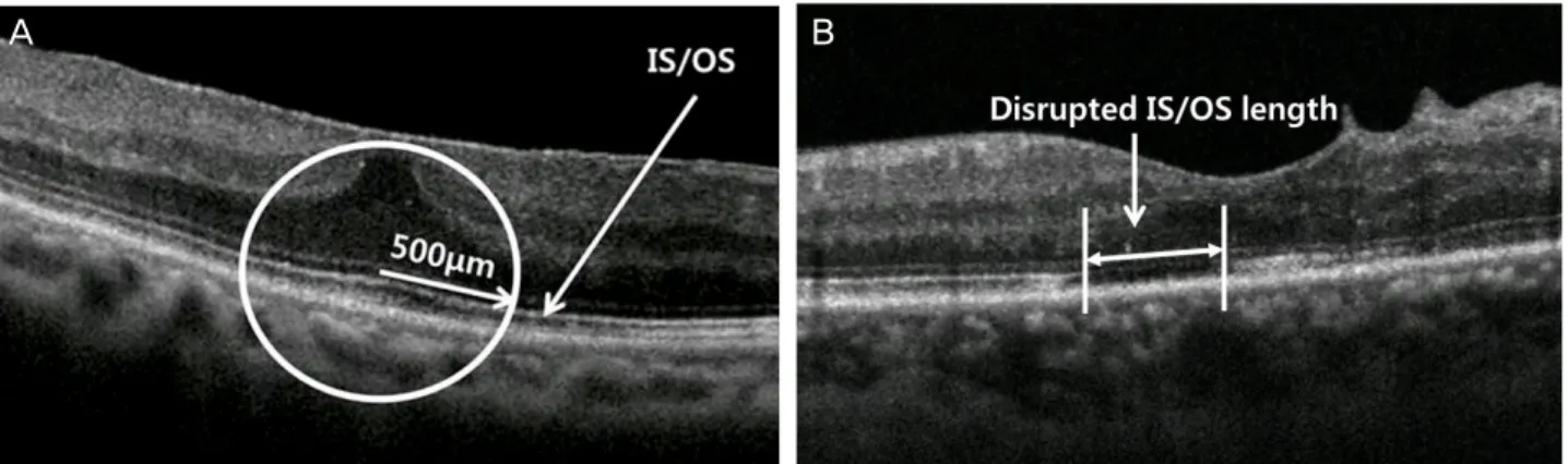
A B
Figure 1. Representative figures of groups according to the integrity of junction between photoreceptor inner and outer segment
(IS/OS) within 500 μm from the center of fovea. (A) Group 1 (Preserved IS/OS). IS/OS is well preserved within the 500 μm from the center of fovea. (B) Group 2 (Disrupted IS/OS). There is partially disrupted area of IS/OS (arrow). The disrupted IS/OS length was measured manually with caliper built in the spectral domain optical coherence tomography.간의 상관관계를 알아보고자 하였다.
대상과 방법
2012년 1월부터 2012년 7월까지 특발성 망막전막에 대 해 수술 받은 환자 20명 20안을 전향적으로 분석하였다. 망 막전막 환자 중 교정시력 0.4 이하이며, 교정시력이 0.5 이 상이더라도 변시증을 호소하는 경우 수술을 시행하였다. 술 후 1개월 이상 추적 관찰된 경우만을 포함하여 수술 후 시 간경과에 따른 시력 및 망막민감도, 시세포층의 변화를 관 찰하였다. 당뇨, 혈관폐쇄, 포도막염, 외상 등의 기왕력으로 인한 속발성 망막전막 환자는 제외하였으며, 다른 황반부 질환을 동반한 경우, LOCS (Lens Opacities Classification System) III NO4 이상의 백내장을 동반한 경우, 6디옵터 이상의 굴절이상이 있는 경우는 제외하였다.
본 연구는 임상시험심사위원회(IRB)의 승인을 받았으며, 모든 환자에게 충분한 설명 및 동의를 구하여 시행하였다.
술 전 모든 환자에게 최대교정시력, 전안부 검사 및 안저 검사, 빛간섭단층촬영(3D OCT-2000, Topcon, Tokyo, Japan), 미세시야계 검사(MAIA, Macular integrity assessment, Centervue, Padoba, Italy)를 시행하였다.
빛간섭단층촬영(SD-OCT)은 황반부 6 mm ×6 mm에 해당하는 부위를 512 A-scan을 구성하는 128개의 hori- zontal scan lines로 측정하는 방식인 cube scan 모드를 이 용하였다. 촬영은 한 명의 숙련된 기사에 의해 피검자에 대 한 사전 정보 없이 이루어졌다. 황반중심오목 500 μm내의 IS/OS 결손여부에 따라 IS/OS junction이 완전히 보존되어 있는 안을 그룹 1로, 부분 또는 전체 IS/OS junction에 결손 이있는 안을 그룹 2로 분류하였다(Fig. 1) (Table 3). IS/OS junction의 결손은 망막색소상피층(retinal pigment epi-
thelium)에 인접한 2개의 고반사층 중 위쪽 고반사층의 전체 또는 부분이 소실되는 것으로 정의하였다. 또한 SD-OCT 내 장 프로그램의 caliper를 이용하여 IS/OS junction 결손부 위의 길이를 측정하였으며, 망막 후극부 중심 10도에 해당 하는 범위 내의 각 horizontal scan line에서 IS/OS 결손길 이를 합산하여 IS/OS defect diameter를 구하였다.
미세시야계 검사에 이용한 MAIA기기는 근적외선을 이 용한 레이저검안경으로 혈관, 시신경 등 망막의 주요 해부 학적 표지를 근거로 안구의 움직임을 감시하는 시선 추적 기술을 갖추고 있다. 망막 후극부 중심 10도 부위에서 자극 의 크기를 Goldmann III로, 세기는 0 dB에서 36 dB까지 총 61개의 부위에 자극을 가하여 망막민감도를 측정하였다.
망막민감도의 역치는 61개 자극부위 역치 평균으로 하였으 며, 주시 안정성은 중심에서 2도 이내에 주시점이 존재하는 비율을 계산하였다(Fig. 2).
수술은 모든 환자에서 1명의 망막전문의에 시행되었으 며, 수술방법은 3개의 모양체 평면부 공막창을 통한 표준 3 port 평면부 유리체 절제술을 시행, 눈속집게를 이용하여 황반부 망막전막을 제거하였고, 특별한 염색은 시행하지 않 았다. 모든 환자에서 LOCS (Lens Opacities Classification System) III 분류에 따른 중등도의 핵백내장(NO2, NO3) 이 있었으며, 백내장에 대해 초음파유화술 및 인공수정체삽 입술을 먼저 시행한 후 망막전막제거술을 시행하였다. 망막 내경계막제거술은 시행하지 않았다.
술 후 1개월째에 최대교정시력, 망막주름의 호전유무, 망막 전막 재발유무를 확인하였으며, 빛간섭단층촬영으로 IS/OS junction의 변화 유무를 판단하였다. 또한 미세시야계검사 를 시행하여 수술 전 후 망막민감도의 변화를 알아보고, 수술 전후 교정시력 및 IS/OS defect diameter와의 상관관계를 알아보았다. 통계학적 분석은 SPSS 17.0 프로그램(SPSS,
Figure 2. A microperimetry combined with Spectral domain optical coherence tomographic image of a 76 year old male patient with
idiopathic ERM. Preoperative fundus image of the right eye shows an epiretinal membrane (upper left). Optical coherence tomog- raphy (OCT) result shows epiretinal membrane delineated as highly reflective band and globally adherent to the retina. IS/OS dis- ruption (arrowheads) also presented. Microperimetry results show mean sensitivity in colors (upper right), and fixation stability (bottom right).Table 1. Summary of demographic data of all eyes with
Idiopathic epiretinal membraneCharacteristics Value
Number of eyes (subjects) 20 (20)
Sex (M:F) [n(%)] 7 (35%) : 13 (65%)
Age (years) 64.55 ± 2.41
Refraction (S.E) 0.63 ± 1.22
Lens status*
Mild cataract 4
Moderate cataract 16
Severe cataract 0
Values are presented as mean ± SD unless otherwise indicated.
S.E = spherical equivalent
*Cataract classified by lens opacification classification system III, Mild cataract: NO 1-NO 2, Moderate cataract: NO 3-NO 4, Severe cataract: NO 5-NO 6.
Inc, Chicago, IL)을 사용하였고, 술 전과 술 후 비교는 Wilcoxon singed ranked test, 그룹간 비교는 Mann- Whitney test를 이용하였다. 시력, 망막민감도, IS-OS defect diameter간의 관계는 상관분석(spearsman’s rank corre- lation)을 이용하였다. p값이 0.05 미만인 경우에 통계적으 로 유의하다고 판단하였다.
결 과
총 20안(20명) 중 남자가 7명(35%), 여자가 13명(65%) 이었으며, 환자들의 평균나이는 64.55 ±2.41세였다(Table 1). 모든 안에서 성공적으로 유리체절제술을 통해 망막전막 을 제거하였으며, 술 후 경과관찰 기간 동안 합병증이나 망 막전막의 재발은 발생하지 않았다.
술 전 20안의 평균 logMAR 시력은 0.58 ±0.05였으며, 술 후 1개월째 시력은 0.39 ±0.057로 술 전에 비해 유의하 게 호전되었다(p=0.003). 술 전 IS/OS defect diameter는
1247.8 ±988.9 μm였으며, 술 후 1023.4 ±896.2 μm로 감 소하였으나 통계학적으로 유의하지 않았다(p=0.062). 미세 시야계로 검사한 망막민감도 역치는 술 전 16.95 ±6.60dB
Table 2. Preoperative data and outcomes during follow up
Characteristics Base line After surgery p-value*
BCVA (log MAR) 0.58 ± 0.05 0.39 ± 0.057 0.003
IS/OS defect diameter (μm) 1247.8 ± 988.9 1023.4 ± 896.2 0.062
Macular sensitivity (dB) 16.95 ± 6.60 22.35 ± 4.29 <0.001
Fixation stability (%) 61.0 ± 18.2 67.4 ± 23.5 0.059
Values are presented as mean ± SD unless otherwise indicated.
*Statistical significance was calculated by Wilcoxon signed rank test.
Table 3. Comparison of preoperative characteristics of patients with IS/OS preserved and disrupted epiretinal membrane
Characteristics Group 1 (IS/OS preserved) Group 2 (IS/OS disrupted) p
Number of eyes (%) 8 (40%) 12 (60%)
Sex (M:F) 5:3 8:4 0.85
Age (years) 64.25 ± 9.59 64.75 ± 10.79 0.79
BCVA (log MAR) 0.46 ± 0.27 0.63 ± 0.23 0.11
IS/OS defect diameter (μm) 288.69 ± 365.81 1887.33 ± 699.27 <0.01
Macular sensitivity (dB) 20.64 ± 2.70 14.49 ± 7.37 0.02
Values are presented as mean ± SD unless otherwise indicated.
Group 1 (preserved) Group 2 (disrupted) IS/OS integrity
Group 1 (preserved) Group 2 (disrupted) IS/OS integrity
Preoperative retinal sensitivity (dB) Preoperative VA (log MAR)
Postoperative VA (log MAR)
Group 1 (preserved) Group 2 (disrupted) IS/OS integrity
25
20
15
10
5
0
1.00
0.80
0.60
0.40
0.20
1.00
0.80
0.60
0.40
0.20
0.00
*
* **
Figure 3. (A) Preoperative macular sensitivity (dB) of the
group 1 and 2. Preoperative macular sensitivity in the group 1 is significantly better than that in the group 2. (B) Preoperative best-corrected visual acuity (BCVA) (log MAR) of the group 1 and 2. However, there is no significant difference between the group 1 and the group 2. (C) Postoperative BCVA (log MAR) of the group 1 and 2. Postoperative BCVA in the group 1 is significantly better than that in the group 2. (p val- ue related to Kruskall-Wallis test). (*p < 0.05, **p > 0.05) The box and whisker plots: horizontal lines within the box represent medians, the ends of the boxes represent the first and third quartiles, and the whiskers represent the smallest and largest non-outlier values.A B
C
에서 술 후 22.35 ± 4.29 dB으로 유의하게 상승하였다 (p<0.001). 주시 안정도는 술 후 상승하는 소견을 보였으
나 유의하지 않았다(p=0.059)(Table 2).
술 전 시력은 백내장의 영향을 배제하기 위한 편상관분
Preoperative IS/OS defect diameter ( m)µ
0 1000 2000 3000
Preoperative retinal sensitivity (dB)
25
20
15
10
5
0
IS/OS integrity Group 1 (preserved) Group 2 (disrupted)
Preoperative IS/OS defect diameter ( m)µ
0 1000 2000 3000
Postoperative retinal sensitivity (dB) 30
25
20
15
10
Postoperative IS/OS defect diameter ( m)µ
0 1000 2000 3000
Postoperative retinal sensitivity (dB)
IS/OS integrity Group 1 (preserved) Group 2 (disrupted)
IS/OS integrity Group 1 (preserved)
Group 2 (disrupted)
Figure 4. Correlation between macular sensitivity (dB) and diame-
ter of disrupted photoreceptor inner and outer segment junction (IS/OS defect diameter) (μm) in the group 1 and 2 (marked with black triangle and white circle respectively) at the preoperative (A) and postoperative (B) visits. (A) shows that there is significant negative correlation between the preoperative IS/OS defect diame- ter (μm) and macular sensitivity (dB) (r = -0.474, p = 0.035). (B) shows that there is significant negative correlation between the preoperative IS/OS defect diameter (μm) and postoperative mac- ular sensitivity (dB) (r = -0.589, p = 0.006). In accordance with an increase in the IS/OS defect diameter at the preoperative visit, the postoperative macular sensitivity worsened. (C) shows there is significant negative correlation between the postoperative IS/OS defect diameter (μm) and macular sensitivity (dB) (r = -0.647, p= 0.002). p-value related to linear logistic regression analysis.
A B
C
석에서 술 전 망막민감도와 유의한 상관관계를 보였으며 (p=0.033), 술 전 IS/OS defect diameter와도 유의한 상관 관계를 보였다(p=0.032). 술 후 시력과 망막민감도 모두 상승하였으나, 술 후 시력은 술 후 망막민감도와 유의한 상 관관계를 보이지 않았다(p=0.053).
술 전 IS/OS junction이 완전히 보존되어 있는 경우는 8 안(그룹 1, 40%), 결손이 있는 경우는 12안(그룹 2,60%) 였다(Table 3). 그룹 1과 그룹 2의 술 전 logMAR시력은 각각 0.46 ±0.27, 0.63 ±0.23로 유의한 차이가 없었고, 술 전 망막민감도는 20.64 ±2.70 dB, 14.49 ±7.37 dB로 그룹 1에서 유의하게 높았다(p=0.025). 술 후 logMAR시 력은 그룹 1에서 0.22 ±0.24, 그룹 2에서 0.47 ±0.24로 술 전 IS/OS junction이 보존되어 있는 경우 술 후 시력예 후가 좋은 것으로 나타났다(p=0.032) (Fig. 3).
술 전 IS/OS defect diameter는 술 전 망막민감도와 유의
한 상관관계를 보였으며(r=-0.474, p=0.035), 술 후 1개월 째의 망막민감도와도 유의한 상관관계를 보였다(r=-0.589, p=0.006). 술 후 IS/OS defect diameter와 술 후 망막민감 도 또한 유의한 상관관계를 보였다(r=-0.647, p=0.002) (Fig. 4). 주시안정도는 IS/OS defect diameter와 유의한 연관관계를 보이지 않았다(p=0.142).
고 찰
망막전막은 1865년 Iwanoff가 처음으로 보고하였으며, 후 유리체 박리로 인해 야기된 신경교세포 등의 증식이 내 경계막 표면에 세포막을 형성하여 발생된다.24,25Machemer 가 유리체절제술을 이용한 망막전막의 제거술에 성공한 이 후 현재까지 유리체절제술이 효과적인 치료로 활발히 시행 되고 있다. 대부분의 망막전막 환자는 술 후 70-80%에서
시력호전을 보이나 일부에서는 색각의 저하와 광각의 변화, 변시증 등을 호소하기도 한다.3 망막전막에 의한 망막견인 이 오래 지속되면서 일으키는 황반부 뒤틀림과 시세포의 왜곡이 술 후 시력개선을 저해하는 것으로 추정되나 생체 현미경으로는 이를 관찰하기 어렵다.26
SD-OCT의 발달로 망막의 미세구조와 시력예후와의 연 관관계에 대한 연구가 활발히 이루어졌으며, 많은 연구에서 IS/OS junction의 보존여부가 술 후 시력에 중요함을 보고
하였다.14,27그러나 이전의 연구들에서 그 결과가 다양하여
논란의 여지가 있다. 일부 연구에서는 IS/OS junction의 결 손이나 IS/OS경계부에 해당하는 IS/OS line의 손상이 술 후 시력개선을 저해한다는 보고가 있으며, 일부에서는 연관관 계가 없다는 보고도 있다.15,16이러한 차이는 IS/OS junc- tion의 측정방법이나 측정 시점에서의 차이가 영향을 주었 을 것으로 생각한다.
또한 기존의 연구들은 망막의 기능 평가를 주로 시력에 의존하고 있다. 시력은 망막 외에 시신경 등의 전달경로와 공간해상도에 영향을 받기 때문에 신뢰성이 떨어지며, 중심 망막의 기능을 선택적으로 평가하기 어렵다는 한계가 있다.
또한 최근의 연구에서 시력은 수술 직후 망막구조 변화에 비하여 황반 기능의 변화를 저평가한다는 보고가 있어, 망 막전막 수술 후 초기 평가방법으로 이용하기 어렵다.28
본 연구에서는 미세시야계 검사를 이용하여 망막 구조와 기능간의 상관관계를 밝히고자 하였다. 미세시야계 검사는 다양한 질환에서 유용성이 보고되어 있다. Okada et al21은 당뇨망막병증에서 미세시야계를 이용하여 측정한 망막민감 도가 시력과 황반부 두께와 유의한 연관관계가 있음을 보 고하였고, Springer et al22은 중심장액맥락망막병증 환자에 서 미세시야계에서 민감도 감소부위가 망막두께 증가부위 와 일치함을 보고하였다. 황반원공 환자에서도 수술 후 시 기능 회복에 있어 시력보다 망막민감도가 더 중요한 예측 인자임이 보고된 바 있다.23
망막 후극부 중심 10도의 망막민감도 역치의 평균을 구 하여 IS/OS junction 결손과의 연관관계를 살펴보았다. 술 전 망막민감도는 술 전 IS/OS defect diameter와 유의한 연 관관계를 보였으며, 술 후에도 망막민감도와 IS/OS defect diameter간 유의한 연관관계가 있는 것으로 나타났다. 또한 술 전 망막민감도는 술 전 시력과도 유의한 연관관계를 보 였다. 망막민감도는 시력과 연관성을 가지며, 구조 손상의 정도에 따른 망막기능의 변화를 측정하는데 있어서 유의한 지표임을 보여준다. 그러나 술 전 IS/OS defect diameter와 술 전 망막민감도는 상관계수 0.474, 술 후 망막민감도와는 상관계수 0.589로 높은 연관성을 가진다고 보기는 어렵다 는 한계가 있다.
망막민감도는 망막 중심두께와도 유의한 상관관계를 보 이며, 망막 기능 예측에 있어 망막 중심두께와 동등한 예측 력을 보인다는 보고가 있다. 또한 본 연구에서 확인한 바와 같이 IS/OS junction을 반영하므로 망막중심두께만으로는 설명되지 않는 망막전막의 기능 평가에 있어 유용하다고 생각한다.6,11
이전의 연구에서 망막민감도는 시력보다 더 민감하게 치 료효과를 반영한다는 보고가 있으며, 수술 후 시기능 회복 예측과 평가에 유용한 지표로 제시되었다.23,29특히 망막부 종 및 망막기능 저하의 경우 중심 외 주시 등으로 시력은 보존되어 나타나기도 하므로 망막민감도가 기능 평가에 유 용하게 사용될 수 있다.21
술 전 IS/OS junction이 보존되어 있는 그룹 1 (8안)은 IS/OS junction이 손상된 그룹 2 (12안)에 비하여 술 전 망 막민감도가 높게 나타났으며, 그룹간의 차이는 유의하였다.
두 그룹 모두 수술 후 IS/OS junction의 결손범위는 유의한 변화를 보이지 않았다. 6안에서 술 후 IS/OS junction의 결 손범위가 증가하는 양상을 보였으나, 유의한 차이는 아니었 다. 이전의 연구에서도 수술 후 가역적 또는 비가역적인 IS/OS 결손이 발생한 예들이 있으며, 유리체절제술로 인한 안내 염증, 혈액망막장벽의 파괴 등에 의한 것으로 추정된 다.30,31
술 후 IS/OS defect diameter는 술 전에 비하여 감소하였 으나 의미있게 줄어들지는 않은 것으로 나타난다. 이는 성 공적인 수술 후에도 구조의 회복이 첫 1개월 내에는 일어나 지 않음을 의미하며, 망막전막의 수술 후 망막구조의 완전 한 회복과 최고교정시력의 도달은 술 후 1년 이내에 서서히 이루어지는 것으로 알려졌다.32망막전막을 대상으로 한 다 른 연구에서는 IS/OS junction의 결손이 있는 안에서 술 후 1년까지도 결손이 회복되지 않음을 보고하였다.33 이는 망 막전막의 견인으로 인하여 시세포에 물리적인 분열을 일으 켜 비가역적인 변화를 초래한 것으로 생각한다.
술 후 망막민감도와 시력은 술 전에 비하여 유의하게 상 승을 보였다. 술 후 IS/OS junction의 결손이 회복되지 않았 음에도 시력과 망막민감도가 상승한 이유는 망막전막의 차 폐효과가 소실되고 망막전막의 견인력이 사라지면서 망막 부종이 줄고 망막두께가 감소하면서 야기된 것으로 보인다.
본 연구에서는 망막전막 술 후 예측인자로 SD-OCT로 측정한 IS/OS junction의 결손 범위의 유용성을 망막민감도 와의 연관성을 통해 확인하였다. 망막전막에서 IS/OS junc- tion의 결손여부와 술 후 시력과의 연관관계는 많은 연구를 통해 밝혀져 있다.12,27,34시력은 망막 외 시신경 전달경로나 공간해상도 등에 영향을 받는 인자이나, 망막민감도는 근적 외선을 이용하므로 매질혼탁 등의 영향이 적어 망막의 기
능을 보다 선택적으로 평가 가능하다. 또한 원하는 범위 내 의 망막 중심 기능을 평가 가능하며, 각 부위별 역치를 제 공하므로 구조와의 연관관계를 연구함에 있어 시력보다 더 유용한 지표로 활용 가능하다.35,36본 연구에서는 각 부위별 역치를 활용하지는 않았으나 망막전막에서 망막중심부의 망막민감도가 IS/OS junction 결손 정도와 연관성을 가짐을 확인하였다.
본 연구는 전향적 연구이나 대상환자 수가 적어 통계적 유의성을 충분히 높이지 못하였고, 대조군을 설정하지 않은 것이 제한점이라 할 수 있다. 또한 망막전막의 수술 후 망 막구조의 완전한 회복과 최고교정시력의 도달은 술 후 1년 이내에 이루어지는 것으로 알려졌으나, 본 연구에서는 1개 월의 비교적 짧은 기간 변화만을 관찰했다는 점에서 한계 를 가지고 있다. 미세시야계 검사와 SD-OCT결과를 망막 의 구역별로 대응시켜 비교하지 않았다는 것 또한 본 연구 의 제한점이다. 망막전막의 형태에 따라 예후에 차이를 보 임은 이전의 다양한 연구에서 알려졌으나, 본 연구에서는 대 상환자 수가 적어 형태에 따른 분류는 고려하지 않았다.37,38 모든 환자가 백내장 수술을 망막전막제거술 시 동시에 시 행 받았으며, 연구결과에 영향을 미칠 것으로 고려하여 NO4 이상의 백내장 환자는 연구대상에서 제외하였다. 모 든 안에서 피질경화도가 3 이하로 백내장이 시력에 큰 영향 을 미치지 않았을 것으로 가정하였으나 백내장에 의한 차 폐효과가 망막민감도에 미치는 영향을 배제할 수 없다는 점에서 본 연구의 제한점이라 할 수 있다.
결론적으로 미세시야계 검사는 망막의 미세구조 변화에 따른 망막기능을 보여주는 비침습적인 검사로서 SD-OCT 와 미세시야계 검사를 통하여 망막전막 술 후 구조와 기능 의 변화를 확인하였다. 술 전 IS-OS junction의 결손은 시 세포층의 구조적 손상과 기능을 반영하는 지표로 술 후 망 막민감도를 예측하는데 있어 중요한 인자이다. 망막민감도 는 망막의 기능을 원하는 부위를 선택적으로 평가 가능하 며, 망막 미세구조 손상과 의미있는 연관성을 가지므로 구 조와 기능의 상관관계 연구에 있어 망막민감도의 측정이 도움이 될 것이다.
REFERENCES
1) McDonald HR, Verre WP, Aaberg TM. Surgical management of idiopathic epiretinal membranes. Ophthalmology 1986;93:978-83.
2) Choi YK, Yoo JS, Kim MH. Result of surgery for epiretinal mem- brane and their recurrence. J Korean Ophthalmol Soc 2000;41:
2357-62.
3) Margherio RR, Cox MS Jr, Trese MT, et al. Removal of epimacular membranes. Ophthalmology 1985;92:1075-83.
4) Kwon SI, Ko SJ, Park IW. The clinical course of the idiopathic epi-
retinal membrane surgery. Korean J Ophthalmol 2009;23:249-52.
5) Rice TA, De Bustros S, Michels RG, et al. Prognostic factors in vi- trectomy for epiretinal membranes of the macula. Ophthalmology 1986;93:602-10.
6) Vujosevic S, Midena E, Pilotto E, et al. Diabetic macular edema:
correlation between microperimetry and optical coherence tomog- raphy findings. Invest Ophthalmol Vis Sci 2006;47:3044-51.
7) Hwang DJ, Na KI, Kwon SI, Park IW. Long term changes in visual acuity and foveal thickness after vitrectomy for idiopathic epi- retinal membrane. J Korean Ophthalmol Soc 2012;53:434-9.
8) van Velthoven ME, Faber DJ, Verbraak FD, et al. Recent develop- ments in optical coherence tomography for imaging the retina.
Prog Retin Eye Res 2007;26:57-77.
9) Ko TH, Fujimoto JG, Schuman JS, et al. Comparison of ultrahigh- and standard-resolution optical coherence tomography for imaging macular pathology. Ophthalmology 2005;112:1922.e1-15.
10) Schmidt-Erfurth U, Leitgeb RA, Michels S, et al. Three-dimen- sional ultrahigh-resolution optical coherence tomography of mac- ular diseases. Invest Ophthalmol Vis Sci 2005;46:3393-402.
11) Karacorlu M, Ozdemir H, Senturk F, et al. Correlation of retinal sensitivity with visual acuity and macular thickness in eyes with idiopathic epimacular membrane. Int Ophthalmol 2010;30:285-90.
12) Oster SF, Mojana F, Brar M, et al. Disruption of the photoreceptor inner segment/outer segment layer on spectral domain-optical co- herence tomography is a predictor of poor visual acuity in patients with epiretinal membranes. Retina 2010;30:713-8.
13) Lee JE, Kim EH, Oum BS. Relationship between visual acuity and photoreceptor layer or foveal thickness on optical coherence to- mography after macular hole surgery. J Korean Ophthalmol Soc 2006;47:1966-71.
14) Oh J, Smiddy WE, Flynn HW Jr, et al. Photoreceptor inner/outer segment defect imaging by spectral domain OCT and visual prog- nosis after macular hole surgery. Invest Ophthalmol Vis Sci 2010;51:1651-8.
15) Chang LK, Koizumi H, Spaide RF. Disruption of the photoreceptor inner segment-outer segment junction in eyes with macular holes.
Retina 2008;28:969-75.
16) Chung H, Shin CJ, Kim JG, et al. Correlation of microperimetry with fundus autofluorescence and spectral-domain optical coher- ence tomography in repaired macular holes. Am J Ophthalmol 2011;151:128-36.e3.
17) Ooto S, Hangai M, Takayama K, et al. Photoreceptor damage and foveal sensitivity in surgically closed macular holes: an adaptive optics scanning laser ophthalmoscopy study. Am J Ophthalmol 2012;154:174-86.e2.
18) Kim YG, Baek SH, Moon SW, et al. Analysis of spectral domain optical coherence tomography findings in occult macular dystrophy.
Acta Ophthalmol 2011;89:e52-6.
19) Lim JW, Cho JH, Kim HK. Assessment of macular function by multifocal electroretinography following epiretinal membrane sur- gery with internal limiting membrane peeling. Clin Ophthalmol 2010;4:689-94.
20) Kang JH, Choi ES, Yoon JM, Yoon HS. Multifocal electroretino- gram before and after epiretinal membrane surgery. J Korean Ophthalmol Soc 2008;49:104-10.
21) Okada K, Yamamoto S, Mizunoya S, et al. Correlation of retinal sensitivity measured with fundus-related microperimetry to visual acuity and retinal thickness in eyes with diabetic macular edema.
Eye (Lond) 2006;20:805-9.
22) Springer C, Völcker HE, Rohrschneider K. [Central serous cho- rioretinopathy--retinal function and morphology: microperimetry and optical coherence tomography]. Ophthalmologe 2006;103:791-7.
23) Amari F, Ohta K, Kojima H, Yoshimura N. Predicting visual out- come after macular hole surgery using scanning laser ophthalmo- scope microperimetry. Br J Ophthalmol 2001;85:96-8.
24) Klein R, Klein BE, Wang Q, Moss SE. The epidemiology of epi- retinal membranes. Trans Am Ophthalmol Soc 1994;92:403-25;
discussion 425-30.
25) Gastaud P, Bétis F, Rouhette H, Hofman P. [Ultrastructural findings of epimacular membrane and detached posterior hyaloid in vitre- omacular traction syndrome]. J Fr Ophtalmol 2000;23:587-93.
26) Michels RG. Vitreous surgery for macular pucker. Am J Ophthalmol 1981;92:628-39.
27) Inoue M, Morita S, Watanabe Y, et al. Preoperative inner seg- ment/outer segment junction in spectral-domain optical coherence tomography as a prognostic factor in epiretinal membrane surgery.
Retina 2011;31:1366-72.
28) Frisén L, Frisén M. Micropsia and visual acuity in macular edema.
A study of the neuro-retinal basis of visual acuity. Albrecht Von Graefes Arch Klin Exp Ophthalmol 1979;210:69-77.
29) Ehrlich R, Mawer NP, Mody CH, et al. Visual function following photodynamic therapy for central serous chorioretinopathy: a com- parison of automated macular microperimetry versus best-cor- rected visual acuity. Clin Experiment Ophthalmol 2012;40:e32-9.
30) Suh MH, Seo JM, Park KH, Yu HG. Associations between macular findings by optical coherence tomography and visual outcomes af- ter epiretinal membrane removal. Am J Ophthalmol 2009;147:
473-80.e3.
31) Tso MO. Animal modeling of cystoid macular edema. Surv Ophthalmol 1984;28 Suppl:512-9.
32) Pesin SR, Olk RJ, Grand MG, et al. Vitrectomy for premacular fibroplasia. Prognostic factors, long-term follow-up, and time course of visual improvement. Ophthalmology 1991;98:1109-14.
33) Inoue M, Morita S, Watanabe Y, et al. Inner segment/outer segment junction assessed by spectral-domain optical coherence tomog- raphy in patients with idiopathic epiretinal membrane. Am J Ophthalmol 2010;150:834-9.
34) Mitamura Y, Hirano K, Baba T, Yamamoto S. Correlation of visual recovery with presence of photoreceptor inner/outer segment junc- tion in optical coherence images after epiretinal membrane surgery.
Br J Ophthalmol 2009;93:171-5.
35) Rohrschneider K, Bültmann S, Springer C. Use of fundus peri- metry (microperimetry) to quantify macular sensitivity. Prog Retin Eye Res 2008;27:536-48.
36) Sunness JS, Schuchard RA, Shen N, et al. Landmark-driven fun- dus perimetry using the scanning laser ophthalmoscope. Invest Ophthalmol Vis Sci 1995;36:1863-74.
37) Michalewski J, Michalewska Z, Cisiecki S, Nawrocki J. Mor- phologically functional correlations of macular pathology con- nected with epiretinal membrane formation in spectral optical coherence tomography (SOCT). Graefes Arch Clin Exp Ophthalmol 2007;245:1623-31.
38) Kinoshita T, Kovacs KD, Wagley S, Arroyo JG. Morphologic dif- ferences in epiretinal membranes on ocular coherence tomography as a predictive factor for surgical outcome. Retina 2011;31:1692-8.
=ABSTRACT=
Morphologic and Functional Evaluation before and after Vitrectomy in Idiopathic Epiretinal Membrane Patients Using Microperimetry
Sam Seo, MD, Han Woong Lim, MD, Yong Un Shin, MD, Min Ho Kang, MD, Min Cheol Seong, MD, Hee Yoon Cho, MD
Department of Ophthalmology, Hanyang University College of Medicine, Seoul, Korea
Purpose: To investigate the relationship between photoreceptor inner/outer segment (IS/OS) integrity and macular sensi- tivity after epiretinal membrane (ERM) surgery using spectral-domain optical coherence tomography combined with microperimetry.
Methods: 20 eyes of 20 patients with idiopathic ERM who underwent pars plana vitrectomy for ERM removal were pro- spectively reviewed. The IS/OS defect diameter, BCVA, macular sensitivity, and fixation stability were measured using spectral-domain optical coherence tomography combined with microperimetry. The correlation of these factors was analyzed.
Results: The macular sensitivity improved after successful ERM surgery (p < 0.001), but the IS/OS defect diameter has not improved. The preoperative and postoperative macular sensitivity were negatively correlated with preoperative IS/OS de- fect diameter (p = 0.035, p = 0.006). The fixation stability was not correlated with preoperative IS/OS defect diameter.
Conclusions: Macular sensitivity showed significant correlation with IS/OS defect diameter. Macular sensitivity is vital func- tional parameter allows subjective quantification of the retinal function and reflects morphologic changes.
J Korean Ophthalmol Soc 2013;54(6):893-901
Key Words: Idiopathic epiretinal membrane, Macular sensitivity, Microperimetry, Spectral domain optical coherence tomography
Address reprint requests to Hee Yoon Cho, MD
Department of Ophthalmology, Hanyang University Guri Hospital
#153 Gyeongchun-ro, Guri 471-701, Korea
Tel: 82-31-560-2350, Fax: 82-31-564-9479, E-mail: [email protected]