The anti-oxidative and anti-inflammatory effect of Psoralea corylifolia on Ulcerative Colitis Induced by
Dextran Sulfate Sodium in Mice
Ahn Sang Hyun
1, Kim Ki Bong
21Department of Anatomy, college of Korean Medicine, Semyung University
2Department of Pediatrics, Korean Medicine Hospital, Pusan National University Original Article
⋅Received:17 November 2016 ⋅Revised:15 December 2016 ⋅Accepted:15 December 2016
⋅Correspondence to:Kibong Kim
Department of Pediatrics, Korean Medicine Hospital, Pusan National University
20, Geumo-ro, Mulgeum-eup, Yangsan-si, Gyeongsangnam-do, 50612, Republic of Korea Tel:+82-55-360-5952, Fax:+82-55-360-5952. E-mail:[email protected]
Objectives: This study was to investigate the anti-oxidative and anti-inflammatory effect of Psoralea corylifolia water extract (PE) on ulcerative colitis which was induced by dextran sulfate sodium (DSS) in mice.
Methods: Ulcerative colitis was induced by DSS in male BALB/c mice. The mice were divided into 3 groups. The control group (Ctrl) was not induced ulcerative colitis. The pathological group (CE) was induced the colitis. The experimental group (PT) was administered PE after inducing the colitis. The effects of the PE on ulcerative colitis were evaluated by morphological change in the colon tissue and cells, substance P production, activity of tumor necrosis factor (TNF)-α and nuclear factor (NF)-κB, cyclooxygenase (COX)-2 production, and anti-oxidative activity.
Results: In the PT group, PE alleviated hemorrhagic erosion in colon mucosa and infiltration of inflammatory cells in lamina propria mucosae. In the colon of the PT group, COX-2 production was inhibited via regulating the activity of TNF-α and NF-κB p65. PE also had an anti-oxidative effect via activating nuclear factor (erythroid-derived 2)-like 2 (Nrf2).
Conclusions: In this study, we found the utility of treatment with PE and the potential of developing a medicine for ulcerative colitis by applying our results. Further investigations for the anti-inflammatory mechanism of PE may be needed.
Key Words : Psoralea corylifolia, ulcerative colitis, anti-inflammatory, anti-oxidative.
Introduction
Ulcerative colitis (UC) is a chronic cryptogenic inflammatory bowel disease (IBD) which was localized in the mucosa or submucosa of the colon.
It accompanies with recurrent bloody diarrhea and the patient complains of rectal urgency and abdominal pain
1). UC is caused by the complex effect of genetic and environmental factors. Although the patients are distributed worldwide, UC is the
most frequent in the North America and Northen Europe. Ethnically, UC more occurs in the Jewish and the Caucasian but relatively rare in the Asian
2,3). However, UC patient is increasing as the incidence increases in the Asian countries including Korea
3,4).
The objective of UC treatment is inducing
remission by alleviating symptoms and inflammation
in mucosa and improving the quality of life by
maintaining the remission as long as possible. About
15% of the UC patients arrive at the remission by
just a placebo
5). However, adequate treatment is advisable because most patients continue bloody stool and diarrhea if not treated
6). 5-aminosalicylic acid (5-ASA), steroids, or immunosuppressants have been used for the standard medication of UC. But 20-40% of the patients fail to conventional medication or have colectomy due to side effects
7). Recently, there are some efforts to find candidate drugs which is effective for UC and have less side effects from natural products including Korean medicine
8). There have been studies on herbal medication for IBD and experimental effectiveness of herbal medicine for colitis animal model induced with dextran sulfate sodium (DSS) in Korea
8). However, the medicine have not been developed because studies on the mechanism of herbal medicine are insufficient.
Psoralea semen, which is originated from Psoralea corylifolia L. (Leguminosae), has been used as external medicine for vitiligo in Korean medicine and it has melanogenesis activity
9). According to Dongeuibogam, it invigorates yang by tonifying the kidney, secures essence, and warm the spleen. Thus, it cures impotence, ganacratia, urinary frequency, and diarrhea.
Based on its known effectiveness, Psoralea corylifolia may be effective for IBD which has main symptoms such as chronic abdominal pain and diarrhea. However, studies on the anti-oxidative activity and inhibitory effect for cyclooxgenase (COX)-2 expression of Psoralea corylifolia are insufficient yet. To provide the basis of utilizing it for UC, we investigated the anti-oxidative and anti-inflammatory effect of Psoralea corylifolia water extract (PE). Thus, we found the anti-oxidative activity, inhibitory effect for COX-2 expression, and anti-inflammatory effect in UC mice induced by DSS.
Materials and Methods
1. Materials 1) Animal Model
Male BALB/c mice with 6 weeks of gestational age were obtained from Orient (Seongnam, Gyeonggi-do, Republic of Korea). These mice were adapted to the aseptic rearing area for 2 weeks and then mice who have 20 g of body weight were selected. The mice were divided into 3 groups: the control group (Ctrl), the DSS-treated group (CE), and both DDS and PE-treated group (PT). 10 mice were allocated to each group. This study was approved by Institutional Animal Care and Use Committee (IACUC) of Dongguk University (IACUC number: DGU-2015 -0006). The management and use of experimental animal were executed according to the guideline of the National Institutes of Health (NIH).
2) Preparing Psoralea corylifolia Water Extract 100 g of Psoralea corylifolia was decocted in 500 mL of distilled water for 2 hours. After filtration, the filtrate was concentrated under low pressure by rotary evaporator and then freeze-dried. The yield of PE was 13.1%.
2. Methods
1) Inducing UC by DSS and PE administration To induce UC, we voluntary adminiseterd 5%
(weight/volume) DSS (molecular weight: 40,000;
ICN, Aurora, OH, USA) to the CE and PT group for 5 days. For the PT group, 20 mg/kg/day of PE was orally administered for 5 days after inducing UC.
For the Ctrl and the CE group, 100 μL/day of saline solution was orally administered during the same period.
2) Preparing Tissue Sample
After 5 days from DSS treatment, the mice were
anesthetized by sodium pentobarbital solution.
Cardiac perfusion fixation were conducted with vascular rinse and 10% neutral buffered formalin (NBF). Descending colon was separated and fixed by 10% NBF for 24 hours at room temperature. Fixed tissue was embedded in the paraffin and made into 5 μ m-thick serial sections.
3) Histochemistry
Phloxine-tartrazine staining was used to observe the change in apical surface of mucous epithelium caused by hemorrhagic abrasion. After nuclear staining with Mayer's hematoxylin for 5 minutes, the tissue reacted with phloxine solution for 30 minutes.
Then, the tissue was differential-stained by tartrazine solution. Observation was performed with optical microscope (BX60, Olympus, Tokyo, Japan).
Masson trichrome staining was used to observe the change in mucus secreting cell. At first, the tissue was mordanted by 50-60 ℃ Bouin solution for 1 hour and then picric acid was removed by 70%
ethanol. Next, the tissue reacted with Weigert iron hematoxylin to stain nucleus for 10 minutes. Then, the tissue reacted with Biebrich scarlet-acid fuchsin and phosphomolybdic-phosphotungstic acid for 15 minutes respectively. After reacting it with aniline blue for 5 minutes, we observed the change in mucus secreting cell.
4) Immunohistochemistry
Colon tissue was proteolyzed by 20 μg/mL proteinase K for 5 minutes and then reacted with 10% normal goat serum, a blocking serum, for 4 hours at room temperature. Next, the tissue was reacted with primary antibodies including goat anti-substance P (1:100, Santa Cruz Biotechnology, USA), goat anti-nuclear factor (erythroid-derived 2)-like 2 (Nrf2) (1:50, Santa Cruz Biotechnology, USA), goat anti-tumor necrosis factor (TNF)-α (1:200, Santa Cruz Biotechnology, USA), goat anti-nuclear factor (NF)-κB p65 (1:500, Santa Cruz Biotechnology, USA), goat anti-p-IκB (1:250, Santa
Cruz Biotechnology, USA), and goat anti-COX-2 (1:100, Santa Cruz Biotechnology, USA) for 72 hours in the 4 ℃ humidified chamber. Then, the tissue was reacted with biotinylated rabbit anti-goat immunoglobulin (Ig) G (1:100, Santa Cruz Biotechnology, USA) for 24 hours at room temperature. After that, the tissue was reacted with avidin-biotin complex kit (Vector Lab, Burlingame, CA, USA) for 1 hour at room temperature. Prepared tissue was developed in 0.05 M tris-HCl buffer solution (pH 7.4) composed of 0.05%
3,3'-diaminobenzidine and 0.01% HCl. Hematoxylin was used for counter-staining.
5) Image Analysis
The result of immunohistochemistry were quantified as ‘mean ± standard error’ by Image Pro Plus (Media cybernetics, Rockville, MD, USA). Mucosa samples were randomly selected from each groups were taken as 400×-magnified photos and then analyzed as positive pixels/20 million pixels.
6) Statistical Analysis
Statistical analysis was performed by SPSS software (SPSS 20, SPSS Inc., Chicago, IL, USA).
One-way analysis of variance (ANOVA) was used for significance test (P<0.05) and Duncan’s multiple range test was used for follow-up test.
Results
1. Alleviating UC in the Colon Mucosa 1) Morphological Change in the Cross Section of
Colon
In the CE group, hemorrhagic erosion
accompanying loss of intestinal cells and glands was
observed in many areas of the mucous superficial
epithelium. Infiltration of many inflammatory cells
such as lymphocyte, fibroblast, or granulocyte was
found in the lamina propria mucosae. In some areas
Fig 1. The alleviating effect of PE on DSS-induced UC in BALB/c mice. Abbreviations: P/T, phloxine-tartrazine staining;
M/T, Masson trichrome staining; Ctrl, the mice which was not treated anything; CE, the mice which was treated DSS only; PT, the mice which was treated both DSS and PE; MU, colonic mucosa; SM, submucosa; EM, muscularis externa. Bar size: 100μm. Arrow: hemorrhagic erosion.
of the colon, hemorrhagic erosion started from colon mucosa spread to the submucosa through the muscularis mucosae. Hemorrhagic erosion of the PT group was more alleviated than that of the CE group. In the lamina propria mucosae, the infiltration of inflammatory cells also decreased in the PT group compared with the CE group (Fig. 1).
2) Morphological Change in the Colon Tissue In the CE group, colon cells located at the apical surface of superficial epithelium was almost damaged and brush border was not observed. Except the apical surface of the superficial epithelium, normal cell arrangement was observed in the PT group.
Brush border was also observed in the PT group
(Fig. 1).
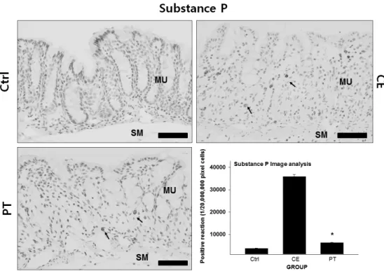
3) Decrease in Substance P Production
To observe the change in substance P distribution, which provokes pain, in the lamina propria mucosae, immunohistochemical staining with goat anti-mouse substance P was used. In the CE group, the distribution of substance P-positive cells more increased in the lamina propria mucosae than that of the Ctrl group. Strong positive reaction was found at the margin of cytoplasm in the positive cell. Positive reaction of the CE group more increased by 881%
than that of the Ctrl group. On the other hand,
positive reaction of the PT group more decreased by
83% than that of the CE group (Fig. 2).
Fig. 2. The inhibitory effect of PE on substance P in the colon mucosa. Abbreviations: Ctrl, the mice which was not treated anything; CE, the mice which was treated DSS only; PT, the mice which was treated both DSS and PE; MU, colonic mucosa; SM, submucosa. Bar size: 100 μm. Arrow: substance P-positive reaction. *: P<0.05 compared with the CE group.
2. Regulating Pro-inflammatory Cytokines 1) TNF-α Inhibitory Effect
To observe the change in TNF-α activity, which is a pro-inflammatory cytokine, in the lamina propria mucosae, immunohistochemical staining with goat anti-mouse TNF-α was used. In the CE group, the distribution of TNF-α-positive cells more increased in the lamina propria mucosae than that of the Ctrl group. Strong positive reaction was found at the cytoplasm of the positive cell. Positive reaction of the CE group more increased by 639% than that of the Ctrl group. On the other hand, positive reaction of the PT group more decreased by 73% than that of the CE group (Fig. 3).
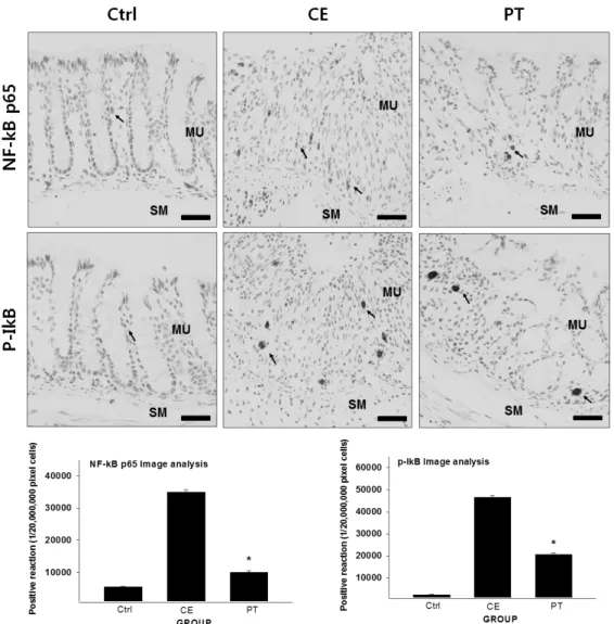
2) NF-κB Inhibitory Effect
To observe the change in NF-κB activity, which is a transcription factor, in the lamina propria mucosae, immunohistochemical staining with goat anti-mouse NF-κB p65 and anti-mouse p-IκB was used. In the CE group, the distribution of NF-κB p65-positive cells more increased in the lamina propria mucosae than that of the Ctrl group. Strong positive reaction was found at the cytoplasm around the nuclear membrane of the positive cell. Positive reaction of the CE group more increased by 530%
than that of the Ctrl group. On the other hand, positive reaction of the PT group more decreased by 71% than that of the CE group (Fig. 4).
For p-IκB, the distribution of the positive cells in
the CE group more increased in the lamina propria
mucosae than that of the Ctrl group. Strong positive
Fig. 3. The inhibitory effect of PE on TNF-α in the colon mucosa. Abbreviations: Ctrl, the mice which was not treated anything; CE, the mice which was treated DSS only; PT, the mice which was treated both DSS and PE. Bar size:
100 μm. Arrow: TNF-α-positive reaction. *: P<0.05 compared with the CE group.
reaction was found at the cytoplasm around the nuclear membrane of the positive cell. Positive reaction of the CE group more increased by 1919%
than that of the Ctrl group. On the other hand, positive reaction of the PT group more decreased by 56% than that of the CE group (Fig. 4).
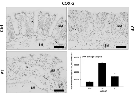
3. Inhibitory Effect on COX-2
To observe the change in COX-2 production, which is an inflammatory enzyme, in the lamina propria mucosae, immunohistochemical staining with goat anti-mouse COX-2 was used. In the CE group, COX-2-positive cells more increased in the lamina propria mucosae than that of the Ctrl group. Strong positive reaction was found at the margin of cytoplasm in the positive cell. Positive reaction of
the CE group more increased by 375% than that of the Ctrl group. On the other hand, positive reaction of the PT group more decreased by 27% than that of the CE group (Fig. 5).
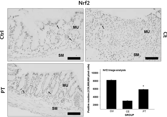
4. Anti-oxidative Effect via Increasing Nrf2
To observe the change in Nrf2, which has an
anti-oxidative activity, in the lamina propria
mucosae, immunohistochemical staining with goat
anti-mouse Nrf2 was used. In the CE group,
Nrf2-positive cells decreased in the lamina propria
mucosae. Strong positive reaction was found at the
cytoplasm of the positive cell. Positive reaction of
the CE group more decreased by 64% than that of
the Ctrl group. On the other hand, positive reaction
Fig. 4. The inhibitory effect of PE on NF-κB p65 and p-IκB in the colon mucosa. Abbreviations: Ctrl, the mice which was not treated anything; CE, the mice which was treated DSS only; PT, the mice which was treated both DSS and PE; MU, colonic mucosa; SM, submucosa. Bar size: 100 μm. Arrow: immunohistochemically positive reaction.
*: P<0.05 compared with the CE group.
of the PT group more increased by 94% than that of the CE group (Fig. 6).
Discussion
Proliferation of westernized diet increased the prevalence of IBD such as UC and Crohn's disease.
These diseases are recently recognized as one of the
causes for increase in the colon cancer mortality of
Korean people. Many studies for the pathogenesis of
IBD are ongoing. Most of all, therapies such as
regulating pro-inflammatory cytokines, transcription
factors, arachidonic acid metabolites, and reactive
oxygen and nitrogen species (RONS) are attracting
Fig. 5. The inhibitory effect of PE on COX-2 in the colon mucosa. Abbreviations: Ctrl, the mice which was not treated anything; CE, the mice which was treated DSS only; PT, the mice which was treated both DSS and PE; MU, colonic mucosa; SM, submucosa. Bar size: 100 μm. Arrow: COX-2-positive reaction. *: P<0.05 compared with the CE group.
much attention
10-12).
The clinical symptoms of US are inflammation, ulcer, shortness of rectum, and infiltration of immune cells to wounds. For UC, aminosalicylates such as sulfasalazine and mesalazine and steroids are currently in use but they are not able to anticipate complete recovery. Long-term administration of these drugs can cause various side effects such as nausea, vomiting, dyspepsia, anorexia, and headache or tolerance
13). Thus, developing novel medicine which has high efficacy and safety is desperately needed.
For the UC medicine, attention to Korean medicine has been recently increasing
14).
Psoralea semen is the dried seeds of Psoralea corylifolia L. (Leguminosae)
9). According to Dongeuibogam, it invigorates yang by tonifying the
kidney, secures essence, and warm the spleen. Based on such literature, this study was to find the potential of PE for UC.
5% DSS was used to induce UC in this study. To identify protective effect of PE on the colon, we observed the change in the colon mucosa and cells.
Mucous epithelium of gastrointestinal tract acts as a
protective barrier against various stimulation. DSS
treatment decreases the distribution of zonula
occudin (ZO)-1, one of the occluding junction
proteins. This is because of the damage of zonula
occludens located at the top of the junctional
complex between mucous epithelial cells. Increase in
intestinal permeability caused by the damage of
zonula occludens induces intestinal inflammation and
provokes UC
15). Also, mice colon treated with DSS
Fig. 6. The promotive effect of PE on Nrf2 in the colon mucosa. Abbreviations: Ctrl, the mice which was not treated anything; CE, the mice which was treated DSS only; PT, the mice which was treated both DSS and PE; MU, colonic mucosa; SM, submucosa. Bar size: 100 μm. Arrow: Nrf2-positive reaction. *: P<0.05 compared with the CE group.