Case Report
https://doi.org/10.5049/EBP.2020.18.2.44
Liver Infarction and Venous Thromboembolism after Tamoxifen Use in an ADPKD Patient with Encapsulating Peritoneal Sclerosis: A Case Report
Kyoung Min Kwak*, Gwang Ho Choi*, Kwang Eon Shim, Ho Yong Jin, Seok Hyung Kim, Jong Woo Yoon, Hyunsuk Kim
Department of Internal Medicine, Hallym University Medical Center, Chuncheon Sacred Heart Hospital, Hallym University College of Medicine, Chuncheon, Korea
Received: October 23, 2020 Revised: November 11, 2020 Accepted: November 12, 2020
Corresponding Author: Hyunsuk Kim, MD, PhD Department of Internal Medicine, Hallym Uni- versity Medical Center, Chuncheon Sacred Heart Hospital, Hallym University College of Medicine, Chuncheon 24253, Korea
Tel: +82-33-240-5196 Fax: +82-33-255-6244
E-mail: [email protected]
*These two authors were equally contributed to this manuscript.
Encapsulating peritoneal sclerosis (EPS) is a potentially fatal complication after long-term peritoneal dialysis, and tamoxifen can be used for its prevention and treatment. However, tamoxifen is known to increase the risk of venous thrombo- embolism. A 49-year-old woman was admitted with sudden abdominal pain. The patient had received peritoneal dialysis for 20 years and switched to hemodialysis after the diagnosis of EPS. Tamoxifen (10 mg) and prednisolone (20 mg) had been administered for 8 months. On computed tomography, the left hepatic lobe was hardly illuminated, leading to a diagnosis of liver infarction. A month later, she was re-admitted due to abdominal pain and extensive deep vein thrombosis of the leg. The administration of tamoxifen was stopped and prednisolone was re- duced to 10 mg. As her malnutrition progressed, she succumbed to death of gram negative sepsis. The patient was concluded to have liver infarction and extensive venous thrombosis as a side effect of tamoxifen.
Key Words: Encapsulating peritoneal sclerosis, Liver infarction, Tamoxifen, Venous thromboembolism
This is an Open Access article distributed under the terms of the Creative Commons Attribution Non-Commercial License (https://creativecommons.org/licenses/by-nc/4.0/) which permits unrestricted non-commercial use, distribution, and reproduction in any medium, provided the original work is properly cited.
INTRODUCTION
Encapsulating peritoneal sclerosis (EPS) is uncommon, but could be the most critical complication of long-term peri- toneal dialysis. It is defined as a clinical syndrome characte- rized by a persisting intestinal obstruction, intermittent or recurrent, associated with peritoneal thickening, sclerosis, calcification, or encapsulation, with or without the presence of inflammatory markers1). Both surgical and medical op- tions exist to treat EPS, but conservative treatment is the current standard approach, although it may be insufficient2).
Tamoxifen, which is used to treat malignancies such as breast cancer, increases the risk of thromboembolism in addition to thromboembolic effect of malignancy itself3).
Although it is not clear how tamoxifen increases the risk of thromboembolism, its thromboembolic effect seems to be related to the factor V Leiden mutation and other mech- anism4,5). Currently, tamoxifen is a component of the main- stream approach to the medical prevention and treatment of EPS6). Despite its promising efficacy, tamoxifen is known to increase the risk of thromboembolism7). Many cases of tamoxifen-related thromboembolism have been reported in cancer patients, but no case reports have described the extent of a thromboembolic event after tamoxifen use to treat EPS.
Herein, we describe our experience with an ambulatory case of hepatic artery thrombosis and extensive venous th- rombosis of the leg that occurred during the use of tamox- ifen to treat EPS.
Table 1. Laboratory findings of the patients at the time of admission
Variables Value Reference
White blood cell count 6,910 3,400-9,500/µL
Hemoglobin 10.6 12.3-15.5 g/dL
Platelet count 37,000 140,000-380,000/µL C-reactive protein 9.3 0.00-3.0 mg/L Blood urea nitrogen 35 10-26 mg/dL
Creatinine 4.2 0.6-1.2 mg/dL
AST 37 8-39 IU/dL
ALT 16 5-45 IU/dL
Total bilirubin 0.75 0.4-1.3 mg/dL
LDH 371 106-211 IU/L
PT, INR 1.13 0.87-1.14
aPTT 32.5 25.0-40.0 sec
AT-III 40 83-128%
Fibrinogen 198 235-465 mg/dL
FDP 6.19 0-2.01 ug/mL
D-dimer 1,176 0-243 ng/mL
AST, aspartate aminotransferase; ALT, alanine aminotransfe- rase; LDH, lactate dehydrogenase; PT, prothrombin time; INR, international normalized ratio; aPTT, activated partial proth- rombin time; AT, antithrombin; FDP, Fibrinogen degradation production.
Fig. 1. Initial abdominal imaging. (A) Abdominal CT shows EPS accompanied with massive amounts of ascites in an ADPKD patient with numerous renal cysts. (B) Abdominal X-ray shows mechanical ileus and ascites with calcification along the peri- toneal membrane.
CASE REPORT
A 49-year-old end-stage renal disease (ESRD) female pa- tient was admitted with abdominal pain that suddenly oc- curred during hemodialysis. Her etiology of ESRD was poly- cystic kidney disease, and she had received peritoneal dialy- sis for 20 years and switched to hemodialysis after the diag- nosis of EPS. Since gross hematuria caused by hemorrhagic cysts frequently occurred, heparin was not used during hemodialysis. For conservative management of EPS, tamox- ifen (10 mg) and prednisolone (20 mg) had been adminis- tered for 8 months. During the period when these two me- dications were administered, the patient’s abdominal pain and ileus were controlled to some degree and she main- tained the ability to carry out routine household tasks and oral feeding. Her abdominal pain mostly continued in the right upper quadrant and periumbilical area, and abdominal computed tomography (CT) showed tenderness in this re- gion. Abdominal CT shows EPS accompanied with massive amounts of ascites in an ADPKD patient with numerous renal cysts. Abdominal X-ray shows mechanical ileus and ascites with calcification along the peritoneal membrane (Fig. 1). Due to EPS, there was a feeling of touching dough.
The patient’s blood pressure at the time of hospitalization was 130/60 mmHg, her heart rate was 86 beats per minute, her respiratory rate was 20 breaths per minute, and her temperature was 36.7℃. Patient’s family has provided in- formed consent for publication of the case. Patient has
provided informed consent for publication of the case. This study was approved by Institutional Review Board (IRB ap- proval number: 2017-11-106) and procedures were in ac- cordance with the ethical standards with the Helsinki De- claration of 1975 (and as revised in 1983).
1. Investigations
Her white blood cell (6,910/µL) count and C-reactive pro- tein (9.3 mg/L) level were almost in the normal range. Pre- viously, her platelet count was in the normal range. Addi- tionally, the patient had no known chronic liver disease or cirrhosis. However, thrombocytopenia (37,000/µL) was pre- sent at this admission, which is known to be a rare compli- cation of tamoxifen. Her prothrombin time, and activated partial prothrombin time were in the normal range. How- ever, the antithrombin-III (40%) and fibrinogen (198 mg/dL) were decreased, and fibrinogen degradation production (6.19 ug/mL) and D-dimer (1,176 ng/mL) were markedly in- creased (Table 1).
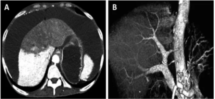
Fig. 2. Further liver imaging. (A) Abdominal CT shows infarction of the left lobe of the liver and massive ascites. The patient’s EPS can be visualized, with no significant difference compared to previous CT findings. (B) Reconstructed angiography shows diffuse narrowing of the left hepatic artery and branch of the left portal vein with non-visualization of the left lobe, sugges- ting a liver infarction.
Because the patient complained of sustained and worse- ning abdominal pain, CT angiography was performed. On contrast CT (Fig. 2A), the left hepatic lobe was barely illumi- nated, leading to a diagnosis of liver infarction. The abdomi- nal cavity had massive ascites, and calcification and thicken- ing of the peritoneal membrane were observed, but this finding was due to the patient’s EPS, and there was no significant difference compared to previous CT findings.
In a 3-dimensional reconstructed view of the vessels, dif- fuse luminal narrowing of the left hepatic artery and branch of the left portal vein was seen. Moreover, the left side of the liver was not visualized (Fig. 2B). Finally, the patient was diagnosed with both hepatic artery thrombosis and portal vein thrombosis, as well as liver infarction.
2. Differential diagnosis
The patient did not have clear risk factors for arterial and venous thromboembolism, as she had no underlying liver disease and no heart abnormality that could be the source of an embolism. Moreover, she was not taking any medications that could induce a thrombus other than tamo- xifen and prednisolone. Therefore, she was deemed to have experienced liver infarction as a side effect of tamoxifen.
After liver infarction was diagnosed based on CT imaging, the levels of liver enzymes, lactate dehydrogenase, and pro- thrombin time were serially measured to monitor ischemic changes of the liver, but there were no abnormal findings.
Echocardiography and electrocardiography were performed
to identify a potential source of the embolism, but no sig- nificant findings were obtained on echocardiography, and electrocardiography showed a normal sinus rhythm.
Furthermore, because she did not have fever, and had a normal white blood cell count and C-reactive protein level, we excluded infection and considered her abdominal pain to have been caused by the liver infarction.
3. Treatment
The patient’s abdominal pain gradually improved with analgesics. Tamoxifen and prednisolone were maintained after discharge because ileus due to EPS was a major prob- lem that needed to be corrected.
4. Outcome and follow-up
After a month, the patient was re-admitted with the same pattern of abdominal pain and swelling of the left leg. The patient suffered from frequent abdominal pain and was unable to take medication orally; therefore, all medications were discontinued and the patient was observed over time.
As she experienced continuous swelling in her leg during her hospital stay, Doppler sonography was performed and extensive proximal deep vein thrombosis involving the right common femoral vein, saphenous vein, and popliteal vein was identified. Malnutrition developed because of her poor oral intake. Thereafter, fever occurred, but as she refused to receive parenteral nutrition and antibiotics, she had an acute downhill course and finally died of Gram-negative sepsis.
DISCUSSION
The patient in the present case experienced liver infarc- tion and venous thrombosis of the leg that occurred during the 8 months-use of tamoxifen for the treatment of EPS.
Although an alternative explanation for the patient’s venous thrombosis could be her progressive immobility, the throm- bosis was too extensive to be explained only by the pa- tient’s immobility. Moreover, the patient was not com- pletely immobile, because she was able to engage in basic household tasks such as laundry and cooking before the second admission. Additionally, the occurrence of liver in-
farction due to hepatic arterial and portal venous throm- boembolism without an underlying heart problem could not be explained. Therefore, we conclude that liver infarction and venous thrombosis were highly likely to have been side effects of tamoxifen in this case.
Tamoxifen is a selective estrogen receptor modulator with both estrogenic and antagonistic potential. In a long- term trial, deep-vein thrombosis and pulmonary embolism occurred during active treatment statistically significantly more often in patients who received tamoxifen than in the placebo group8). Additionally, case reports have shown that tamoxifen-related thromboembolic disorders could affect almost any vessel, including the tibial artery, superior sagit- tal sinus, cerebral vein, portal vein, and even the digital artery. In the current report, we identified tamoxifen as the cause of arterial and venous thrombosis. However, it was used together with prednisolone, which is commonly known to cause venous thrombosis. Thus, we cannot rule out the possibility that venous thrombosis was caused by prednisolone. However, there are more reports mentioning the risks of arterial thromboembolism associated with ta- moxifen than with steroid, so we considered tamoxifen as a more significant causative agent than prednisolone9-13).
The precise mechanism through which tamoxifen incre- ases the risk of thromboembolic events has yet to be elu- cidated. However, in previous studies, tamoxifen has been found to lead to changes in anticoagulant proteins (anti- thrombin III, protein C, and protein S). In one study, both arterial and venous thromboembolism increased when ta- moxifen was administered in breast cancer patients, partic- ularly affecting the carotid and artery of extremities14). This is explained by the paradoxical estrogen effect of tamoxifen and they are for instance, reduced levels of antithrombin III, and protein C and S15). Paradoxically, tamoxifen therapy is related to myocardial infarct reduction, probably because the pathway to atherogenesis and thrombogenesis are af- fected in different ways. A certain study revealed the effect of tamoxifen on arterial microvascular anastomosis, pre- senting the occurrence of intimal hyperplasia when the anastomosed femoral artery of rats is administered with tamoxifen. The study mentioned the possibility that this was related to arterial thrombosis16-19).
CT angiography is necessary for an accurate diagnosis of
hepatic artery thrombosis and remains the diagnostic gold standard20). Color Doppler ultrasonography also can diag- nose the disease, but CT angiography has higher accuracy, and requires a short examination time21). However, dis- advantages of CT angiography include both concerns about radiation exposure and renal injury caused by the contrast material. In this case, though, the risk of renal injury was not deemed to be relevant because the patient already had no remnant renal function.
Treatment options for hepatic arterial thrombosis inclu- de intra-arterial thrombolysis, percutaneous transluminal angioplasty and stent implantation22). Furthermore, antico- agulation is the treatment of choice for both deep vein thrombosis and portal vein thrombosis23,24). We administe- red dalteparin at 100 IU/kg and discontinued tamoxifen, which was considered to have been the cause of hepatic arterial thrombosis. However, the patient died shortly beca- use of a combination of progressive malnutrition and sepsis.
This case illustrates the need to consider tamoxifen as the root cause of thromboembolic disease in patients taking tamoxifen. The patient had no known liver disease or cir- rhosis and was only taking tamoxifen and prednisolone for EPS treatment. Hepatic artery thrombosis and deep-vein thrombosis occurred twice over the course of a month. Since the patient had no specific risk factors, these two events are considered to have been side effects of tamoxifen25).
In conclusion, clinicians should bear in mind that such events can occur in patients who use tamoxifen, and careful observation is therefore needed. Moreover, additional re- search should investigate the mechanism underlying the association between tamoxifen and thromboembolic risk.
Disclosure
The authors have no potential conflicts of interest to disclose.
Funding
This research was financially supported by National Re- search Foundation of Korea (NRF-2016R1D1A1B03934173, data collection and organization).
REFERENCES
1. Mihalache O, Bugă C, Doran H, et al.: Encapsulating peri- toneal sclerosis: A rare and serious complication of perito- neal dialysis: case series. J Med Life 7 Spec No. 3(Spec Iss 3):8-12, 2014
2. Kawanishi H, Watanabe H, Moriishi M, Tsuchiya S: Success- ful surgical management of encapsulating peritoneal scle- rosis. Perit Dial Int 25(Suppl 4):S39-S47, 2005
3. Hendrick A, Subramanian VP: Tamoxifen and thromboem- bolism. JAMA 243(6):514-515, 1980
4. Kovac M, Kovac Z, Tomasevic Z, et al.: Factor V leiden muta- tion and high FVIII are associated with an increased risk of VTE in women with breast cancer during adjuvant tamo- xifen: results from a prospective, single center, case control study. Eur J Intern Med 26(1):63-67, 2015
5. Eroglu A: Tamoxifen-associated thromboembolism in breast cancer. Thromb Res 131(6):566, 2013
6. Summers AM, Clancy MJ, Syed F, et al.: Single-center expe- rience of encapsulating peritoneal sclerosis in patients on peritoneal dialysis for end-stage renal failure. Kidney Int 68(5):2381-2388, 2005
7. Meier CR, Jick H: Tamoxifen and risk of idiopathic venous thromboembolism. Br J Clin Pharmacol 45(6):608-612, 1998 8. Cuzick J, Forbes JF, Sestak I, et al.: Long-term results of tamo-
xifen prophylaxis for breast cancer--96-month follow-up of the randomized IBIS-I trial. J Natl Cancer Inst 99(4):272-282, 2007
9. Goggin C, Power S, O'Reilly S: Superior sagittal sinus throm- bosis secondary to Tamoxifen treatment. Breast J 25(3):510- 511, 2019
10. Hsu A, Belkin E, Han S, Pellish R: Tamoxifen-associated por- tal vein thrombosis causing severe oesophageal variceal bleeding. BMJ Case Rep 2015, 2015
11. Kim Y, Kim OJ, Kim J: Cerebral venous thrombosis in a breast cancer patient taking tamoxifen: Report of a case. Int J Surg Case Rep 6c:77-80, 2015
12. Hutchison RL, Rayan GM: Thrombosis of digital arteries as- sociated with tamoxifen use: case report. J Reconstr Micro- surg 28(2):145-148, 2012
13. Deshmukh N, Tripathi SP: Thrombosis of tibial arteries in a patient receiving tamoxifen therapy. Cancer 76(6):1006- 1008, 1995
14. Saphner T, Tormey D, Gray R: Venous and arterial thrombo- sis in patients who received adjuvant therapy for breast cancer. Journal of Clinical Oncology 9(2):286-294, 1991 15. De Pinho Pessoa BBG, Cavalcante BBM, Maia MP, et al.:
Effect of tamoxifen on arterial microvascular anastomosis.
microsurgery: Official Journal of the International Microsur- gical Society and the European Federation of Societies for Microsurgery 27(4):286-288, 2007
16. Love RR, Surawicz TS, Williams EC: Antithrombin III level, fibrinogen level, and platelet count changes with adjuvant tamoxifen therapy. Arch Intern Med 152(2):317-320, 1992 17. Mannucci PM, Bettega D, Chantarangkul V, et al.: Effect of
tamoxifen on measurements of hemostasis in healthy wo- men. Arch Intern Med 156(16):1806-1810, 1996
18. Auger MJ, Mackie MJ: Effects of tamoxifen on blood coagu- lation. Cancer 61(7):1316-1319, 1988
19. Pemberton KD, Melissari E, Kakkar VV: The influence of ta- moxifen in vivo on the main natural anticoagulants and fib- rinolysis. Blood Coagul Fibrinolysis 4(6):935-942, 1993 20. Mourad MM, Liossis C, Gunson BK, et al.: Etiology and man-
agement of hepatic artery thrombosis after adult liver trans- plantation. Liver Transpl 20(6):713-723, 2014
21. Kim SY, Kim KW, Kim MJ, et al.: Multidetector row CT of various hepatic artery complications after living donor liver transplantation. Abdom Imaging 32(5):635-643, 2007 22. Chen J, Weinstein J, Black S, et al.: Surgical and endovas-
cular treatment of hepatic arterial comlications following liver transplant. Clin Transplant 28(12):1305-1312, 2014 23. Intagliata NM, Caldwell SH, Tripodi A: Diagnosis, develop-
ment, and treatment of portal vein thrombosis in patients with and without cirrhosis. Gastroenterology 156(6):1582- 1599, e1, 2019
24. Stubbs MJ, Mouyis M, Thomas M: Deep vein thrombosis.
BMJ (Clinical Research ed) 360:k351, 2018
25. Johannesdottir SA, Horváth-Puhó E, Dekkers OM, et al.: Use of glucocorticoids and risk of venous thromboembolism:
a nationwide population-based case-control study. JAMA Internal Medicine 173(9):743-752, 2013