Received: June 10, 2020•Revised: August 12, 2020•Accepted: August 27, 2020
Corresponding Author: Jonathon S. Jundt, Department of Oral and Maxillofacial Surgery, University of Texas, 7500 Cambridge Suite 6510, Houston, Texas 77056.
E-mail: Jonathon.Jundt@uth.tmc.edu
Copyrightⓒ 2020 Journal of Dental Anesthesia and Pain Medicine
Computed tomography-guided 3D printed patient-specific regional anesthesia
Jonathon S. Jundt
1, Christopher C. Chow
1, Marcus Couey
21Department of Oral and Maxillofacial Surgery, The University of Texas, Houston, Texas, United States
2Head and Neck Oncologic and Microvascular Reconstructive Surgery, Providence Cancer Institute, Houston, Texas, United States
Classic anesthetic techniques for the inferior alveolar nerve, lingual nerve, and long buccal nerve blockade are achieved by estimating the intended location for anesthetic deposition based on palpation, inspection, and subsequent correlation for oral anatomical structures. The present article utilizes computed tomography (CT) data to 3D print a guide for repeatable and accurate deposition of a local anesthetic at the ideal location.
This technical report aims to anatomically define the ideal location for local anesthetic deposition. This process has the potential to reduce patient discomfort, risk of nerve damage, and failed mandibular anesthesia, as well as to reduce the total anesthetic dose. Lastly, as robotic-based interventions improve, this provides the initial framework for robot-guided regional anesthesia administration in the oral cavity.
Keywords: Guided Regional Anesthesia; Local Anesthesia; 3D Printing.
This is an Open Access article distributed under the terms of the Creative Commons Attribution Non-Commercial License (http://creativecommons.org/licenses/by-nc/4.0/) which permits unrestricted non-commercial use, distribution, and reproduction in any medium, provided the original work is properly cited.
BACKGROUND
Mandibular regional anesthesia is a common technique used to facilitate dental and oral surgical procedures.
However, failures are common, occurring in about 20-25% of attempts with the standard IAN block technique [1]. Variations of the IAN block, such as the Gow-Gates and Akinosi blocks, are shown to be more reliable than the traditional Halstead IAN block, with the Akinosi block demonstrating the highest anesthetic success at 95.71% [2]. However, disadvantages may include a longer time of onset and lower comfort level [2].
The ideal location for the deposition of the local anesthetic for mandibular nerve blockade has not been defined. Most sources recommend deposition “as close
to the nerve as possible” without penetrating the nerve [3,4]. In the standard IAN block, the clinician uses surface landmarks to estimate the position of the mandibular foramen, which, if located correctly, will correlate with the entrance of the nerve into the mandibular ramus.
Other techniques similarly utilize surface landmarks to estimate the location of the mandibular nerve in an attempt to deposit the local anesthetic solution in close proximity to allow diffusion into the nerve. These blind approaches, while successful in many instances, are inherently imprecise, as evidenced by a relatively high failure rate.
The reported height of the mandibular foramen above the mandibular occlusal plane ranges from 3.8 to 10 mm, and the relative position of palpable landmarks to estimate the location of the mandibular foramen can vary considerably in different age groups, ethnicities, and
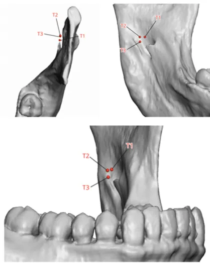
Fig. 1. Target sites T1, T2, T3
craniofacial disease [3-9]. Further, variations in sensory innervation to the mandibular teeth, for example, from branches of the mylohyoid nerve, undoubtedly confound this effort in a subset of patients [10,11]. Communications can occur between the lingual nerve and mylohyoid nerve, which can possibly contribute sensory components through a lingual foramen [12]. Without the ability to determine accessory innervation as the cause of failed injection in real-time, the scenario usually results in repeated attempts using similar techniques, increasing volumes of local anesthetic administered, and perhaps never achieving sufficient anesthesia.
If reliable localization of the anesthetic needle in relation to the inferior alveolar nerve could be ensured, the ability to troubleshoot a failed nerve block would be greatly enhanced. Specifically, if profound saturation of the nerve at its entrance to the mandibular foramen fails to provide
pulpal anesthesia, suspicion of accessory innervation would be warranted rather than a technical error or variant in the patient’s mandibular anatomy. This would allow for a more systematic approach to address anesthetic failure while avoiding unnecessary deposition of larger volumes of local anesthetic and the associated risk of adverse events caused by diffusion outside of the target area.
TECHNIQUE DESCRIPTION
Patients requiring regional anesthesia for surgical intervention require a preoperative computed tomography (CT) scan with a resolution of at least 0.5 mm as well as scanned or shipped accurate maxillary and mandibular impressions. A point 3 mm medial and 6 mm superior
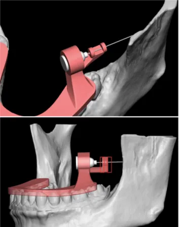
Fig. 3. 3D Printed guide with 3D printed model
Fig. 4. Device application and injection
Fig. 2. 3D Rendering of guide with reconstructed model
to the mandibular foramen was identified at a location along the proximal aspect of the IAN, marked as T2 (Fig.
1). A patient-specific CT-based channeled, tooth-borne guide is then designed using the needle hub diameter to maintain orientation of the needle (Fig. 2). The final design is printed using a 3D printer and tested on the model (Fig. 3). Lastly, the tooth-borne guide is placed
in the patient’s mouth, the needle is inserted into the predetermined hub depth, and the local anesthetic is injected (Fig. 4).
DISCUSSION
Traditional techniques for administering anesthesia in the oral cavity are solely based on the rational application of average anatomical relationships among surface landmarks, bones, and nerves. Conversely, the use of image-guided anesthesia provides a framework for accurate and systematic investigations to define the ideal anatomical location for anesthetic deposition, which to date, has not been rigorously evaluated. The definition of an optimal anatomical location for needle positioning has the potential to maximize local anesthetic efficacy, thereby reducing the total anesthetic dose required.
Further, image-guided anesthesia has significant potential to improve patient comfort by 1) reducing the rate of repeat injection, 2) reducing the risk of the needle penetrating the nerve, and 3) obviating the need to sound bone, thereby eliminating barb formation, a proposed mechanism for permanent paresthesia [13,14]. This
technique can also be used to investigate differences in the ideal location for anesthetic deposition in patients with varying facial/jaw morphologies (i.e., long vs. short face, class III vs. class II malocclusion), which may inform improved techniques for traditional landmark-based nerve blocks.
This method of delivering anesthetic to achieve blockade may help minimize the dose of anesthetic and epinephrine in cardiac patients. In addition, a decrease in total volume may lower the risk of unintended diffusion of the anesthetic, which has demonstrated complications such as ophthalmoplegia or visual impairment [15]. Troubleshooting failed blocks using blind approaches to locate the IAN can result in multiple needle punctures and increase anesthetic volume, which are known risk factors for deep fascial space abscess formation [15,16].
Traditional local anesthetic techniques demonstrate an unacceptably high failure rate. Several groups have attempted to apply current technologies to improve the accuracy of IAN blocks. Won and Kang demonstrated the utility of fusing cone-beam CT images with intraoral photographs to improve the standard landmark-based approach [17]. In contrast, our technique specifically allows for precise placement of the needle tip in three dimensions based on hard-tissue landmarks and a rigid guide for needle positioning. We demonstrate that this image-guided approach provides an objective, repro- ducible method for accurate needle placement and local anesthesia deposition. Furthermore, as robotic technology emerges within the field of regional anesthesia, this technique provides a proof-of-concept for the use of robotic-assisted anesthesia in the oral cavity.
This patient-specific guide, similar to a fully occlusal stabilization splint, could be used repeatedly for dental visits unless or until substantial changes to the occlusion occur. We propose that the currently available techno- logy, CBCT and commercially available 3D printing devices, can be utilized to improve upon the ubiquitous, yet inherently imprecise, traditional IAN block, with implications for current clinical practice and future
research. However, without validation in a clinical study, this technique cannot be recommended for routine use in all patients. CBCT is also associated with increased radiation exposure compared to panoramic radiographs that are often obtained as the standard screening imaging in dental patients [18]. Therefore, CBCT should not be obtained solely for the fabrication of a 3D-printed anesthetic guide unless the patient has a history of failed IAN block or another compelling circumstance requiring increased precision for local anesthetic deposition.
Ultimately, clinical studies are required to evaluate the utility and broader applicability of this new technique.
AUTHOR ORCIDs
Jonathon Jundt: https://orcid.org/0000-0002-3544-6331 Christopher C. Chow: https://orcid.org/0000-0001-5540-0544 Marcus Couey: https://orcid.org/0000-0002-9464-2682
AUTHOR CONTRIBUTIONS
Jonathon Jundt: Conceptualization, Formal analysis, Investigation, Project administration, Resources, Visualization, Writing – original draft, Writing – review & editing
Christopher C. Chow: Investigation, Project administration, Resources, Writing – review & editing
Marcus Couey: Data curation, Formal analysis, Writing – original draft, Writing – review & editing
ACKNOWLEDGMENTS: Katie Weimar with 3D Systems generously donated the time and provided the materials to produce this technical note.
DECLARATION OF INTEREST: The authors declare no conflicts of interest.
REFERENCES
1. Thangavelu K, Kannan R, Kumar NS, RethishnE, Sabitha S, Sayeeganesh N. Significance of localization of mandi- bular foramen in an inferior alveolar nerve block. J Nat Sci Biol Med 2012; 3: 156-60.
2. Ravi Kiran BS, Kashyap VM, Uppada UK, Tiwari P, Mishra A, Sachdeva A. Comparison of efficacy of halstead, vazirani
akinosi and gow gates techniques for mandibular anesthesia. J Maxillofac Oral Surg 2018; 17: 570-5.
3. Kang SH, Byun IY, Kim JH, Park HK, Kim MK.
Three-dimensional anatomic analysis of mandibular foramen with mandibular anatomic landmarks for inferior alveolar nerve block anesthesia. Oral Surg Oral Med Oral Pathol Oral Radiol 2013; 115: e17-23.
4. Malamed S. Handbook of Local Anesthesia. 6th ed.
Mayland heights, Mosby. 2012, pp.157-253.
5. Blacher J, Van DaHuvel S, Parashar V, Mitchell JC.
Variation in location of the mandibular foramen/inferior alveolar nerve complex given anatomic landmarks using cone-beam computer tomographic scans. J Endod 2016;
42: 393-6.
6. Al-Shayyab MH. A simple method to locate mandibular foramen with cone-beam computed tomography and its relevance to oral and maxillofacial surgery: a radio- anatomical study. Surg Radiol Anat Surg Radiol Anat 2018;
40: 625-34.
7. Nicholson ML. A study of the position of the mandibular foramen in the adult human mandible. Anat Rec 1985;
212: 110-2.
8. Hwang TJ, Hsu SC, Huang QF, Guo MK. Age changes in location of mandibular foramen. Zhonghua Ya Yi Xue Hui Za Zhi 1990; 9: 98-103.
9. Chung MT, Levi B, Hyun JS, Lo DD, Montoro DT, Lisiecki J, et al. Pierre Robin sequence and Treacher Collins hypoplastic mandible comparison using three-dimensional morphometric analysis. J Craniofac Surg 2012; 23: 1959-63.
10. Sakamoto Y, Akita K. Spatial relationships between masticatory muscles and their innervating nerves in man with special reference to the medial pterygoid muscle and accessory muscle bundle. Surg Radiol Anat 2004; 26: 122-7.
11. Stein P, Brueckner J, Milliner M. Sensory innervation of mandibular teeth by the nerve to the mylohyoid:
implications in local anesthesia. Clin Anat 2007; 20: 591-5.
12. Kim SY, Hu KS, Chung IH, Lee EW, Kim HJ.
Topographic anatomy of the lingual nerve and variation in communication pattern of the mandibular nerve branches. Surg Radiol Anat 2004; 26: 128-35.
13. Smith MH, and Lung KE. Nerve injuries after dental injection: a review of the literature. J Can Dent Assoc 2006; 72: 559-64.
14. Pogrel MA, Thamby S. Permanent nerve involvement resulting: from inferior alveolar nerve blocks. J Am Dent Assoc 2000; 131: 901-7.
15. Boynes SG, Echeverria Z, Abdulwahab M. Ocular compli- cations associated with local anesthesia administration in dentistry. Dent Clin North Am 2010; 54: 677-86.
16. Leventhal D, Schwartz DN. Infratemporal fossa abscess:
complication of dental injection. Arch Otolaryngol Head Neck Surg 2008; 134: 551-3.
17. Won YJ, Kang SH. Application of augmented reality for inferior alveolar nerve block anesthesia: a technical note.
J Dent Anesth Pain Med 2017; 17: 129-34.
18. Li G. Patient radiation dose and protection from cone-beam computed tomography. Imaging Sci Dent 2013;
43: 63-9.