• 검색 결과가 없습니다.

Mesenchymal Stem Cell-derived Exosomes: Applications in Cell-free Therapy

N/A
N/A
Protected

Academic year: 2021

Share "Mesenchymal Stem Cell-derived Exosomes: Applications in Cell-free Therapy"

Copied!
8
0
0

로드 중.... (전체 텍스트 보기)

전체 글

(1)

pISSN 1738-3544 eISSN 2288-1662

Mesenchymal Stem Cell-derived Exosomes:

Applications in Cell-free Therapy

June Seok Heo 1 , Jinkwan Kim 2

1

Department of Integrated Biomedical and Life Sciences, Graduate School, Korea University, Seoul, Korea

2

Department of Biomedical Laboratory Science, College of Health Science, Jungwon University, Goesan, Korea

중간엽줄기세포유래 엑소좀: 비세포치료제로서의 활용

허준석 1 , 김진관 2

1

고려대학교 대학원 의생명융합과학과,

2

중원대학교 의료보건대학 임상병리학과

Mesenchymal stem cells (MSCs) are an attractive resource for refractory patients because of their anti-inflammatory/immunomodulatory capability and multi-lineage differentiation potential. The transplantation of MSCs has led to positive results in preclinical and clinical application to various diseases, including autoimmune disease, cardiovascular disease, cancer, liver cirrhosis, and ischemic stroke. On the other hand, studies have shown that paracrine factors, not direct cell replacement for damaged cells or tissue, are the main contributors in MSC-based therapy. More recently, evidence has indicated that MSC-derived exosomes play crucial roles in regulating the paracrine factors that can mediate tissue regeneration via transferring nucleic acids, proteins, and lipids to the local microenvironment and cell-to-cell communication. The use of these exosomes is likely to be beneficial for the therapeutic application of MSCs because their use can avoid harmful effects, such as tumor formation involved in cell transplantation. Therefore, therapeutic applications using MSC-derived exosomes might be safe and efficient strategies for regenerative medicine and tissue engineering. This review summarizes the recent advances and provides a comprehensive understanding of the role of MSC-derived exosomes as a therapeutic agent.

Key words: Exosome, Mesenchymal stem cell, Stem cell, Therapy

Corresponding author: Jinkwan Kim Department of Biomedical Laboratory Science, College of Health Science, Jungwon University, 85 Munmu-ro, Goesan-eup, Goesan-gun, Goesan 28024, Korea

Tel: 82-43-830-8860 Fax: 82-43-830-8115 E-mail: jkkim@jwu.ac.kr

This is an Open Access article distributed under the terms of the Creative Commons Attribution Non-Commercial License (http://creativecommons.org/licenses/by-nc/4.0) which permits unrestricted non-commercial use, distribution, and reproduction in any medium, provided the original work is properly cited.

Copyright © 2018 The Korean Society for Clinical Laboratory Science. All rights reserved.

Received: September 4, 2018 Revised 1

st

: September 17, 2018 Revised 2

nd

: October 1, 2018 Accepted: October 1, 2018

INTRODUCTION

Mesenchymal stem cells (MSCs) are one of the most extensively employed somatic stem cells for cell-based therapy because of their excellent immunomodulatory and multi-lineage properties [1, 2]. Approximately 800 clinical trials utilizing MSCs, listed at www.clinicaltrials.

gov, support that MSCs are a very promising cell source for the treatment of various human diseases. Over the last

decade, the potential of MSC-based cell therapy has been

demonstrated by its successful application in clinical trials

for a wide variety of diseases [3]. However, the precise

therapeutic mechanisms of MSCs in human patients have

not been clearly elucidated. Accumulative evidence has

shown that MSCs exert therapeutic impact through

paracrine-mediated effects but not direct cell replace-

ment [4]. Previous studies have also demonstrated that

transplanted MSCs have very low survival rates, indicating

(2)

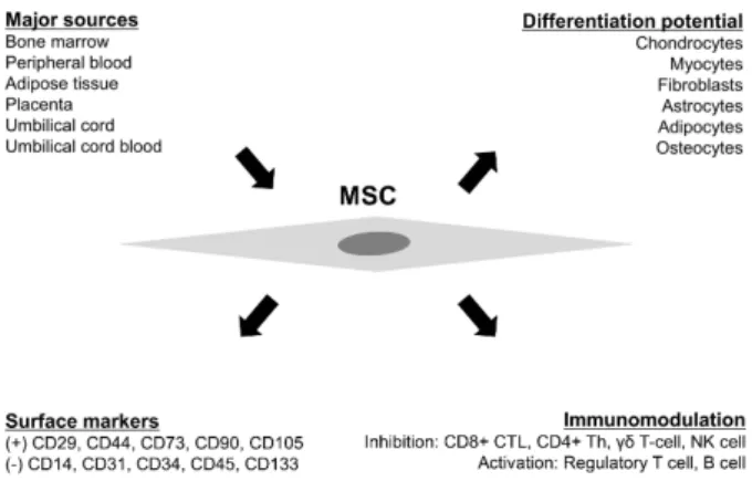
Figure 1. Schematic representation of mesenchymal stem cell (MSC). MSCs can be obtained from various sources in the body, including bone marrow, peripheral blood, adipose tissue, placenta, umbilical cord, and umbilical cord blood. They have the ability to differentiate into myocytes, chondrocytes, fibroblasts, astrocytes, adipocytes, and osteocytes under specific in vitro conditions. In general, MSCs are positive for CD29 (

1

-integrin), CD44 (HCAM), CD73 (5’-nucleotidase), CD90 (Thy-1), and CD105 (endoglin), but negative for CD14, CD31 (PECAM-1), CD34, CD45, and CD133 (prominin-1). Soluble factors secreted from MSCs including transforming growth factor-1 (TGF-1), hepatocyte growth factor (HGF), nitric oxide (NO), prostaglandin E2 (PGE2), and indoleamine 2,3-dioxygenase (IDO) affect various subsets of lymphocytes including CD8+ cytotoxic T-lymphocytes (CTL), regulatory T-lymphocytes (Treg), B-lymphocytes, natural killer (NK) cells, gamma delta T-cells, and CD4+ helper T-lymphocytes (Th).

that their mechanisms of action are not through the MSCs themselves [5].

More recently, MSC-derived exosomes have been described as essential mediators in the therapeutic functions of MSCs [6]. Several studies have revealed that exosomes secreted by MSCs perform diverse functions, and they have been successfully applied in graft-versus- host disease, allogenic skin grafts, and myocardial ischemia/reperfusion injury [7-9]. Although the exact mechanisms of action of exosomes are not fully understood, exosomes have been described as more stable and reservable cell-derived vesicles compared with cells.

MSC-derived exosomes play a critical role in exerting beneficial effects because they pose no risk of immune rejection and aneuploidy [6]. For these reasons, exosomes may provide an ideal alternative therapy for diverse diseases.

Based on recent advances, the therapeutic mechanisms of MSCs can be divided into two types: 1) a paracrine factor-mediated mechanism involving cytokines and hormones and 2) an exosome-mediated mechanism containing RNA and other molecules. We herein summarize the therapeutic potential of MSC-mediated exosomes to better understand them as cell-free agents.

MAIN ISSUE

1. Mesenchymal stem cell

In 1966, MSCs were first reported by Friedenstein et al [10]. At the beginning, they were isolated from rat bone marrow, but since then, many investigators have isolated MSCs from various tissues, including amniotic fluid, cartilage, tonsil, synovial tissue, skin, tooth, peripheral blood, umbilical cord blood, liver, lung, spleen, adipose tissue, and placenta (Figure 1). It is easier to obtain MSCs from these sources, and cells with higher quality than those derived from bone marrow could be isolated [11, 12]. MSCs are generally positive for CD29 ( 1 -integrin), CD44 (hyaluronate receptor), CD73 (SH-3), CD90 (Thy-1), and CD105 (SH-2), while it is negative for hematopoietic surface markers (CD14, CD34, CD45), CD133 (AC133), and CD31 (Figure 1) [13]. It was also reported that MSCs

express major histocompatibility complex (MHC) class I, whereas they do not express human leukocyte antigen class II antigens [14]. The described surface proteins could be expressed differently depending on tissue source, species, and method of isolation and cultivation. MSCs possess high proliferative and self-renewal capacity when maintained in classical culture conditions, but there are variations depending on their surroundings or cell donors, as MSCs are highly sensitive to external environments or stimuli, especially in in vitro culture.

MSCs with multi-differentiation potential are maintained in an undifferentiated state under proper culture conditions.

However, MSCs could be easily differentiated into

osteoblasts, adipocytes, and chondrocytes when placed

under certain environments (Figure 1). Differentiation

capacity is another index for the identification of MSCs in

addition to morphological observation and surface

phenotyping [15]. It has been previously reported that

mesoderm-derived MSCs could be trans-differentiated

into endodermal and ectodermal lineages, suggesting that

(3)

Figure 2. The biogenesis and function of MSC-derived exosomes.

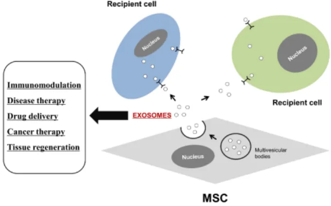
Secreted exosomes through fusion of multi-vesicular bodies with the cell membrane are mediators of paracrine effects. Exosomes can elicit cell-to-cell communication through intracellular uptake or membrane receptors. Exosomes, which are bilipid membrane vesicles with diverse proteins and RNAs, have the potential to exert various effects such as immunomodulation, disease therapy, and tissue repair in local recipient cells.

MSCs might be widely utilized and could improve the functions of damaged tissues or organs [16].

MSCs express a number of surface proteins but are negative for the co-stimulatory surface proteins CD40, CD80, CD86, and MHC II. The immunophenotypic characteristics of MSCs enable their successful trans- plantation because of the absence of immunorejection.

MSCs can also modulate and inhibit the functions of immune cells such as dendritic cells and B cells (Figure 1) [17]. These features make MSCs an attractive candidate for cell-based therapy. Therefore, application of non- engineered MSCs to various diseases such as autoimmune disorders may potentially be suitable for enhanced treatment.

2. Clinical application of mesenchymal stem cells Stem cell therapy is the medical treatment of patients with various disorders using autologous or allogeneic stem cells through local or systemic administration depending on the patient’s conditions or damaged tissues. A number of experiments have been performed to verify the effects of infused MSCs using various animal models. From these studies, it has been proposed that primary MSCs can be effectively used as organ grafts in mice, while cultivated MSCs in vitro lost homing property easily because of the absence of adhesion molecules [18]. These results indicated that infused MSCs are difficult to engraft on host cells or tissues [19].

Nevertheless, other studies have demonstrated the beneficial therapeutic effects of MSCs since the first clinical trial was performed in 1995 [20]. Recently, data from clinical trials have shown numerous clinical applications of MSCs for various diseases, including acute myocardial ischemia, lung fibrosis, spinal cord injury, Alzheimer’s disease, stroke, liver cirrhosis, and amyotrophic lateral sclerosis [21]. Previous studies have shown that hepatic regeneration was promoted in a carbon tetrachloride-injured liver model by human placenta-derived MSCs [22]. More recently, a meta- analysis of 950 patients demonstrated that MSCs can be beneficial in enhancing the heart function of patients with

myocardial infarction [23]. These data support the use of MSCs in human applications.

Therapies using MSCs are novel emerging strategies that are undergoing rapid development, even though MSC-based therapies have adverse effects such as tumorigenicity and utilization of animal-derived reagents.

Thus, researchers are aiming to find more optimal, advanced, and safe conditions or methods for MSC therapy in the future.

3. MSC-derived exosomes

Exosomes, one of several types of cell-derived vesicles, were first discovered in mammalian reticulocytes [24].

They are lipid bilayer vesicles that can be characterized

based on their morphology (cup- or saucer-shaped) and

size (30∼100 nm) by electron microscopy [25]. Exosomes

can be confirmed by surface markers such as CD9, CD63,

CD81 of tetraspanins, heat-shock proteins such as HSP60,

HSP70, HSP90, Alix, and tumor susceptibility gene 101,

which distinguish them from microvesicles and apoptotic

bodies of various extracellular vesicles [26]. Exosomes are

secreted directly from the plasma membrane or from

multi-vesicular bodies fused with the plasma membrane

(Figure 2) [27] and can be isolated by various methods such

as ultracentrifugation, chromatography, and commercial

(4)

kits based on polymer precipitation. Purification by ultracentrifugation at 100,000×g is the most widely established method [25].

Exosomes released from various types of cells including stem cells, B cells, mast cells, platelets, cancer cells, dendritic cells, T cells, and Schwann cells as well as physiological fluids including media of cultivated cells, plasma, urine, and bronchial fluid can travel through biological fluids to diverse tissues or organs [28]. Although the exact functions of exosomes are not well known, increasing evidence has suggested that secreted exosomes can communicate with recipient cells in nearby or remote areas by signaling through transferring proteins, RNAs, and other molecules, resulting in the alteration of various signaling activities in affected cells (Figure 2). In this regard, there is growing interest in their use as a therapeutic target or diagnostic marker.

Recently, the use of MSC-derived exosomes has been suggested as a novel strategy in regenerative medicine and tissue engineering because there is no immune rejection and tumor formation based on MSC properties [29].

Furthermore, the use of exosomes is appropriate in terms of manufacturing as undifferentiated MSCs have a long lifespan compared to that of other types of cells [30].

Generally, MSC-derived exosomes carry complex biologically active molecules including proteins, mRNAs, and microRNAs. The exosomal contents are associated with diverse cellular and biochemical processes, such as cell-to-cell communication, tissue regeneration, metabolism, and immune modulation (Figure 2). Thus, MSC-derived exosomes have been considered as a promising tool to detect various molecules resulting from various cellular responses via interaction with diverse cell types.

Moreover, MSC-derived exosomes play a crucial role in maintaining and restoring homeostasis within the body by mediating the function of MSCs. Particularly, exosomes have a key role in maintaining normal tissues when the homeostasis of the microenvironment is impaired by internal/external stimuli, injury, or disease [31].

4. Therapy using MSC-derived exosomes

Direct transplantation of viable MSCs poses risks such as tumor formation in vivo, resulting in adverse terminal outcomes. Transplantation of MSCs is relatively safe in terms of immune rejection, but the large size of MSCs has led to occlusion in the distal vasculature after intravascular administration [32]. Furthermore, some studies have reported potential risks of MSC transplantation due to calcification and/or ossification [33]. Exosomes can circumvent the risks involved in cell transplantation because cultured MSCs themselves are not used in clinical application.

Membrane molecules of exosomes such as lactadherin, transferrin receptors, tetraspanin proteins, tumor necrosis factor receptors, and integrins play key roles in the homing of exosomes to specific cells or tissues. In addition, the contents of exosomes, including RNA and proteins, can be protected from toxic chemicals or internal/external stimuli as exosomes bear a bilipid membrane [34, 35].

Exosomes containing biological molecules have the potential to participate in diverse biochemical and cellular interactions and alter the activities of cells via various signaling pathways.

Several studies have applied MSC-derived exosomes to reduce cardiac fibrosis in ischemic heart disease, inhibit cancer cell growth, and enhance functional recovery in stroke [36-38]. Amarnath et al [39] also reported that MSC-derived exosomes effectively inhibited graft-versus- host disease by repressing Th1 cells, which resulted in promoted adenosine-based immune suppression, suggesting that MSC-derived exosomes are an ideal therapeutic tool to treat a variety of conditions, especially untreatable diseases.

1) MSC-derived exosomes in autoimmune diseases Exosomes derived from MSCs have been shown to mediate immune response in autoimmune models [40].

The immunological potential of exosomes affects certain

processes in innate and adaptive immunity, including

antigen presentation, immune modulation such as T cell

(5)

activation, and anti-inflammation, as demonstrated by several studies showing the crucial involvement of exosomes in immunoregulation [41]. Recent studies have reported that MSC-derived exosomes have potential novel therapeutic effects on autoimmune uveitis by inhibiting the migration of inflammatory cells [42]. Moreover, Stella Cosenza et al showed that MSC-derived exosomes are efficient in suppressing inflammation and exhibited an anti-inflammatory role by decreasing the percentage of CD4 and CD8 T cells and increasing regulatory T cell populations [43]. Therefore, it is suggested that MSC- derived exosomes could be used as new and safe therapeutic agents for a variety of autoimmune disorders such as rheumatoid arthritis, systemic lupus erythematosus, inflammatory bowel disease, multiple sclerosis, type 1 diabetes mellitus, Hashimoto’s thyroiditis, and Graves’

disease.

2) MSC-derived exosomes in cardiovascular diseases The favorable effects of exosomes have been demonstrated in animal models relevant to cardiac disorders in human. In an acute myocardial infarction model, exosomes improved cardiac repair by increasing cardiomyocyte growth, inhibiting fibrosis, and reducing apoptosis [44]. Studies have also shown that exosomes collected from MSCs significantly reduced infarct size and protected cardiac tissues from ischemic injury by enhancing blood vessel formation in a rat myocardial infarction model [45, 46]. Furthermore, Feng et al demonstrated that miRNAs such as miR-22 in exosomes secreted from MSCs played a role in reducing infarct size and cardiac fibrosis via anti-apoptotic effects in a myocardial infarction model [47]. Recent data have also demonstrated that MSC-derived exosomes markedly enhanced cell survival, improved cardiac protective effects, reduced cardiac fibrosis, and restored cardiac function after myocardial infarction in a rat model [48], proposing that exosomes derived from MSCs could be potential therapeutic agents for the treatment of cardiovascular diseases.

3) MSC-derived exosomes in liver diseases

The therapeutic effects of MSC-derived exosomes have been reported in preclinical data, demonstrating that the exosomes can be applied for regenerative and reparative medicine in liver diseases, including liver fibrosis and acute liver injury [49, 50]. MSC-derived exosomes were found to promote hepatic regeneration in drug-induced liver injury models and suppress hepatocellular carcinoma growth in a rat model [51, 52]. A recent study has also shown that MSC-derived exosomes containing miR-125b suppressed the activation of hedgehog signaling, which reduced fibrosis and suggested that microRNAs in exosomes released from MSCs contributed to liver regeneration [53]. In a mouse model of acute liver injury that investigated the functions of MSC-derived exosomes, the exosomes evoked hepatoprotective effects by promoting high cell viability and inducing an increase in hepatocyte proliferation [51]. The beneficial effects of exosomes led to high expression of anti-apoptotic and proliferation genes, which eventually upregulated genes and proteins involved in liver regeneration and reduced aspartate aminotransferase, alanine aminotransferase, and proinflammatory cytokines such as interleukin-6, interleukin-1, and interferon-, tumor necrosis factor-.

These results imply that exosome-mediated therapy offers a novel strategy for treating diverse types of liver diseases.

CONCLUSION

Over the last decades, paracrine effects have been

found to be the therapeutic mechanism of MSCs, but not

cell replacement or differentiation in damaged cells or

tissues. Exosomes secreted from MSCs are crucial

mediators and messengers of paracrine signaling action

and delivery. In other words, exosomes from MSCs have

the potential ability to modify the function of damaged

cells and tissues back to the normal state via signaling

pathways based on the transfer of microRNAs, proteins,

and mRNAs. One of the advantages exosome-based

therapy is that cells themselves are not used, as large MSCs

do not circulate easily, while small exosomes readily

(6)

circulate if a high dose is administered in the body.

Although exosomes have many potential advantages as therapeutic or diagnostic targets in clinical or research settings as mentioned above, some limitations and disadvantages have also been reported. One of the major limitations is that there is no standardized method to characterize heterogeneous MSC-derived exosomes. For this reason, effective and standardized methods of isolating exosomes from different MSCs should be developed in the future. Recently, clinical trials using exosomes are under way, but issues on the effectiveness and safety of exosomes still exist. First, mass production of exosomes should be possible at good manufacturing practice grade for safe and efficient clinical applications.

Moreover, efficient guided delivery of exosomes into targeted cells or tissues is critical for successful therapeutic efficacy. Understanding cell-to-cell communication via microvesicles including exosomes is also crucial to maximize the effects of exosome-mediated approaches in the field of stem cells and regenerative medicine.

Accumulating technology and experience through clinical testing using MSC-based exosomes have advanced one step closer to more rapid development of stem cell-based therapies. In conclusion, the therapeutic potential of MSC-derived exosomes presents promising new stem cell-based cell-free agents for many intractable and rare diseases.

요 약

중간엽줄기세포는 항염증능, 면역조절능 뿐만 아니라 다계 통으로의 분화능 때문에 난치성 환자 치료를 위한 매력적인 대 안적 치료방법으로 알려져 왔다. 지금까지 중간엽줄기세포의 이식 치료법은 면역질환, 심혈관질환, 암, 간질환 및 뇌졸중을 비롯한 다양한 질병의 전임상 및 임상적용에 긍정적인 결과를 가져왔다. 여러 연구들에 의하면, 중간엽줄기세포를 이용한 치 료는 손상된 세포나 조직에 중간엽줄기세포가 이동하여 직접 세포를 대체하거나 분화시키는 작용이 아니라 중간엽줄기세포 에서 분비하는 여러 인자들 즉, 주변분비 효과(paracrine effect)에 의한 것으로 확인되고 있다. 최근에 중간엽줄기세포 유래 엑소좀은 핵산, 단백질, 지질 등을 손상된 세포나 조직의

국소 미세환경으로 전달함으로써 세포간 상호작용을 통해 조직 재생을 중재할 수 있는 중요한 역할을 하는 것으로 알려졌다. 엑 소좀의 이용은 세포이식으로부터 발생할 수 있는 종양형성과 같은 다양한 위험성을 피할 수 있으므로 줄기세포 기반 치료 적 용에 유용성이 매우 높다. 이러한 이유에서 중간엽줄기세포 유 래 엑소좀은 재생의학 및 조직공학에서 안전하고 효율적인 치 료적 도구(tool)가 될 수 있다. 여기에서 우리는 치료제로서의 중간엽줄기세포 유래 엑소좀의 정의와 역할에 대한 최신 지견 과 함께 포괄적인 이해를 제공하고자 한다.

Acknowledgements: None Conflict of interest: None

Author’s information (Position): Heo JS 1 , M.T.; Kim J 2 , Professor.

REFERENCES

1. Heo JS. Chondrogenic differentiation of human mesenchymal stem cells on a patterned polymer surface. Korean J Clin Lab Sci. 2015;47:117-124.

2. Wang LT, Ting CH, Yen ML, Liu KJ, Sytwu HK, Wu KK, et al.

Human mesenchymal stem cells (MSCs) for treatment towards immune- and inflammation-mediated diseases: review of cur- rent clinical trials. J Biomed Sci. 2016;23:76.

3. Lee PH, Lee JE, Kim HS, Song SK, Lee HS, Nam HS, et al. A randomized trial of mesenchymal stem cells in multiple system atrophy. Ann Neurol. 2012;72:32-40.

4. Hodgkinson CP, Bareja A, Gomez JA, Dzau VJ. Emerging con- cepts in paracrine mechanisms in regenerative cardiovascular medicine and biology. Circ Res. 2016;118:95-107.

5. Toma C, Wagner WR, Bowry S, Schwartz A, Villanueva F. Fate of culture-expanded mesenchymal stem cells in the micro- vasculature: in vivo observations of cell kinetics. Circ Res.

2009;104:398-402.

6. Yu B, Zhang X, Li X. Exosomes derived from mesenchymal stem cells. Int J Mol Sci. 2014;15:4142-4157.

7. Kordelas L, Rebmann V, Ludwig AK, Radtke S, Ruesing J, Doeppner TR, et al. MSC-derived exosomes: a novel tool to treat therapy-refractory graft-versus-host disease. Leukemia.

2014;28:970-973.

8. Zhang B, Yin Y, Lai RC, Tan SS, Choo AB, Lim SK. Mesenchymal stem cells secrete immunologically active exosomes. Stem Cells Dev. 2014;23:1233-1244.

9. Cho BS, Kim JO, Ha DH, Yi YW. Exosomes derived from human adipose tissue-derived mesenchymal stem cells alleviate atopic dermatitis. Stem Cells Res Ther. 2018;9:187.

10. Friedenstein AJ, Piatetzky-Shapiro II, Petrakova KV. Osteogenesis in transplants of bone marrow cells. J Embryol Exp Morphol.

1966;16:381-390.

11. Campagnoli C, Roberts IA, Kumar S, Bennett PR, Bellantuono I,

(7)

Fisk NM. Identification of mesenchymal stem/progenitor cells in human first-trimester fetal blood, liver, and bone marrow.

Blood. 2001;98:2396-2402.

12. In‘t Anker PS, Noort WA, Scherjon SA, Kleijburg-van der Keur C, Kruisselbrink AB, van Benzooijen RL, et al. Mesenchymal stem cells in human second-trimester bone marrow, liver, lung, and spleen exhibit a similar immunophenotype but a heteroge- neous multilineage differentiation potential. Haematologica.

2003;88:845-852.

13. Haynesworth SE, Baber MA, Caplan AI. Cell surface antigens on human marrow-derived mesenchymal cells are detected by monoclonal antibodies. Bone. 1992;13:69-80.

14. Le Blanc K, Ringdén O. Immunobiology of human mesen- chymal stem cells and future use in hematopoietic stem cell transplantation. Biol Blood Marrow Transplant. 2005;11:321- 334.

15. Majumdar MK, Thiede MA, Haynesworth SE, Bruder SP, Gerson SL. Human marrow-derived mesenchymal stem cells (MSCs) express hematopoietic cytokines and support long-term hema- topoiesis when differentiated toward stromal and osteogenic lineages. J Hematother Stem Cell Res. 2000;9:841-848.

16. Subramanian K, Geraerts M, Pauwelyn KA, Park Y, Owens DJ, Muijtjens M, et al. Isolation procedure and characterization of multipotent adult progenitor cells from rat bone marrow.

Methods Mol Biol. 2010;636:55-78.

17. Jiang XX, Zhang Y, Liu B, Zhang SX, Wu Y, Yu XD, et al. Human mesenchymal stem cells inhibit differentiation and function of monocyte-derived dendritic cells. Blood. 2005;105:4120-4126.

18. Rombouts WJ, Ploemacher RE. Primary murine MSC show highly efficient homing to the bone marrow but lose homing ability following culture. Leukemia. 2003;17:160-170.

19. Cilloni D, Carlo-Stella C, Falzetti F, Sammarelli G, Regazzi E, Colla S, et al. Limited engraftment capacity of bone mar- row-derived mesenchymal cells following T-cell-depleted hematopoietic stem cell transplantation. Blood. 2000;96:

3637-3643.

20. Lazarus HM, Haynesworth SE, Gerson SL, Rosenthal NS, Caplan Al. Ex vivo expansion and subsequent infusion of human bone marrow-derived stromal progenitor cells (mesenchymal pro- genitor cells): implications for therapeutic use. Bone Marrow Transplant. 1995;16:557-564.

21. Wang S, Qu X, Zhao RC. Clinical applications of mesenchymal stem cells. J Hematol Oncol. 2012;5:19.

22. Jung J, Choi JH, Lee Y, Park JW, Oh IH, Hwang SG, et al. Human placenta-derived mesenchymal stem cells promote hepatic re- generation in CCl4-injured rat liver model via increased auto- phagic mechanism. Stem Cells. 2013;31:1584-1596.

23. Jeong H, Yim HW, Park HJ, Cho Y, Hong H, Kim NJ, et al.

Mesenchymal stem cell therapy for ischemic heart disease: sys- tematic review and meta-analysis. Int J Stem Cells. 2018;11:

1-12.

24. Harding C, Heuser J, Stahl P. Receptor-mediated endocytosis of transferrin and recycling of the transferrin receptor in rat reticulocytes. J Cell Biol. 1983;97:329-339.

25. Théry C, Amigorena S, Raposo G, Clayton A. Isolation and char- acterization of exosomes from cell culture supernatants and bi- ological fluids. Curr Protoc Cell Biol. 2006;3:22.

26. Kahlert C, Melo SA, Protopopoy A, Tang J, Seth S, Koch M, et al.

Identification of double-stranded genomic DNA spanning all chromosomes with mutated KRAS and p53 DNA in the serum exosomes of patients with pancreatic cancer. J Biol Chem.

2014;289:3869-3875.

27. Booth AM, Fang Y, Fallon JK, Yang JM, Hildreth JE, Gould SJ.

Exosomes and HIV Gag bud from endosome-like domains of the T cell plasma membrane. J Cell Biol. 2006;172:923-935.

28. Lai RC, Chen TS, Lim SK. Mesenchymal stem cell exosome: a novel stem cell-based therapy for cardiovascular disease.

Regen Med. 2011,6:481-492.

29. Nooshabadi VT, Mardpour S, Yousefi-Ahmadipour A, Allahverdi A, Izadpanah M, Daneshimehr F, et al. The extracellular vesi- cles-derived from mesenchymal stromal cells: a new ther- apeutic option in regenerative medicine. J Cell Biochem.

2018;119:8048-8073. https://doi.org/10.1002/jcb.26726.

30. Yeo RW, Lai RC, Zhang B, Tan SS, Yin Y, The BJ, et al.

Mesenchymal stem cell: an efficient mass producer of exo- somes for drug delivery. Adv Drug Deliv Rev. 2013;65:336-341.

31. Dioufa N, Clark AM, Ma B, Beckwitt CH, Wells A. Bi-directional exosome-driven intercommunication between the hepatic ni- che and cancer cells. Mol Cancer. 2017;16:172.

32. Furlani D, Ugurlucan M, Ong L, Bieback K, Pittermann E, Westien I, et al. Is the intravascular administration of mesen- chymal stem cells safe? Mesenchymal stem cells and intravital microscopy. Microvasc Res. 2009;77:370-376.

33. Breitbach M, Bostani T, Roell W, Xia Y, Dewald O, Nygren JM, et al. Potential risks of bone marrow cell transplantation into in- farcted hearts. Blood. 2007;110:1362-1369.

34. Mathivanan S, Simpson RJ. ExoCarta: a compendium of exoso- mal proteins and RNA. Proteomics. 2009;9:4997-5000.

35. Chen TS, Lai RC, Lee MM, Choo AB, Lee CN, Lim SK.

Mesenchymal stem cell secretes microparticles enriched in pre-microRNAs. Nucleic Acids Res. 2010;38:215-224.

36. Feng Y, Huang W, Wani M, Yu X, Asharf M. Ischemic pre- conditioning potentiates the protective effect of stem cells through secretion of exosomes by targeting Mecp2 via miR-22.

PLoS One. 2014;9:e88685.

37. Katakowski M, Buller B, Zheng X, Lu Y, Rogers T, Osobamiro O, et al. Exosomes from marrow stromal cells expressing miR- 146b inhibit glioma growth. Cancer Lett. 2013;335:201-204.

38. Xin H, Li Y, Liu Z, Wang X, Shang X, Cui Y, et al. MiR-133b pro- motes neural plasticity and functional recovery after treatment of stroke with multipotent mesenchymal stromal cells in rats via transfer of exosome-enriched extracellular particles. Stem Cells. 2013;31:2737-2746.

39. Amarnath S, Foley JE, Farthing DE, Gress RE, Laurence A, Eckhaus MA, et al. Bone marrow-derived mesenchymal stromal cells harness purinergenic signaling to tolerize human Th1 cells in vivo. Stem Cells. 2015;33:1200-1212.

40. Shigemoto-Kuroda T, Oh JY, Kim DK, Jeong HJ, Park SY, Lee HJ, et al. MSC-derived extracellular vesicles attenuate immune responses in two autoimmune murine models: type 1 diabetes and uveoretinitis. Stem Cell Reports. 2017;8:1214-1225.

41. Zhang B, Yin Y, Lai RC, Lim SK. Immunotherapeutic potential of extracellular vesicles. Front Immunol. 2014;5:518.

42. Bai L, Shao H, Wang H, Zhang Z, Su C, Dong L, et al. Effects of

(8)

mesenchymal stem cell-derived exosomes on experimental au- toimmune uveitis. Sci Rep. 2017;7:4323.

43. Cosenza S, Toupet K, Maumus M, Luz-crawford P, Blanc-Brude O, Jorgensen C, et al. Mesenchymal stem cells-derived exo- somes are more immunosuppressive than microparticles in in- flammatory arthritis. Theranostics. 2018;8:1399-1410.

44. Shao L, Zhang Y, Lan B, Wang J, Zhang Z, Zhang L, et al.

MiRNA-sequence indicates that mesenchymal stem cells and exosomes have similar mechanism to enhance cardiac repair.

Biomed Res Int. 2017;2017:4150705.

45. Bian S, Zhang L, Duan L, Wang X, Min Y, Yu H. Extracellular vesicles derived from human bone marrow mesenchymal stem cells promote angiogenesis in a rat myocardial infarction model. J Mol Med. 2014;92:387-397.

46. Teng X, Chen L, Chen W, Yang J, Yang Z, Shen Z. Mesenchymal stem cell-derived exosomes improve the microenvironment of infarcted myocardium contributing to angiogenesis and an- ti-inflammation. Cell Physiol Biochem. 2015;37:2415-2424.

47. Feng Y, Huang W, Wani M, Yu X, Asharf M. Ischemic pre- conditioning potentiates the protective effect of stem cells through secretion of exosomes by targeting Mecp2 via miR-22.

PLoS One. 2014;9:e88685.

48. Zhang Z, Yang J, Yan W, Li Y, Shen Z, Asahara T. Pretreatment

of cardiac stem cells with exosomes derived from mesenchymal stem cells enhances myocardial repair. J Am Heart Assoc.

2016;5:e002856.

49. Wang Y, Wang S, Wu J, Jiang Y, Zhang H, Li S, et al. Hepatitis E vi- rus infection in acute non-traumatic neurophathy: a large pro- spective case-control study in China. EBioMedicine. 2018;36:

122-130. https://doi.org/10.1016/j.ebiom.2018.08.053.

50. Fiore EJ, Mazzolini G, Aquino JB. Mesenchymal stem/stromal cells in liver fibrosis: recent findings, old/new caveats and fu- ture perspectives. Stem Cell Rev. 2015;11:586-597.

51. Tan CY, Lai RC, Wong W, Dan YY, Lim SK, Ho HK.

Mesenchymal stem cell-derived exosomes promote hepatic re- generation in drug-induced liver injury models. Stem Cell Res Ther. 2014;5:76.

52. Ko SF, Yip HK, Zhen YY, Lee CC, Lee CC, Huang CC, et al.

Adipose-derived mesenchymal stem cell exosomes suppress hepatocellular carcinoma growth in a rat model: apparent dif- fusion coefficient, natural killer T-cell responses, and histo- pathological features. Stem Cells Int. 2015;2015:853506.

53. Hyun J, Wang S, Kim J, Kim GJ, Jung Y. MicroRNA125b-medi- cated hedgehog signaling influences liver regeneration by cho- rionic plate-derived mesenchymal stem cells. Sci Rep. 2015;

5:14135.

수치

Figure  1.    Schematic  representation  of  mesenchymal  stem  cell  (MSC).  MSCs  can  be  obtained  from  various  sources  in  the  body,  including  bone  marrow,  peripheral  blood,  adipose  tissue,  placenta,  umbilical  cord,  and  umbilical  cord
Figure  2.  The  biogenesis  and  function  of  MSC-derived  exosomes.

참조

관련 문서

The study derived the problems according to results of field-centered synthetic analysis on yellow croakers of fishery system currently used in Chujado, Korea

The purpose of this study was to evaluate the effect of these substances on bone regeneration by applying these materials to bone defects after cyst

Our results also showed that pigment epithelium-derived factor concentration in vitreous was higher than that in serum, suggesting that it is mainly derived from

In this Thesis, an empirical study on the Influence of Consumer's Preferences and Satisfaction on Purchase Intention of Korean Automobile in Vietnamese Consumers

Hesperetin belongs to the class of flavonoids, called flavanones, which are abundant in Citrus fruits. Hesperetin is derived from the hydrolysis of its

This study derived the financial risk elements of the sports circle and suggested the methods to decrease subsidy support and increase financial independence

In this study, platelet-derived growth factors-BB (PDGF-BB), vascular endothelial growth factor (VEGF), and insulin-like growth factor-1 (IGF-1) were quantified in the PRF

– Water-filling solution in the frequency-time domain is derived – Theoretical limit of data rate in single user OFDM. • Transmit power allocation