•교신저자 : 이병렬, 대전 동구 용운동 96-3 대전대학교 한의과대학 침구학교실, Tel. 042-280-2641, Fax. 042-274-2600,
E-mail : [email protected]
∙접수 : 2004/05/31 ∙수정 : 2004/06/16 ∙채택 : 2004/6/16
太衝(LR3)․合谷(LI4) 電鍼刺戟이 腦活性 變化에 미치는 影響
- fMRI를 利用한 硏究 -
박태균2․김영일2․홍권의2․임윤경1․이 현2․이병렬2
대전대학교 한의과대학 1경혈학교실 2침구학교실
A study on Brain activity induced by electro-acupuncture on Taechung(LR3) and Hapkok(LI4) using functional
Magnetic Resonance Imaging
Tae-Gyoon Park2, Young-Il Kim2, Kwon-Eui Hong2, Yun-Kyoung Yim1, Hyun Lee2, Byung-Ryul Lee2
Dept. of 1Meridian & Acupoint, 2Acupuncture & Moxibustion, College of Oriental Medicine, Daejeon University
Abstract
Objectives and Methods : This study was performed to investigate the effect of electro -acupuncture at four gates(bilateral LR3 and LI4) on brain activity in normal subjects using fMRI.
Results and Conclusions :
1. fMRI signal increase by electro-acupuncture at Lt. LR3 was observed in Rt. Middle frontal gyrus in group average as well as more than half of the subjects.
2. fMRI signal decreases by electro-acupuncture at Lt. LR3 were observed in Rt. Superior frontal gyrus, Rt. Middle temporal gyrus, Rt. Cingulate gyrus in group average as well as more than half of the subjects.
3. fMRI signal increases by electro-acupuncture at Lt. LI4 were observed in Lt. Superior frontal gyrus, Lt. Middle frontal gyrus, Lt. Inf. Semi-Lunar Lobule(cerebellum), Rt. Middle frontal gyrus, Rt.
Cingulate gyrus in group average as well as more than half of the subjects.
4. fMRI signal decreases by electro-acupuncture at Lt. LI4 were observed in Lt. Middle frontal gyrus, Lt. Inferior frontal gyrus, Lt. Precentral gyrus and Rt. Middle frontal gyrus, Rt. Middle temporal gyrus, Rt. Precuneus, Rt. Inferior frontal gyrus, Rt. Postcentral gyrus in group average as well as more than half of the subjects.
5. fMRI signal increase by electro-acupuncture at Lt. LR3 and Lt. LI4 in group average as well as more than half of the subjects was not observed.
6. fMRI signal decreases by electro-acupuncture at Lt. LR3 and Lt. LI4 were observed in Lt.
culmen(cerebellum), Lt. Cingulate gyrus와 Rt. Middle frontal gyrus, Rt. Cingulate gyrus, Rt. Inferior frontal gyrus in group average as well as more than half of the subjects.
The Korean Journal of Meridian & Acupoint
Ⅰ. 緖 論
經絡은 人體의 氣‧血‧津液이 運行하는 주요 通 路로, 臟腑‧器官‧孔竅 및 皮毛‧骨格 등의 組織을 連結하여 하나의 統一體를 이루며, 이 反應系統 線上의 一定한 點의 反應點을 經穴이라고 한다1).
鍼療法은 經絡을 이용하여 人體의 病理的 現 狀을 緩和시키고 疾病을 豫防하며 治療하는데 活用되어 왔으며2), 오랜 歷史와 臨床적인 檢證 으로 經絡과 經穴의 實體가 認知되고 있으나 보 다 客觀的이고 可視的인 解釋이 要求되는 狀態 로, 現代에 이르러 經絡에 대해 神經 刺戟說, 疼 痛 機轉說, 關門 調節 理論 등의 仮說 등이 擡頭 되며2) 經絡과 鍼의 機轉에 대한 客觀的 根據를 찾기 위해 여러 가지 硏究가 試圖되고 있는 實 情이다.
最近들어 映像化技法의 發展과 腦의 機能에 따른 區劃 方法(functional brain mapping)이 視覺이나 運動 등의 刺戟에 대한 大腦皮質活動 의 生理 變化를 可視化시킬 수 있게 되어 刺鍼 으로 인한 大腦皮質活動의 機能變化를 觀察할 수 있게 되었다3). 특히 여러 映像化技法 중에서 Magnetic resonance imaging(MRI), Positron emission tomography(PET)가 많이 쓰이고 있 고, 이 중 腦機能에 隨伴되는 酸素要求量, 容積 量등을 測定하는 Functional MRI가 刺鍼으로 因한 腦의 機能的 變化를 觀察하는데 有用하다
4).
刺鍼과 腦機能에 關한 硏究로 Yang 등5)이 rat의 鎭痛效果를, Wu 등6) 이 足三里 및 合谷 을, 尹 등7)이 照海穴을 硏究하여 中樞神經과 經 穴의 相關關係에 대하여, Takashi Yoshida8) 등은 fMRI와 腦活性과의 關係를, Hui9)는 合谷 과 fMRI와의 關聯性을, Hsieh10)는 合谷과 PET 와의 關聯性을, Kong11)은 合谷과 fMRI上에서 活性變化를 報告하였지만 四關에 對한 硏究는 없는 實情이다.
이에 著者는 太衝, 合谷12)에 電鍼2)刺戟을 加 하고 fMRI로 腦 活性變化를 觀察하여 유의한 結果를 얻었기에 보고하는 바이다.
Ⅱ. 對象 및 方法
1. 對 象
身體 健康한 21-25세의 成人男女 34명을 對 象으로 하였다.
2. 方 法
1) 施術前 處置
본 試驗에서 被驗者를 30분간 安靜 후 fMRI 장치 안의 표준 頭部 코일(standard head coil) 안에 머리를 두고 누워 實驗室 環境에 適應시킨 후 움직이지 않도록 머리 양측에 스폰지로 고정 시킨 다음 試驗에 임하게 하였다.
7. fMRI signal increases by electro-acupuncture at four gates (bilateral LR3 and LI4) were observed in Lt. Middle temporal gyrus and Lt. Postcentral gyrus in group average as well as more than half of the subjects.
8. fMRI signal decrease by electro-acupuncture at four gates (bilateral LR3 and LI4) were observed in Lt. Middle frontal gyrus, Lt. Precentral gyrus, Lt. Inferior frontal gyrus, Lt. Middle temporal gyrus, Lt. Frontal sub-gyral and Rt. Tuber(cerebellum) in group average as well as more than half of the subjects.
Key words : fMRI, electro-acupuncture, LR3(Taechung), LI4(Hapkok)
2) 試驗群 分類
試驗群을 左側 太衝(LR3)電鍼刺戟群(8명), 左 側 合谷(LI4)電鍼刺戟群(10명), 左側 太衝(LR3)‧
合谷(LI4)電鍼刺戟群(8명), 兩側 太衝(LR3)‧合谷 (LI4)電鍼刺戟群(8명) 등 4群으로 나누어 實驗을 施行하였다. 合谷(LI4)은 第1中手骨과 第2中手 骨의 手背部 岐骨間에서 第2中手骨側으로 母指 와 次指를 伸張하고 取穴하였고, 太衝(LR3)은 足第 1‧2趾岐骨間 本節後에서 取穴하였다.
3) 刺針方法
鍼은 seirin acupuncture needle {size No1 (0.16)×30 mm, Japan}을 사용하였으며, 鍼刺의 깊이는 10 mm 정도로 하였다.
刺鍼후 電鍼器(PG306, Suzuki iryoki, Japan)를 사용하여 1Hz의 電氣刺戟을 加하였 다. 電氣刺戟器의 兩 channel 중 한 쪽은 鍼體 에 連結하고 다른 한쪽은 패드에 連結하여 접지 용으로 使用하였다. 太衝에 連結한 電鍼은 膝內 踝下方에, 合谷에 連結한 電鍼은 肘窩外側部에 접지하였다.
4) 刺戟方法
block design方式으로 30초간 5회 加하였으 며, 총 15분 15초동안 施行하였고, 처음 15초간 dummy scan을 5scan 찍었다. Motor task는 30초간 오른손 주먹을 쥐었다 폈다 하는 運動을 5회 하였으며, 이 때 head coil 위에 있는 LCD 모니터창을 통해 fist 라는 단어를 1초 간격으로 提示하였다. 電鍼刺戟과 Motor task 사이에 不 規則的으로 30초의 Rest를 5회 두었다(Scheme 1).
5) 測定裝置
fMRI는 한국과학기술원의 뇌과학연구동에 위 치한 3.0T Forte (Isol Technology. Co.)를 이용
하였으며, 부속물로 head coil, LCD monitor 등 을 이용하였다.
6) 映像獲得 및 데이터 처리 방법 (1) fMRI 映像 獲得
fMRI 映像은 超高速 映像技法인 Echo Planar Imaging(EPI)技法을 使用했다. 이 때 image protocol은 Flip Angle( FA) : 9 0 ° , R epetition T ime( T R ) : 3 0 0 0 msec, Echo Time(TE): 35 msec, slice thickness: 5 ㎜, number of slices: 25, matrix size: 64×64, field of view: 220×220 ㎜로 하여 영상을 획득 하였다.
(2) Data processing
特定 刺戟에 對한 腦活性 變化를 알아보기 위 해, 現在 fMRI 試驗에서 가장 많이 사용되는 分 析用 software 인 SPM99를 使用하였다.
① Realignment를 통한 motion cor- rection
Affine transform을 利用하여 空間 座標上(X Y Z)에서 rotation과 translation 된 정도를 計 算하여 움직임만큼 再整列해주어 MRI scan 중 에 발생할 수 있는 試驗 對象者의 微細한 머리 움직임을 補整하였다.
② Normalization을 통한 Talairach 空 間으로의 平準化
試驗을 통해 얻은 機能 Data映像은 解剖學的 分析을 위한 Data 映像과는 空間 해상도가 다르
R M A M R A R A M A R M A M R
0 10 20 30 40 50 60 70 80 90 100 110 120 130 140 150 (scan)
R: Rest (30초)
A: Acupuncture stimulation (30초) M: Motor task (30초)
Scheme 1. Stimulation paradigm
므로 이를 解決하기 위해 共同 座標로 合成하였 다. 本 實驗에서는 Talairach과 Tournoux에 의 해 提案되었고 現在 SPM99에서 使用되고 있는 Standard anatomical space로 實驗에서 얻은 MRI data를 transformation 하였다.
③ Smoothing을 통한 Data 映像의 非格 子化
Hemodynamic response에 의한 신호 변화는 일정한 spatial scale 범주 내에서 表現되며, 이 範圍를 벗어난 것은 fMRI 試驗 중 발생된 high spatial frequency의 noise이므로 data 영상에 서도 이것을 除去하여야 한다. 이를 위하여, Gaussian kernel을 이용하여 spatial smooth- ing을 통해 영상을 비격자화 하였으며, Full within half maxium(FWHM)값은 7 ㎜로 하였 다.
④ fMRI data 統計分析
data의 統計的인 分析을 위해 試驗 패러다임 에 根據한 design matrix를 作成하고, 그에 따 른 reference model을 指定하였다. 그 후, pa- rameter estimation을 통해 最適의 parameter 에 맞는 計算을 遂行하고 機能 地圖를 出力하여 分析하였다.
Ⅲ. 結 果
1. 左側 太衝 電鍼刺戟群
1) 腦機能 活性化 領域
左側 太衝 電鍼刺戟群의 group average에서는 左側(ipsilateral side)의 Insula(BA 13), 右側 (contralateral side)의 Copus callosum, Middle frontal gyrus, Parahippocampal gyrus (BA 27) 에서 腦活性이 增加되었다(Table 1).
個體別 data를 分析한 結果, 鍼刺戟과 同側인
左側 腦에서는 Middle frontal gyrus, Inferior parietal lobule, Postcentral gyrus에서 8명중 4 명이 活性增加를 보였고, 對側인 右側 腦에서는 Superior frontal gyrus, Middle frontal gyrus, Frontal sub-gyral, Inferior parietal lobule, Superior temporal gyrus, Middle occipital gyrus, Lingual gyrus, Cingulate gyrus, Cere- bellum에서 8명중 4명이 活性增加를 보였다.
2) 腦機能 活性低下 領域
左側(ipsilateral side)의 Celebellar tonsil, Pons, 右側(contralateral side)의 Middle occi- pital gyrus(BA 18), Cerebellum (tuber), Mid- dle temporal gyrus(BA 39), Cingulate gyrus, Superior frontal gyrus(BA 8)에서 腦活性이 減 少되었다(Table 2).
個體別 data를 分析한 結果, 鍼刺戟과 同側인 左側 腦에서는 Middle frontal gyrus에서 8명중 7명이, Superior frontal gyrus에서 8명중 6명 이, Precentral gyrus, Cuneus, cingulate gy- rus, Parahippocampal gyrus에서 8명중 4명이 活性 減少를 보였고, 對側인 右側 腦에서는 Superior frontal gyrus, Middle frontal gyrus, 에서 8명중 6명이, Superior temporal gyrus에 서 8명중 5명이, Precentral gyrus, Middle
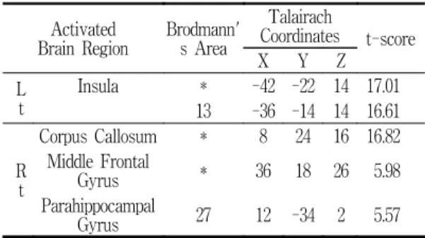
Table 1. Averaged brain activation by EA on Lt. LR3
Activated
Brain Region Brodmann' s Area
Talairach
Coordinates t-score
X Y Z
Lt
Insula * -42 -22 14 17.01
13 -36 -14 14 16.61
R t
Corpus Callosum * 8 24 16 16.82 Middle Frontal
Gyrus * 36 18 26 5.98
Parahippocampal
Gyrus 27 12 -34 2 5.57
P<0.001
temporal gyrus, cingulate gyrus에서 8명중 4 명이 活性 減少를 보였다.
2. 左側 合谷 電鍼刺戟群
1) 腦機能 活性化 領域
左側 合谷 電鍼刺戟群의 Group Average에서 腦活性 增加 領域은 左側(ipsilateral side)의 Precentral gyrus, Insula(BA13), Cerebellum (Inf. semi-lunar lobule), Superior frontal gyrus (BA8), Superior parietal lobule, Middle frontal gyrus(BA 6), Lingual gyrus, 右側(contralateral side)의 Middle frontal gyrus(BA10) Postcentral gyrus, Parahippocampal gyrus(BA36), Cingu- late gyrus(BA24)에서 腦活性이 增加되었다 (Table 3).
個體別 data를 分析한 結果, 鍼刺戟과 同側인 左側 腦에서는 inferior semi-lunar lobule (ce- rebellum)에서 10명중 7명이, Superior frontal gyrus, Middle frontal gyrus, Cuneus, Cingu- late gyrus에서 10명중 6명이, Postcentral gy- rus, Precuneus, Superior temporal gyrus,
Middle temporal gyrus, Middle occipital gyrus에서 10명중 5명이 活性增加를 보였고, 對 側인 右側 腦에서는 Frontal sub-gyral에서 10 명중 7명이, Superior frontal gyrus, Middle frontal gyrus, Medial frontal gyrus, Cuneus 에서 10명중 6명이, Precentral gyrus, cingu- late gyrus에서 10명중 5명이 活性增加를 보였 다.
2) 腦機能 活性 低下 領域
左側 合谷 電鍼刺戟群의 Group Average에서 腦活性 減少 領域은 左側(ipsilateral side)의 Uncus(BA20), Cerebellum(Inf. semi-lunar lo- bule), Cerebellum(tuber), Precentral gyrus, Inferior frontal gyrus, Middle frontal gyrus, Table 2. Averaged brain deactivation by EA on
Lt. LR3.
Activated Brain Region
Brodma nn's Area
Talairach
Coordinates t-score
X Y Z
L t
Celebellar
Tonsil * -32 -54 -40 6.73
Pons * -10 -18 -24 5.82
Rt
Middle
Occipital Gyrus 18 44 -88 12 16.86 Cerebellum
(Tuber) * 24 -84 -30 15.23
Middle
Temporal Gyrus 39 50 -76 24 14.93 Cingulate
Gyrus * 2 22 30 6.70
Superior
Frontal Gyrus 8 16 38 50 6.07 P<0.001
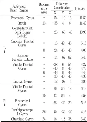
Table 3. Averaged brain activation by EA on Lt. LI4.
Activated Brain Region
Brodma nn's Area
Talairach
Coordinates t-score
X Y Z
Lt
Precentral Gyrus * -54 -10 16 11.50
Insula 13 -38 4 6 11.40
Cerebellum(Inf.
Semi-Lunar
Lobule) * -26 -68 -40 10.95 Superior Frontal
Gyrus * -16 42 46 6.15
8 -24 46 40 4.06
Superior
Parietal Lobule * -14 -62 62 5.45 Middle Frontal
Gyrus *
* 6*
-38-40 -48-30
68 408
5446 4840
4.874.76 4.454.33 Lingual Gyrus * -12 -92 -4 3.86
R t
Middle Frontal
Gyrus * 36 56 12 6.13
10 42 56 4 4.53 Postcentral
Gyrus * 68 -22 20 5.16
Parahippocampa
l Gyrus 36 40 -32 -20 4.16 Cingulate Gyrus 24 16 -18 38 3.49 P<0.001
Precuneus(BA19)와, 右側(contralateral side)의 Cerebellum(tuber), Cerebellum(uvula), Infer-
ior frontal gyrus(BA46), Middle frontal gyrus (BA6), Precuneus(BA7, BA19), Thalamus (ventral lateral nuleus), Cerebellum(culmen), Middle temporal gyrus, Temporal sub-gyral, Postcentral gyrus에서 腦活性이 減少되었다 (Table 4).
個體別 data를 分析한 結果, 同側인 左側 腦 에서는 Middle frontal gyrus, Inferior frontal gyrus에서 10명중 9명이, 10명중 8명이, Middle temporal gyrus에서 10명중 7명이, Superior frontal gyrus, Precentral gyrus, Inferior parietal lobule, Postcentral gyrus, Lingual gyrus, cingulate gyrus, declive(cerebellum)에 서 10명중 5명이 活性減少를 보였고, 對側인 右 側 腦에서는 Middle frontal gyrus, Superior temporal gyrus에서 10명중 8명이, Superior frontal gyrus, Precentral gyrus, Middle temporal gyrus에서 10명중 7명이, Precuneus, cingulate gyrus, declive(cerebellum)에서 10명 중 6명이, Inferior frontal gyrus, Postcentral gyrus, Middle occipital gyrus에서 10명중 5명 이 活性減少를 보였다.
3. 左側 太衝‧合谷 電鍼刺戟群
1) 腦機能 活性化 領域
左側 太衝‧合谷 電鍼刺戟群의 Group Average 에서 腦活性이 增加된 領域은 左側(Ipsilateral side)의, Cerebellum(Inf. semi-lunar lobule), Precuneus(BA7), Cerebellar tonsil, Cerebell- um(culmen)과 右側(contralateral side)의 Infer- ior frontal gyrus(BA45), Temporal sub- gy- ral, Superior temporal gyrus(BA22)에서 腦活 性이 增加되었다(Table 5).
個體別 data를 分析한 結果, 鍼刺戟과 同側인 左側 腦에서는 Superior frontal gyrus에서 8명 Table 4. Averaged brain deactivation by EA
on Lt. LI4.
Activated Brain Region
Brodma nn's Area
Talairach
Coordinates t-score
X Y Z
Lt
Uncus 20 -26 0 -42 12.01
Cerebellum(Inf.
Semi-Lunar
Lobule) * -34 -80 -36 8.51
Cerebellum(Tube
r) * -26 -86 -30 3.69
Precentral Gyrus * -48 -4 46 7.70
* -42 -12 48 4.86 Inferior Frontal
Gyrus * -52 38 8 6.64
* -44 44 -2 6.09
* -40 26 -18 6.21
* -48 34 0 5.21
Middle Frontal
Gyrus * -38 42 -14 6.59
* -32 34 -16 4.28
* -50 26 22 4.91
Precuneus 19 -14 -82 40 4.91
R t
Cerebellum(Tube
r) * 44 -74 -28 7.02
Cerebellum(Uvul
a) * 10 -88 -26 6.96
Inferior Frontal
Gyrus * 54 36 8 5.99
46 44 38 12 4.50
Middle Frontal
Gyrus * 42 46 16 5.36
6 48 4 44 3.94
Precuneus * 8 -58 54 5.36
7 4 -46 50 4.35
19 32 -80 40 4.50 Thalamus(Ventra
l Lateral Nuleus) * 14 -16 10 5.32 Cerebellum(Culm
en) * 10 -56 -10 4.81
Middle Temporal
Gyrus * 60 -10 -8 4.65
Temporal
Sub-Gyral * 48 -16 -10 3.82
Postcentral
Gyrus * 50 -28 52 4.24
P<0.001
중 6명이, Middle frontal gyrus에서 8명중 5명 이, Inferior frontal gyrus, Precentral gyrus에 서 8명중 4명이 活性增加를 보였고, 對側인 右 側 腦에서는 cingulate gyrus에서 8명중 5명이, Middle frontal gyrus, declive(cerebellum)에서 8명중 4명이 活性增加를 보였다.
2) 腦機能 活性 低下 領域
左側 太衝‧合谷 電鍼刺戟群의 Group Average 에서는 左側(Ipsilateral side) Cerebellum(Cul- men), Cerebellum(Inf. Semi-lunar lobule), Ce- rebellar tonsil, Middle occipital gyrus, Cingu- late gyrus와 右側(Contralateral side)의 Infer- ior occipital gyrus, Middle frontal gyrus, In- ferior frontal gyrus, Cingulate gyrus에서 腦活 性이 減少되었다(Table 6).
個體別 data를 分析한 結果, 鍼刺戟과 同側인 左側 腦에서는 Superior frontal gyrus, Middle frontal gyrus에서 8명중 6명이, Precentral gy- rus, Superior temporal gyrus, Middle tempo- ral gyrus, Cuneus, cingulate gyrus, culmen (cerebellum)에서 8명중 5명이, Inferior frontal gyrus, Medial frontal gyrus, Inferior parietal
lobule, Precuneus, Parahippocampal gyrus에 서 8명중 4명이 活性減少를 보였고, 對側인 右 側 腦에서는 Superior frontal gyrus, Middle frontal gyrus, Precentral gyrus, cingulate gy- rus, culmen(cerebellum)에서 8명중 5명이, In- ferior frontal gyrus, Medial frontal gyrus, Frontal sub-gyral, Superior temporal gyrus, Middle temporal gyrus, putamen에서 8명중 4 명이 活性減少를 보였다.
4. 兩側 太衝‧合谷 電鍼刺戟群
1) 腦機能 活性化 領域
兩側 太衝‧合谷 電鍼刺戟群의 Group Average 에서는 左側의 Middle temporal gyrus(BA19), Cuneus와 右側의 Postcentral gyrus, Tempo- ral sub-gyral, Inferior frontal gyrus, Cerebell- um(declive)에서 腦活性이 增加되었다(Table 7).
個體別 data를 分析한 結果, 左側 腦에서는 Middle frontal gyrus, Inferior frontal gyrus, Precuneus, Superior temporal gyrus에서 8명 Table 5. Averaged brain activation by EA on
Lt. LR3 and LI4 Activated
Brain Region
Brodma nn's Area
Talairach Coordinates t-score
X Y Z
Lt
Cerebellum(Inf.
Semi-Lunar Lobule) * -32 -70 -36 11.54
Precuneus 7 -4 -60 60 7.47
Cerebellar Tonsil * -22 -60 -36 5.57 Cerebellum(Culmen) * -4 -40 -2 4.06
R t
Inferior Frontal
Gyrus 45 56 36 4 14.81
* 48 46 2 13.64 Temporal Sub-Gyral * 40 -12 -18 6.63
Superior Temporal
Gyrus 22 58 -6 2 4.94
P<0.001
Table 6. Averaged brain deactivation by EA on Lt. LR3 and LI4.
Activated Brain Region
Brodm ann's
Area
Talairach
Coordinates t-score
X Y Z
L t
Cerebellum(Culm
en) * -32 -60 -26 8.06
* -44 -48 -28 7.68 Cerebellar Tonsil * -42 -58 -34 5.52 Angular Gyrus * -36 -70 32 6.28
Cerebellum(Inf.
Semi-Lunar
Lobule) * -38 -78 -38 6.08
Temporal
Sub-Gyral * -22 -54 18 4.79
Rt
Inferior Occipital
Gyrus * 36 -88 -14 12.52
Pons * 0 -20 -30 6.93
P<0.01
중 6명이, Middle temporal gyrus에서 8명중 5 명이, Superior frontal gyrus, Medial frontal gyrus, Precentral gyrus, Temporal sub-gyral, cingulate gyrus, cerebellar tonsil(cerebellum) 에서 8명중 4명이 活性增加를 보였고, 右側 腦 에서는 Middle frontal gyrus에서 8명중 7명이, cingulate gyrus에서 8명중 6명이, Superior frontal gyrus, , Inferior parietal lobule, Pre- cuneus, Superior temporal gyrus, 에서 8명중 5명이, Postcentral gyrus, Middle temporal gyrus, cerebellar tonsil(cerebellum)에서는 8명 중 4명이 活性增加를 보였다.
2) 腦機能 活性 低下 領域
兩側 太衝‧合谷 電鍼刺戟群의 Group average 에서 腦活性이 低下된 領域은 左側의 Uncus, Superior occipital gyrus(BA19), Middle tem- poral gyrus(BA19), Inferior occipital gyrus, Fusiform gyrus(19), Cerebellum(Tuber), Pre- central gyrus(BA6, BA4), Postcentral gyrus, Inferior parietal lobule(40), Frontal sub- gy- ral, Middle frontal gyrus, Inferior frontal
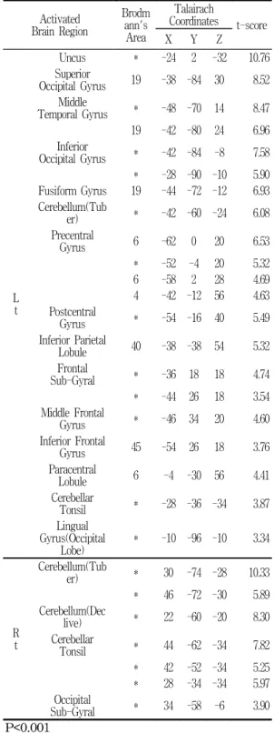
Table 8. Averaged Brain Deactivation by EA on LR3 and LI4 Bilaterally.
Activated Brain Region
Brodm ann's
Area
Talairach
Coordinates t-score
X Y Z
L t
Uncus * -24 2 -32 10.76
Superior
Occipital Gyrus 19 -38 -84 30 8.52 Middle
Temporal Gyrus * -48 -70 14 8.47 19 -42 -80 24 6.96 Inferior
Occipital Gyrus * -42 -84 -8 7.58
* -28 -90 -10 5.90 Fusiform Gyrus 19 -44 -72 -12 6.93 Cerebellum(Tub
er) * -42 -60 -24 6.08
Precentral
Gyrus 6 -62 0 20 6.53
* -52 -4 20 5.32
6 -58 2 28 4.69
4 -42 -12 56 4.63 Postcentral
Gyrus * -54 -16 40 5.49
Inferior Parietal
Lobule 40 -38 -38 54 5.32
Frontal
Sub-Gyral * -36 18 18 4.74
* -44 26 18 3.54
Middle Frontal
Gyrus * -46 34 20 4.60
Inferior Frontal
Gyrus 45 -54 26 18 3.76
Paracentral
Lobule 6 -4 -30 56 4.41
Cerebellar
Tonsil * -28 -36 -34 3.87
Lingual Gyrus(Occipital
Lobe) * -10 -96 -10 3.34
Rt
Cerebellum(Tub
er) * 30 -74 -28 10.33
* 46 -72 -30 5.89 Cerebellum(Dec
live) * 22 -60 -20 8.30
Cerebellar
Tonsil * 44 -62 -34 7.82
* 42 -52 -34 5.25
* 28 -34 -34 5.97 Occipital
Sub-Gyral * 34 -58 -6 3.90
P<0.001 Table 7. Averaged brain activation by EA on
LR3 and LI4 Bilaterally.
Activated Brain Region
Brodm ann's Area
Talairach
Coordinates t-score
X Y Z
L t
Middle Temporal
Gyrus 19 -50 -76 20 5.69
Cuneus * -8 -96 6 4.67
Rt
Postcentral
Gyrus * 60 -16 18 12.74
Temporal
Sub-Gyral * 38 -12 -14 12.62 Inferior
Frontal Gyrus * 38 18 -6 12.08 Cerebellum(D
eclive) * 30 -78 -16 4.64
P<0.001
gyrus(BA45), Paracentral lobule(BA6), Cere- bellar tonsil, Lingual gyrus와, 右側의 Cere- bellum(tuber), Cerebellum(declive), Cerebe- llar tonsil, Occipital sub-gyral에서 腦活性이 減少되었다(Table 8).
또한, 個體別 data를 分析한 結果, 左側 腦에 서는 Middle frontal gyrus, Precentral gyrus에 서 8명 모두가, Inferior frontal gyrus에서 8명 중 7명이, Middle temporal gyrus, 8명중 6명 이, Superior frontal gyrus, Superior temporal gyrus, declive(cerebellum)에서 8명중 5명이, Medial frontal gyrus, Frontal sub-gyral, Precuneus, Middle occipital gyrus, Lingual gyrus, inf. semi-lunar lobule(cerebellum)에서 8명중 4명이 活性減少를 보였고, 右側 腦에서는 Middle temporal gyrus,에서 8명중 6명이, Su- perior frontal gyrus, 에서 8명중 5명이, Medi- al frontal gyrus, Precentral gyrus, Precuneus, cingulate gyrus, tuber(cerebellum)에서 8명중 4명이 活性減少를 보였다.
5. Motor task에 의한 腦機能 活性化 領域
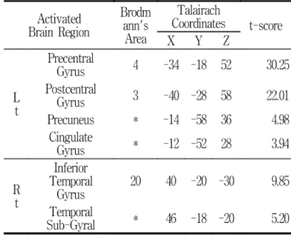
右側 Hand motor task에 의해 fMRI상 腦活性 이 增加된 部位는 左側(contralateral side)의 Precentral gyrus(BA4), Postcentral gyrus (BA4), Precuneus, Cingulate gyrus와, 右側 (ipsilateral side)의 Inferior temporal gyrus (BA 20), Temporal sub-gyral로 나타났다(Table 9).
Ⅳ. 考 察
經絡은 “經脈者 所以行血氣而榮陰陽 濡筋骨 利關節”13)라 하여 氣血運行의 通路로 認識하였 으며, ≪難經‧二十二難≫14)에서 “氣主照之 血之 濡之”라 하여 生理的으로 身體를 滋養하는 作用
을 가지고 있다. 그러므로 經絡은 人體異常이 나타날 경우 이를 反影하는데, 人體에 만약 어 떤 發病因子가 侵犯하여 臟腑의 正常機能이 損 傷되어 疾病이 發生한 경우, 經絡은 人體의 각 部分과 特別한 관계를 맺고 있는 經絡으로 通連 된 體表 關聯部位에서 各種의 異常變化를 나타 내게 된다. 이러한 反應을 現代的인 名稱으로 壓通點 혹은 過敏點이라 하는데, ≪千金要方≫
15)에는 “摩膂肉之表 肋間空處 按之自覺牽引胸 中”이라 하였다.
經穴은 疾病의 反應點이며, 또한 治療上의 刺 戟點이 되므로 臨床應用에서는 이 두가지 面이 서로 結合되어 쓰여지게 된다16). 經絡은 侵犯病 邪를 傳導하는 作用을 갖추고 있어 體表에 侵犯 한 病邪가 經絡을 通하여 內臟으로 轉入되고 內 臟間의 經絡의 關係에 의하여 病邪는 하나의 內 臟에서 다른 內臟으로 轉入하는데 이를 “病邪之 傳變” 또는 “傳經”이라고 稱하였고, ≪素門 繆刺 論編≫13)에서는 “夫邪之客于形也 必先舍于皮毛 留而不去 入舍于孫脈 留而不去 入舍于絡脈 留而 不去 入舍于經脈 內運五臟 散于腸胃”이라 하였 다.
Table 9. Averaged brain activation by Motor task.
Activated Brain Region
Brodm ann's Area
Talairach
Coordinates t-score
X Y Z
L t
Precentral
Gyrus 4 -34 -18 52 30.25
Postcentral
Gyrus 3 -40 -28 58 22.01
Precuneus * -14 -58 36 4.98
Cingulate
Gyrus * -12 -52 28 3.94
R t
Inferior Temporal
Gyrus 20 40 -20 -30 9.85
Temporal
Sub-Gyral * 46 -18 -20 5.20
P<0.001
經絡 혹은 內臟이 機能을 失調했을 때, 體表 의 一定部位를 鍼灸 등으로 刺戟함으로써 經絡 이 그 治療性 刺戟을 有關한 部位와 內臟으로 傳導할 수 있다. 그래서 人體의 臟器가 調節機 能을 發揮하여 氣血의 運行을 圓滑하게 하고, 營衛가 調和되어 疾病이 治癒되는 것이다. 刺針 治療와 經絡의 傳導作用과의 關係는 具體的으로 經絡이 順行하고 있는 經路上에서 적당한 經穴 을 選定하여 治療을 進行함으로써 表現된다. 예 컨데 合谷을 取하여 齒痛을 治療하고 內關을 取 穴하여 胃脘痛을 治療하며 頸部捻挫에는 後谿, 中渚 등 穴을 取하고 腸胃疾患에는 足三里, 上 巨虛 등 穴을 取하는 方法은 臨床上 모두 좋은 治療效果가 있다. 이러한 治療效果를 얻을 수 있는 것은 經絡의 傳導機能과 不可分의 關係가 있다2).
最近들어 鍼灸治療의 效果 및 機轉에 대한 硏 究는 經絡과 神經系 機能과의 關係, 體表 및 內 臟과의 關係, 內分泌 호르몬과의 關係, 血球 및 血液學的 方面과의 關係 등으로 硏究되고 있으 며, 아울러 組織學的, 神經系, 心血管系, 泌尿生 殖器系, 精神系統, 運動器系, 消火器系 등의 方 面으로 硏究되고 있는데, 좀더 具體的으로 Ku- rabayash17)와 Palummer18)는 經絡 穴位의 作用 이 周圍神經系統과 有關하며 鍼刺作用은 神經의 反射活動이라고 하였고, Jansen19) 등은 鍼刺에 의한 흰쥐 皮下血管 내의 感覺 neuropeptide의 增加에 대하여 報告하였으며 Thomas20)는 經穴 部位에 多數의 神經纖維의 存在를 報告하였고, Mann21)은 鍼刺戟의 效果를 皮部-筋-神經反射의 經路로 說明하였다.
CT, MRI, PET같은 映像化 技法은 腦機能을 硏究하는데 새로운 指標를 열고 있는데22) 특히 腦의 機能에 따른 區劃方法은 視覺이나 運動 등 의 刺戟에 대한 大腦皮質活動의 生理變化를 可
視化 시킬 수 있게 되었다. 지금까지 大腦에서 나타나는 鍼의 效果를 評價하기 위해 EEG나 PET를 利用한 論文들이 있었으나 腦의 反應機 轉에 關하여 明確히 밝히지는 못했으며 PET의 경우 放射線 物質이 添加된 신호대비용 藥品을 人體에 계속 注入해야 하는 短點으로 사람을 對 象으로 하는 反復的인 試驗을 하기에는 어려움 이 있었다. 그러나 fMRI는 이런 短點을 補完해 줄 수 있을 뿐 아니라 空間과 側頭部의 해상도 가 더욱 뛰어나다는 長點을 갖고 있으므로 腦에 서 일어나는 機能的 變化를 觀察하는데 매우 有 用하며 經穴의 刺鍼 效果를 硏究하는 데에도 有 用하다4).
fMRI의 生理學的 理論은 뇌 뉴런의 反應에 基礎를 두고 있다. 탐침 electrode로 직접 action potential을 測定하거나 赤外線을 이용한 이산 화탄소 濃度 測定을 통해 뇌 뉴런의 反應時間을 測定해 보면 外部 刺戟에 대해 뇌 뉴런이 反應 하는 時間은 약 100 ms 정도의 時間 差를 가지 고 發生된다고 알려져 있다. 이러한 뇌 뉴런들 이 活性化되면 CBF(Cerebral Blood Flow), CBV(Cerebral Blood Volume), 酸素 消費量 등 이 增加하는데 fMRI는 이와 같은 腦 機能에 隨 伴되는 perfusion 關聯 變數들을 測定하는 것이 다23).
이때 必要한 酸素 運搬을 擔當하는 hemog- lobin은 酸素가 조합된 有無를 基準으로 oxyhe- moglobin과 deoxyhemoglobin으로 나뉘는데 腦 의 一部分이 活性化되면 이에 따른 에너지를 生 成하기 위해 部分的 酸素 要求量이 增加하고 이 런 酸素 要求量의 增加를 補充하기 위해 더 많 은 血液이 흐르게 되고, 結果的으로 酸素가 結 合된 oxyhemoglobin의 量이 deoxyhemoglobin 보다 많게 된다24). 이것이 oxygen metabolism 과 BOLD effect의 關係로서 現在 fMRI技法 中
가장 많이 使用되는 技法이며 本 硏究에서도 이 러한 技法들을 이용하였다25).
磁氣場의 크기가 增加할수록 BOLD effect에 의한 신호의 크기는 커진다. 따라서 强한 磁氣場 일수록 BOLD effect를 利用한 fMRI試驗이 優 秀하며 本 試驗에서는 效果的인 試驗 遂行을 위 해 3.0 Tesla의 高磁場 MRI 裝置를 使用하여 試驗을 進行하였다. 뉴런의 activation은 몇 개 의 cluster 나 個別的으로 일어나며 空間的 分布 는 넓게는 centimeter부터 작게는 micrometer 로 廣範圍하게 나타난다. 磁氣場이 강할수록 더 큰 신호대비가 可能하므로 高磁場의 fMRI를 利 用하면 보다 큰 공간 해상도를 갖는 映像을 獲 得할 수 있다.
BOLD effect는 血流와 關係되어 있는데 實質 的으로 뉴런의 activation 後 略 2~3 초 정도의 delay가 생기는데 이는 信號 處理 技術의 發達 로 이를 考慮한 分析이 充分히 可能해 졌다.
어떤 外部 刺戟에 대해 腦의 여러 地域이 順 次的으로 activation되는 것을 解釋하기 위해서 는 빠른 時間 해상도가 必要한데 MRI에서는 高 速 sequence인 EPI(Echo Palanar Imaging)을 使用하여 時間 해상도를 計算한다26). EPI는 slice 당 略 100 msec의 時間이 所要되어 1초 未滿의 時間 해상도를 可能하게 해 준다.
일찍이 Takashi, Yoshida 등8)은 fMRI와 經 穴刺鍼을 연결하여 硏究할 수 있는 可能性을 提 示하였다.
본 試驗에 使用된 電鍼은 傳統的인 鍼과 現代 科學技術이 結合하여 發展된 新鍼療法으로서 1825년 프랑스의 Sarandiere가 처음으로 電鍼 을 應用하여 痛症 및 神經系 疾患을 治療한 結 果를 報告하면서 以後 臨床에 많이 活用되고 있 다2).
明의 馬元臺25)가 肘膝關節 以下 部位를 四關
으로 言及한 후, 李梴27)은≪醫學入門‧雜病六法≫
에서 “四關三部識其處”라 했고 스스로 이 部分 을 註釋하여 四關은 合谷, 太衝이라 하였다. 楊 繼洲28)는≪鍼灸大成 經外奇穴編≫에서 四關四 穴이란 合谷 太衝을 말한다고 指摘하였다.
四關은 大腸經의 原穴인 合谷과 肝經의 原穴 인 太衝과 組合을 이루므로 氣血의 病變을 治療 할 수가 있고 合谷과 太衝을 陰陽論上에서 볼 때 合谷은 陽性으로 氣의 作用이 强하고, 또한 下向性이며, 太衝穴은 陰性으로 穴의 作用이 强 하면서도 上向性이다. 이와 같은 穴性에서 이들 兩穴의 組合은 陰陽으로 兩分되고 있는 氣血의 調整作用이 있음을 推論할 수가 있다.
四關의 穴位에 대해 살펴보면, 合谷穴은 ≪鍼 灸資生經≫29)에서는 “手大指次指岐骨間陷中”이 라 하였으며 ≪鍼灸集成≫30)에서는 “手大指次指 兩骨間宛, 宛中動脈應手處”라 하였고, ≪鍼灸醫 學典論≫31)에서는 “母指와 食指를 벌렸을 때 第 一第二中手骨의 中央에서 若干 食指側”이라 하 였으며 ≪實用鍼灸辭典≫32)에서는 “手背部第一 二掌骨間之間, 近第二掌骨橈側之中點”이라 하였 다. 以上에서 合谷의 位置는 “手大指次指岐骨間 陷中”이 普遍的인 것으로 思料된다.
太衝의 穴位는 ≪鍼灸聚英≫33)에서는 “足大趾 本節後二寸或寸半陷中”이라 하였고 ≪鍼灸學簡 編≫34)에서는 “足大趾本節後二寸”이라 하였다.
以上에서 太衝의 位置는 “足大趾本節後二寸, 骨 間陷中”이라 普遍的인 것으로 思料된다.
合谷穴 및 太衝穴에 대한 穴性과 特徵을 살펴 보면 合谷穴은 爲陽主氣, 發表解熱, 疎風解表, 淸泄肺氣, 通降腸胃, 鎭痛安神, 通經活絡등의 穴 性을 볼 수 있으며12) 또한 合谷穴은 原穴로서 自然治癒力을 增强시키는 要穴이며 四總穴35)의 하나로 「面口合谷收」하며 回陽九鍼穴36)의 하 나이기도 하다. 또한 全身反應의 最大刺戟穴로
病精과 身體의 强弱을 決定하는데 있어서 重要 하며 不注意時 暈鍼이 일어나기도 쉬운 곳이다.
太衝은 爲陰主血하고 淸熄肝火肝陽, 疎泄下焦濕 熱, 舒肝理氣, 通絡活血 등의 穴性을 볼 수 있고 또한 肝經의 原穴이며 調節血量하며 ��女子二 七, 太衝脈盛 月事以時下 故能有子. 診病人 太衝 脈有無로 可以決死生��하는 특징을 보인다37).
한편 四關穴의 特性을 살펴보면12,28,36,37)��
鎭靜作用, 搜風理痺, 開關節, 墮痰瀉火, 順氣鎭 靜神志安, 氣血通行之關, 理氣活血, 淸熱鎭痙, 開竅醒神, 淸瀉陽明, 疎風鎭痛, 降高血壓, 調整 機能, 行氣血而通經消瘀��등의 效能이 있음을 볼 수 있다. 合谷과 太衝은 二穴의 작은 짝이지 만 하나의 處方이라는 것은 穴性을 分析해 보면 알 수 있다. 合谷은 手陽明原穴, 爲陽主氣하고, 太衝은 足厥陰原穴, 爲陰主血이며 이는 氣血通 行之 關門이 되며 다 같이 手足의 岐骨之間에 位置하여 氣血失常疾病의 重要한 方이 된다.
Wu6)는 合谷 자침에 의한 hypothalamus, nucleus accumbens 에서의 活性增加와 anter- ior cingulate cortex의 rostral part, amygdala formation, hippocampal complex 에서의 活性 저하를 fMRI를 이용하여 관찰하였고, Hui9)는 건강한 사람에게 合谷 자침을 하여 fMRI로 관 찰한 결과, nucleus accumbens, amygdala, hi- ppocampus, parahippocampus, hypothalamus, ventral tegmental area, anterior cingulate gyrus, caudate, putamen, temporal pole, in- sula에서 活性 減少를 somatosensory cortex에 서는 活性減少를 관찰하였다고 보고하였다. 한 편, Hsieh10) 는 PET를 이용하여, 合谷 자침에 의한 腦活性 변화를 관찰한 결과, hypothala- mus, midbrain, insula, anterior cingulate cor- tex 그리고 cerebellum에서 活性增加가 나타났 다고 하였고, Kong11)은 合谷에 자침후 수기자극
을 하였을 때에는 fMRI상 posterior cingulate, superior temporal gyrus, putamen, insula에 서 活性減少를 보였고, 3Hz의 電鍼刺戟을 가하 였을 때에는 precentral gyrus, postcentral gy- rus, inferior parietal loule, putamen, insula에 서 活性增加를 보였다고 보고 하였다.
이와 같이, 合谷 자침 수기의 조작방법, 전침 의 강도 등에 따라 뇌活性에 미치는 영향이 다 르게 보고되고 있다.
한편, 太衝 자극에 의한 腦活性변화를 연구한 문헌은 찾아 볼 수 없었다.
本 實驗에서는 四關 鍼治療 機轉硏究의 일환 으로, 健康한 男女 34名의 支援者를 募集하여, 太衝電鍼刺戟群 8名, 合谷 電鍼刺戟群 10名, 太 衝+合谷 電鍼 刺戟群 8名, 四關 電鍼刺戟群 8名 으로 나누고, 各各의 電鍼刺戟이 腦活性變化에 미치는 影響을 fMRI를 利用하여 觀察하였다.
實驗結果, 左側 太衝 電鍼刺戟群의 group average에서는 左側(ipsilateral side)의 Insula (BA 13), 右側(contralateral side)의 Copus cal- losum, Middle frontal gyrus, Parahippocam- pal gyrus(BA 27)에서 腦活性이 增加되었다 (Table 1).
個體別 data를 分析한 結果, 鍼刺戟과 同側인 左側 腦에서는 Middle frontal gyrus, Inferior parietal lobule, Postcentral gyrus에서 8명중 4 명이 活性增加를 보였고, 對側인 右側 腦에서는 Superior frontal gyrus, Middle frontal gyrus, Frontal sub-gyral, Inferior parietal lobule, Superior temporal gyrus, Middle occipital gyrus, Lingual gyrus, cingulate gyrus에서 8 명중 4명이 活性 增加를 보였다.
左側 太衝 電鍼刺戟群의 Group average를 통 한 腦活性 增加 領域과 個體別 data 중 過半數 의 被驗者들에게서 나타난 腦活性 增加 領域의
共通部位는 右側 Middle frontal gyrus로 나타 났다.
左側 太衝 電鍼刺戟群의 Group Average를 통한 腦活性 減少 領域은 左側(ipsilateral side) 의 Celebellar tonsil, Pons, 右側(contralateral side)의 Middle occipital gyrus(BA 18), Cere- bellum (tuber), Middle temporal gyrus(BA 39), Cingulate gyrus, Superior frontal gyrus (BA 8)로 나타났다(Table 2).
個體別 data를 分析한 結果, 電鍼刺戟과 同側 인 左側 腦에서는 Middle frontal gyrus에서 8 명중 7명이, Superior frontal gyrus에서 8명중 6명이, Precentral gyrus, Cuneus, cingulate gyrus, Parahippocampal gyrus에서 8명중 4명 이 活性減少를 보였고, 對側인 右側 腦에서는 Superior frontal gyrus, Middle frontal gyrus, 에서 8명중 6명이, Superior temporal gyrus에 서 8명중 5명이, Precentral gyrus, Middle temporal gyrus, cingulate gyrus에서 8명중 4 명이 活性減少를 보였다.
左側 太衝 電鍼刺戟群의 Group average를 통 한 腦活性 減少 領域과 個體別 data 중 過半數 의 被驗者들에게서 나타난 腦活性 減少 領域의 共通部位는 右側 Superior frontal gyrus, Middle temporal gyrus, Cingulate gyrus로 나 타났다.
이에, 太衝電鍼刺戟의 作用機轉은 右側(對側) Middle frontal gyrus의 活性增加 및 右側(對側) Superior frontal gyrus, Middle temporal gy- rus, Cingulate gyrus의 活性減少와 關聯이 있 을 것으로 推測된다.
左側 合谷 電鍼刺戟群의 Group average에서 는 左側(ipsilateral side)의 Precentral gyrus, Insula(BA13), Cerebellum(Inf. semi-lunar lo- bule), Superior frontal gyrus(BA8), Superior
parietal lobule, Middle frontal gyrus(BA 6), Lingual gyrus, 右側(contralateral side)의 Mi- ddle frontal gyrus(BA10) Postcentral gyrus, Parahippocampal gyrus(BA36), Cingulate gy- rus(BA24)에서 腦活性이 增加되었다(Table 3).
個體別 data를 分析한 結果, 鍼刺戟과 同側인 左側 腦에서는 inferior semi-lunar lobule(cere- bellum)에서 10명중 7명이, Superior frontal gyrus, Middle frontal gyrus, Cuneus, Cingu- late gyrus에서 10명중 6명이, Postcentral gyr- us, Precuneus, Superior temporal gyrus, Mi- ddle temporal gyrus, Middle occipital gyrus에 서 10명중 5명이 活性增加를 보였고, 對側인 右 側 腦에서는 Frontal sub-gyral에서 10명중 7명 이, Superior frontal gyrus, Middle frontal gyrus, Medial frontal gyrus, Cuneus에서 10명 중 6명이, Precentral gyrus, cingulate gyrus에 서 10명중 5명이 活性增加를 보였다.
左側 合谷 電鍼刺戟群의 Group average를 통 한 腦活性 增加 領域과 個體別 data 중 過半數 의 被驗者들에게서 나타난 腦活性 增加 領域과 의 共通部位는 左側 Superior frontal gyrus, Middle frontal gyrus, Inf. semi-lunar lobule (cerebellum)과, 右側 Middle frontal gyrus, Cingulate gyrus로 나타났다.
이는 Kong11)이 3Hz의 電氣刺戟을 合谷에 加 하였을 때와는 다소 다른 結果라고 할 수 있다 左側 合谷 電鍼刺戟群의 Group Average에서 腦活性 減少 領域은 左側(ipsilateral side)의 Uncus(BA20), Cerebellum(Inf. semi-lunar lo- bule), Cerebellum(tuber), Precentral gyrus, Inferior frontal gyrus, Middle frontal gyrus, Precuneus(BA19)와, 右側(contralateral side)의 Cerebellum(tuber), Cerebellum(uvula), Inferi- or frontal gyrus(BA46), Middle frontal gyrus
(BA6), Precuneus(BA7, BA19), Thalamus(ve- ntral lateral nuleus), Cerebellum(culmen), Middle temporal gyrus, Temporal sub-gyral, Postcentral gyrus로 나타났다(Table 4).
個體別 data를 分析한 結果, 鍼刺戟과 同側인 左側 腦에서는 Middle frontal gyrus, Inferior frontal gyrus에서 10명중 9명이, 10명중 8명이, Middle temporal gyrus에서 10명중 7명이, Su- perior frontal gyrus, Precentral gyrus, Infer- ior parietal lobule, Postcentral gyrus, Lingual gyrus, cingulate gyrus, declive(cerebellum)에 서 10명중 5명이 活性減少를 보였고, 對側인 右 側 腦에서는 Middle frontal gyrus, Superior temporal gyrus에서 10명중 8명이, Superior frontal gyrus, Precentral gyrus, Middle tem- poral gyrus에서 10명중 7명이, Precuneus, cingulate gyrus, declive(cerebellum)에서 10명 중 6명이, Inferior frontal gyrus, Postcentral gyrus, Middle occipital gyrus에서 10명중 5명 이 活性減少를 보였다.
左側 合谷 電鍼刺戟群의 Group average를 통 한 腦活性 減少 領域과 個體別 data 중 過半數 의 被驗者들에게서 나타난 腦活性 減少 領域과 의 共通部位는 左側 Middle frontal gyrus, Inferior frontal gyrus, Precentral gyrus, 右側 Middle frontal gyrus, Middle temporal gyrus, Precuneus, Inferior frontal gyrus, Postcentral gyrus로 나타났다.
이에 合谷電鍼刺戟의 작용기전은 左側 Su- perior frontal gyrus, Middle frontal gyrus, Inf. Semi-Lunar Lobule(cerebellum)과, 右側 (對側) Middle frontal gyrus, Cingulate gyrus 의 活性 增加 및, 左側(同側) Middle frontal gyrus, Inferior frontal gyrus, Precentral gy- rus 및 右側(對側) Middle frontal gyrus, Mi-
ddle temporal gyrus, Precuneus, Inferior frontal gyrus, Postcentral gyrus의 活性減少와 관련이 있을 것으로 사료된다.
左側 太衝‧合谷 電鍼刺戟群의 Group Average 에서 腦活性이 增加된 領域은 左側(Ipsilateral side)의, Cerebellum(Inf. semi-lunar lobule), Precuneus(BA7), Cerebellar tonsil, Cerebel- lum(culmen)과 右側(contralateral side)의 In- ferior frontal gyrus(BA45), Temporal sub-gy- ral, Superior temporal gyrus(BA22)로 나타났 다(Table 5).
個體別 data를 分析한 結果, 鍼刺戟과 同側인 左側 腦에서는 Superior frontal gyrus에서 8명 중 6명이, Middle frontal gyrus에서 8명 중 5명 이, Inferior frontal gyrus, Precentral gyrus에 서 8명중 4명이 活性增加를 보였고, 對側인 右 側 腦에서는 cingulate gyrus에서 8명중 5명이, Middle frontal gyrus, declive(cerebellum)에서 8명 중 4명이 活性增加를 보였다.
左側 太衝‧合谷 電鍼刺戟群의 Group average 를 통한 腦活性 增加 領域과 個體別 data 중 過 半數의 被驗者들에게서 나타난 腦活性 增加 領 域과의 共通部位는 觀察되지 않았다.
左側 太衝‧合谷 電鍼刺戟群의 Group Average 에서 腦活性 減少 領域은 左側(Ipsilateral side) Cerebellum(Culmen), Cerebellum(Inf. Semi- lunar lobule), Cerebellar tonsil, Middle occi- pital gyrus, Cingulate gyrus와 右側(Contrala- teral side)의 Inferior occipital gyrus, Middle frontal gyrus, Inferior frontal gyrus, Cingu- late gyrus로 나타났다(Table 6).
個體別 data를 分析한 結果, 同側인 左側 腦 에서는 Superior frontal gyrus, Middle frontal gyrus에서 8명중 6명이, Precentral gyrus, Su- perior temporal gyrus, Middle temporal gy-