Introduction
For the successful root canal treatment, thorough cleaning and shaping of the root canal system is a necessary procedure. C-shaped canals are known to have a complex canal anatomy with numerous fins connecting individual canals, thus require supple- mentary effort to accomplish a successful root canal
treatment.1,2 Generally, anthropologic differences in tooth shape is not an important issue in the root canal treatment, however, mandibular second molar is a matter of concern due to its high incidence of C- shaped root canal configurations especially in Asian populations. In this regard, recent studies3-5reported that more than 30% of Chinese or Koreans have C- shaped mandibular second molar in their arch. This
A retrospective study of the intentionally replanted mandibular second molars with C-shaped root canal configurations
Won-Jun Shon, Kee-Yeon Kum, Seung-Ho Baek, Woo-Cheol Lee*
Department of Conservative Dentistry, Dental Research Institute, Seoul National University School of Dentistry, Seoul, Korea
Objectives: The purpose of this retrospective study was to evaluate the success rate of intentionally replanted mandibular second molar with C-shaped canal configurations and to access the impact of preop- erative periapical lesion on the success of intentional replantation procedure.
Materials and Methods: This retrospective chart review study evaluated 52 intentionally replanted mandibular second molar teeth treated at Seoul National University Dental Hospital Department of Conservative Dentistry from January 2005 to December 2007. Seventeen teeth were lost for the follow-up, and another 6 teeth did not meet inclusion criteria of C-shaped root canal configurations. Healing outcome such as success, uncertain healing, and failure after follow-up was evaluated by clinical criteria and radi- ographs.
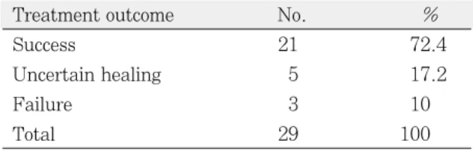
Results: The overall success rate was 72.4% for the 29 intentionally replanted C-shaped mandibular sec- ond molars. The success rate of replanted teeth with preoperative periapical lesions was similar to that of replanted teeth which have no periapical lesions.
Conclusions: Therefore, root canal treatment failure on C-shaped mandibular second molar can be pre- dictably treated by intentional replantation regardless of the presence of periapical lesion. [J Kor Acad Cons Dent 2011;36(1):19-25.]
Key words:C-shaped root canal; Intentional replantation; Mandibular second molar; Preoperative periapi- cal lesions; Success rate
-Received 2 October 2010; revised 27 October 2010; accepted 2 November 2010- ABSTRACT
Shon WJ, DDS, PhD, Assistant Professor; Kum KY, DDS, PhD, Associate Professor; Baek SH, DDS, PhD, Associate Professor; Lee WC, DDS, PhD, Associate Professor, Dept. of Conservative Dentistry, School of Dentistry, Dental Research Institute, Seoul National University, Seoul, Korea
*Correspondence to Woo-Cheol Lee, DDS, PhD.
Associate Professor, Department of Conservative Dentistry, Dental Research Institute, Seoul National University School of Dentistry, 275-1 Yeongeon-dong, Jongno-gu, Seoul, Korea 110-768
TEL, +82-2-2072-1634; FAX, +82-2-2072-3859; E-mail, [email protected]
C-shaped root canal configuration is difficult to exe- cute a successful root canal treatment because of the anatomical variations in the root canal system.6Since the isthmus-like small canals are connecting the C- shaped fin area, perforation or overinstrumentation may occur during the canal instrumentation. The possibility of the strip perforation in this area can be much higher than in the normal tooth with a diver- gent root shape.5,7 Although the success rate of the root canal treatment for this type of tooth is not clar- ified yet, we suspect that there can be much more treatment failure than the other teeth in the arch because the transverse anastomoses connecting the C- shaped root canal configuration can harbor bacte- ria and infected pulp tissue and as a result, their removal can be extremely difficult.8,9
Intentional replantation is a viable treatment option when original trial of root canal treatment or even retreatment was unsuccessful and also in cases where a dental implant or a surgical treatment is not possible. Peer suggested that intentional replantation should be considered more often as a treatment modality in an effort to maintain the natural denti- tion.10Therefore, intentional replantation is now con- sidered as a reliable and predictable procedure.11 In this aspect, C-shaped mandibular second molar can be a good candidate for the intentional replantation procedure due to its conical shape of the root.9 However, there are no reports on the success rate for this type of tooth when it was intentionally replant- ed. Therefore, the purpose of this study is to evalu- ate the success rate of intentionally replanted mandibular second molar with C-shaped canal sys- tem and to access the impact of preoperative periapi- cal lesion on the success of intentional replantation procedure by retrospective chart review.
Materials and Methods
Data for this study were obtained from the chart of the patients treated at Seoul National University Dental Hospital Department of Conservative Dentistry from January 2005 to December 2007. The study protocol was approved by the Institutional Review Board of the Seoul National University Dental Hospital. Records of all patients were
screened retrospectively for intentional replantation procedures performed on mandibular second molar teeth with radiographically diagnosed to have a root with C-shaped canal system. From the charts of patients receiving intentional replantation treatment in this 3-year period, 52 replantation cases fit the preliminary inclusion criteria. Existence of preopera- tive periapical lesion of the tooth and the reason for the failure of the conventional endodontic treatment were recorded from the patient charts and radi- ographs (Table 1).
Detailed clinical procedure of the intentional replantation was as follows. The tooth was intention- ally extracted with forceps under local anesthesia with 2% Lidocaine. Right after the extraction, the apical area of the tooth was carefully inspected under the operating microscope. Apical 3 mm was resected with high speed diamond bur and the granulation tissue attached to the root surface was carefully removed. Root-end cavity was prepared with #330 bur under the microscopic examination, then the cav- ity was filled with mineral trioxide aggregate (ProrootMTA, Dentsply, Johnson city, TN, USA). All this procedure was done within 10 minutes, and the tooth was placed back to its socket.
All the replanted teeth were evaluated 3, 6, 9 and 12 months after the treatment, and then the case was followed twice a year. The teeth were evaluated according to the clinical criteria and radiographically examined at each observation. Evaluation periods ranged from 6 months to 3 years. The radiographic and clinical data from recall visits were collected and evaluated according to the assessment criteria estab- lished by Rud et al.12and Molven et al.13
Success was defined as complete resolution of the former periapical radiolucency during the follow-up period and the tooth was in function with no symp- toms demonstrated by recorded clinical data.
Uncertain healing was assumed if the teeth were present in the mouth with incomplete healing of peri- apical lesion or with certain degree of clinical symp- toms. A case with unresolved periapical lesion or per- sistent clinical symptoms which needed extraction was considered as a failure.
The effect of the existence of previous periapical lesion on the success of intentional replantation pro-
cedure was analyzed by Chi-square test at a signifi- cance level of p = 0.05.
Results
This retrospective chart review study included 52 replanted mandibular second molar teeth. However,
6 teeth did not meet inclusion criteria for C-shaped root canal configuration as Fan et al. had suggested and another 17 teeth were lost for the 1 year follow- up.14Average follow-up period was 18.6 months.
The overall success rate was 72.4% for the 29 intentionally replanted teeth (Table 2). Teeth with preoperative periapical lesions showed similar Table 1.Summary of the cases for the treatment outcome, presence of preoperative periapical lesion and cause of the root canal treatment failure that required intentional replantation
Case Follow-up
Treatment Previous Reason for
number Age/sex Tooth periods
outcome periapical re-treatment failure
(mon) radiolucency which requires IR
1 71/M 37 24 S Y Post
2 61/F 37 15 U Y Block
3 33/F 47 26 S N Pain
4 24/F 47 18 F* N Curved canal
5 29/M 37 12 S Y Fistula
6 41/F 37 12 U N Block
7 20/F 47 12 S Y Post
8 35/F 37 16 S Y Fistula
9 16/F 37 16 S Y Pain
10 59/F 47 6 F* Y Block
11 35/F 37 12 S Y Post
12 30/M 37 16 F* Y Pain
13 33/M 37 28 S Y Post
14 46/F 47 6 F* Y Verforation
15 69/M 47 21 S Y Pain
16 22/F 47 16 U Y Block
17 48/M 37 18 S N Root resorption
18 34/F 37 12 S Y Block
19 35/M 47 36 S Y Pain
20 26/F 47 15 S Y Block
21 22/F 37 24 S Y Post
22 18/F 37 18 S Y Block
23 54/M 47 15 S N Block
24 37/F 37 28 S N Block
25 17/M 47 12 S Y Post
26 34/F 47 24 S Y Post
27 62/F 47 12 U Y Block
28 28/M 37 36 S N Pain
29 26/F 37 32 S Y Overfilling
IR, intentional replantation; M, male; S, success; Y, presence of preoperative periapical lesion; F, female; U, uncertain healing; N, no periapical lesion; F*, failure.
Discussion
The overall success rate of intentional replantation for C-shaped mandibular second molar was 72.4%.
This result was slightly lower than the success rate of intentional replantation reported in other stud-
ies.15,16 This can be explained by the fact that most
studies included all tooth types in the arch. When only molar tooth of intentional replantation was con- sidered, a success rate of 72% was reported by Raghoebar and Vissink,17 and it was comparable to that of our study.
According to Jerome, a root with C-shaped configu- ration is a good candidate for the intentional replan- tation procedure because the extraction of a tooth with this type of root can be relatively easy.9 In regard to this, Cotter and Panzarino reported one intentional replantation case which was proved to be a C-shaped mandibular second molar.18 Super EBA was used as a root-end filling material in their case and it was successfully maintained after 1 year fol- low-up.
In this study, MTA was used for the root end-fill- ing. Although no confirmed protocol was set for the root-end filling material of the intentional replanta- tion upto now, there were multiple reasons for using MTA during the intentional replantation procedure.
Recent prospective clinical study19demonstrated that periapical surgery using MTA as a root-end filling showed high success rate. A meta-analysis20 of root- end filling materials also reported that MTA was
associated with the highest success when it was com- pared with IRM, Super-EBA or amalgam. The fact that the success rate of this study was similar to the weighted average success rate of periapical surgery21 was a good evidence for the use of MTA as a root-end filling during the intentional replantation procedure.
Pre-operative recognition of the C-shaped root canal configuration can facilitate diagnosis and treat- ment decision making process.22 For this purpose, radiographs can be the best practical methods to pro- vide clues about the morphology of the root canal system. Recent study23 suggested that the presence and the configuration of C-shaped root canal system could be predicted by the periapical radiographic appearance. In this respect, Calsen demonstrated that 76.3% of the mandibular second molar with one-root shape showed a good concordance with the actual canal configuration.24 Computed tomography (CT) can be a good diagnostic aid before the inten- tional replantation is performed. In fact, Jin et al.
demonstrated that more than 44.5% of mandibular second molars were found to have C-shaped canals by CT analysis in Korean population.5
Due to its high cost, we could not take CT for each patient in this study. From the 52 teeth which were expected to have C-shaped canal configurations by the preoperative radiographic root morphology, 6 teeth did not meet the inclusion criteria because the tooth qualified as having a C-shaped canal system had to exhibit all three following features: fused roots, a longitudinal groove on the lingual or buccal success rate when it was campared with that of
replanted teeth which have no preoperative periapi- cal lesions (Table 3). The most frequent reason for re-treatment failure which requires intentional replantation was the blockage of the root canals.
Table 3.The influence of preoperative periapical lesion on the success
Treatment outcome Success Uncertain Failure Total
With previous lesion 16 (72.7%) 4 (18.2%) 2 (9.1%) 22
Without lesion 5 (71.4%) 1 (14.3%) 1 (14.3%) 7
Table 2.Outcome of intentional replantation
Treatment outcome No. %
Success 21 72.4
Uncertain healing 5 17.2
Failure 3 10
Total 29 100
surfaces of the root, and at least one cross-section of the canal belong to the C1, C2, or C3 configuration according to the Fan’s classification.14 This was because of the superimposition of the mandibular alveolar bone, even though preoperative radiographs indicated the presence of a C-shaped root.22,25,26
In this study, the main reason for the failure of the intentional replantation of C-shaped root canal seemed to be the periodontal bone breakdown (data not shown). Ricucci et al. demonstrated that peri- odontal involvement was the cause of failure when conventional root canal treatment of C-shaped mandibular molar cases was followed-up for 1 year.27 When intentional replantation procedure was consid- ered, periodontal ligament could be damaged during the extraction and repositioning procedure. In fact, most of the failed cases could not obtain enough sta- bility and in turn, periodontal bone breakdown was detected along the lingual groove. A longitudinal groove located on the lingual surface of this type of the root presumed to transmit periodontal disease from coronal part to the apical area of replanted tooth. Since this groove begins 3.8 mm below the cementoenamel junction,28 if the replanted tooth could not get enough stability, then this groove act as a highway to transport periodontal inflammation directly to the periapical tissue of the replanted tooth.
In the present study, blockage of the C-shaped root canal system was the most frequent reason for the conventional root canal treatment failure requiring additional surgical procedure such as intentional replantation. Canal bifurcation under mesial orifice or MB-D orifice was the main cause of the calcifica- tion and canal blockage.2 Complete negotiation of C- shaped canal system was impossible due to this canal bifurcation especially in the apical third. In fact, 36.4% of the canal bifurcation was present within 2 mm from the apex and the 90% of the canal below the bifurcation was blocked.2 Because of this complex anatomy, it would be extremely difficult to negotiate and debride the root canal system com- pletely. Moreover, NiTi rotary file instrumentation is not useful in shaping fins and isthmus area of the C- shaped canal system.28,29
In conclusion, C-shaped canal exhibits multiple
canal irregularities and provides a challenge in root canal treatment. If the treatment failure occurs on this type of tooth because of the bifurcation or canal blockage, then the intentional replantation can be a good alternative treatment option.
When indicated, root canal treatment failure on C- shaped mandibular second molar tooth can be pre- dictably treated by intentional replantation regard- less of the presence of periapical lesion.
References
1. Jafarzadeh H, Wu Y-N. The C-shaped root canal con- figuration: a review, J Endod 2007;33:517-523.
2. Fan B, Min Y, Lu G, Yang J, Cheung GSP, Gutmann Jl. Negotiation of C-shaped canal systems in mandibu- lar second molars. J Endod 2009;35:1003-1008.
3. Yang ZP, Yang SF, Lin YC, Shay JC, Chi CY. C- shaped root canals in mandibular second molars in a Chinese population. Endod Dent Traumatol 1988;4:
160-163.
4. Seo MS, Park DS. C-shaped root canals of mandibular second molars in a Korean population: clinical observa- tion and in vitro analysis. Int Endod J 2004;37:139- 144.
5. Jin GC, Lee SJ, Roh BD. Anatomical study of C- shaped canals in mandibular second molars by analysis of computed tomography. J Endod 2006;32:10-13.
6. Melton DC, Krell KV, Fuller MW. anatomical and his- tological features of C-shaped canals in mandibular second molars. J Endod 1991;17:384-388.
7. Gao Y, Fan B, Cheung GSP, Gutmann Jl, Fan M. C- shaped canal system in mandibular second molars Part
Ⅳ-3D morphological analysis and transverse measure- ment. J Endod 2006;32:1062-1065.
8. Ingle J, Bakland L: Endodontics. 5th ed. Philadelphia:
Lea & Febiger; 2002, p558.
9. Jerome CE. C-shaped root canal systems: diagnosis, treatment, and restoration. Gen Dent 1994;42:424- 427.
10. Peer M. Intentional replantation - a “last resort”treat- ment or a conventional treatment procedure: Nine case reports. Dent Traumatol 2004;2:48-55.
11. Wolcott J, Rossman LE. Intentional replantation of endodontically treated teeth: an update. Compend Contin Educ Dent 2003;24:68-74.
12. Rud J, Andreasen JO, Jensen JE. Radiographic criteria for the assessment of healing after endodontic surgery.
Int J Oral Surg 1972;1:195-214.
13. Molven O, Halse A, Grung B. Observer strategy and the radiographic classification of healing after endodon- tic surgery. Int J Oral Maxillofac Surg 1987;16:432- 439.
14. Fan B, Cheung GSP, Fan M, Gutmann JL, Bian Z. C- shaped canal system in mandibular second molars:
Part I-anatomical features. J Endod 2004;30:899-903.
15. Bender IB, Rossman LE. Intentional replantation of endodontically treated teeth. Oral Surg Oral Med Oral Pathol 1993;76:623-630.
16. Kingsbury BC Jr, Wiesenbaugh JM Jr. Intentional
replantation of mandibular premolars and molars. J Am Dent Assoc 1971;83:1053-1057.
17. Raghoebar GM, Vissink A. Results of intentional replantation of molars. J Oral Maxillofac Surg 1999;
57:240-244.
18. Cotter MR, Panzarino J. Intentional replantation: a case report. J Endod 2006;32:579-582.
19. Saunders WP. A prospective clinical study of periradic- ular surgery using mineral trioxide aggregate as a root- end filling. J Endod 2008;34:660-665.
20. Fernandez-Yanez SA, Leco-Berrocal MI, Martinez- Gonzalez JM. Metaanalysis of filler materials in peri- apical surgery. Med Oral Patol Oral Cir Bucal 2008;
13:E180-185.
21. Torabinejad M, Corr R, Handysides R, Shabahang S.
Outcomes of nonsurgical retreatment and endodontic surgery: a systematic review. J Endod 2009;35:930- 937.
22. Fan W, Fan B, Gutmann JL, Cheung GSP. Identification of C-shaped canal in mandibular second molars: Part I - radiographic and anatomical features revealed by intraradicular contrast medium. J Endod 2007;33:806- 810.
23. Fan B, Cheung GSP, Fan M, Gutmann JL, Fan W. C- shaped canal system in mandibular second molars:
Part II - radiographic features. J Endod 2004;30:904- 908.
24. Calsen O. Root complex and root canal system: a cor- relation analysis using one-rooted mandibular second molars. Scand J Dent Res 1990;98:273-285.
25. Fan W, Fan B, Gutmann JL, fan M. Identification of C-shaped canal in mandibular second molars - Part III: Anatomical features revealed by digital subtraction radiography. J Endod 2008;34:1187-1190.
26. Fan B, Gao Y, Fan W, Gutmann JL. Identification of C-shaped canal in mandibular second molars - Part II:
the effect of bone image superimposition and intraradicular contrast medium on radiograph interpre- tation. J Endod 2008;34:160-165.
27. Ricucci D, Pascon EA, Langeland K. Long-term follow- up on C-shaped mandibular molars. J Endod 1996;
22:185-187.
28. Cheung LHM, Cheung GSP. Evaluation of a rotary instrumentation method for C-shaped canals with micro-computed tomography. J Endod 2008;34:1233- 1238.
29. Rodig T, Hulsmann M, Muhge M, Schafer F. Quality of preparation of oval distal root canals in mandibular molars using nickel-titanium instruments. Int Endod J 2002;35:919-928.
국문초록
C-형 근관계를 가진 하악 제2대구치의 의도적 재식술 결과에 대한 후향적 연구
손원준∙금기연∙백승호∙이우철*
서울대학교 치의학대학원 치과보존학교실
연구목적: 인류지질학적으로 동양인에게서 많이 발견되는 C-형 근관계를 가진 하악 제2대구치는 근관치료나 비외과적 재 치료에 실패하는 경우 외과적 재치료 방법으로 의도적 재식술을 시행하게 되는데 그 성공률에 대한 보고가 없었다. 따라서 본 연구의 목적은 C-형 근관계를 가진 하악 제2대구치에 의도적 재식술을 시행한 환자의 기록을 후향적으로 조사하여 치 료 결과에 따른 성공률을 확인할 뿐 아니라 치료전 존재하던 치근단 병소가 성공률에 영향을 미치는 바에 대하여 알아보는 것이다.
연구 재료 및 방법: 연구를 위해 2005년 1월부터 2007년 12월에 걸쳐 서울대학교치과병원 치과보존과에 내원하여 하악 제 2대구치에 의도적 재식술을 받은 52명의 환자 기록을 조사하였으며 7개의 치아는 정기적인 내원검진 기록이 없어서 조사 대 상에서 제외하였고 다른 6개의 치아는 C-형 근관계의 조건에 해당되지 않아 연구에서 제외하였다. 정기 내원후 시행한 임상 검사와 방사선 사진 기록을 후향적으로 검토하여 성공이나 불완전한 치유 또는 실패등의 치료 결과에 대하여 판단하였다.
결과: C-형 근관계를 가진 29개의 하악 제2대구치에 시행한 의도적 재식술의 성공률은 72.4%로 관찰되었으며 치료전 치근 단 병소가 존재한 경우의 성공률은 치근단 병소가 없었던 경우와 유사한 것으로 확인되었다.
결론: 본 연구결과 C-형 근관계를 가진 하악 제2대구치의 근관치료가 실패한 경우 시행하게 되는 의도적 재식술은 술전 병소 의 존재 유무에 상관없이 치근단 수술의 성공률과 유사한 결과를 얻을 수 있는 치료라고 사료된다.
주요단어: 성공률; 술전 병소; 의도적 재식술; 하악 제2대구치; C-형 근관계