Background and Purpose This study compared the muscle thickness (MT) and echo in- tensity (EI) of the abdominal, thigh, and lower leg muscles between the paretic and nonparetic sides in chronic stroke survivors.
Methods Thirty-two stroke survivors living in the community participated in this study. The MT and EI, which are indicators of muscle mass and intramuscular fat or connective tissue, were assessed in the rectus abdominis, external oblique, internal oblique, transversus abdomi- nis, rectus femoris, vastus intermedius, vastus lateralis, vastus medialis, tibialis anterior, gastroc- nemius, and soleus via transverse ultrasound imaging. In addition, a possible indicator of physi- cal activity—the frequency of going out per week—was evaluated.
Results All quadriceps muscles and the tibialis anterior were significantly thinner and the EI values of the vastus intermedius, vastus lateralis, vastus medialis, and soleus were significantly higher in the paretic limb than the nonparetic limb. The MT and EI values of abdominal mus- cles did not differ significantly between the two sides. The MT values of the paretic rectus femoris, vastus lateralis, and vastus medialis were significantly associated with the frequency of going out after adjusting confounding factors. The MT of the nonparetic vastus lateralis was significantly associated with latency from stroke onset after adjusting confounding factors.
Conclusions Our results indicate that quantitative and qualitative changes on the paretic side in stroke survivors were the most robust in the thigh muscles, whereas such changes might not occur in the abdominal muscles.
Key Words muscle thickness, echo intensity, ultrasound, stroke survivors.
Muscle Thickness and Echo Intensity of the Abdominal and Lower Extremity Muscles in Stroke Survivors
INTRODUCTION
Several recent studies have found that stroke induces sarcopenia and other changes in skeletal muscle.1,2 A meta-analysis of muscle atrophy revealed that the muscle mass is sig- nificantly lower in paretic limbs than nonparetic limbs.3 A loss of muscle mass in the lower limbs following a stroke leads to a decrease in muscle strength of the knee extensors,4,5 and so it is important to prevent any such loss of muscle mass.
Paretic limbs reportedly exhibit not only a decrease in muscle mass (i.e., change in mus- cle quantity) but also an increase in intramuscular fat and other noncontractile tissue (i.e., change in muscle quality). The increase in intramuscular fat is related to a reduction in mus- cle strength independent of the loss of the muscle mass,6,7 which indicates the importance of investigating both the muscle quantity and quality in stroke patients. Previous studies utilizing computed tomography4,8 and MRI9 found that there is more intramuscular fat in the thigh and lower leg on the paretic side than the nonparetic side. Several recent studies investigated the qualitative changes in muscle in stroke survivors using ultrasound imaging as a noninvasive and safe method.10,11 A higher echo intensity (EI) indicates increased in- Hiroki Monjoa
Yoshihiro Fukumotob Tsuyoshi Asaib Hisato Shuntohb
a Graduate School of Medical Rehabilitation, Kobe Gakuin University, Kobe, Japan
b Faculty of Rehabilitation, Kobe Gakuin University, Kobe, Japan
pISSN 1738-6586 / eISSN 2005-5013 / J Clin Neurol 2018;14(4):549-554 / https://doi.org/10.3988/jcn.2018.14.4.549
Received March 12, 2018 Revised June 24, 2018 Accepted June 27, 2018 Correspondence Hiroki Monjo, MS Graduate School of Medical Rehabilitation, Kobe Gakuin University, 518 Ikawadanicho, Arise, Nishi-ku, Kobe 651-2180, Japan
Tel +81-78-9742461 Fax +81-78-9742461 E-mail [email protected]
cc This is an Open Access article distributed under the terms of the Creative Commons Attribution Non-Com- mercial License (https://creativecommons.org/licenses/by-nc/4.0) which permits unrestricted non-commercial use, distribution, and reproduction in any medium, provided the original work is properly cited.
JCN
Open Access ORIGINAL ARTICLEMuscle Thickness and Echo Intensity in Stroke
JCN
tramuscular fat and connective tissues,12 and so ultrasound imaging represents an easily accessible and suitable tech- nique for assessing the characteristics of individual muscles at various anatomical sites. Lee et al.10 found that the EI of the biceps brachii was higher in the paretic limb than the nonparetic limb in stroke survivors. More recently, Berenpas et al.11 reported that the EI values of the biceps brachii, fore- arm flexors, and medial head of the gastrocnemius were higher in paretic limbs than nonparetic limbs in stroke sur- vivors. However, while qualitative changes in the upper and lower extremities in stroke survivors have been reported, to the best of our knowledge the qualitative changes in ab- dominal muscles in stroke survivors have not been exam- ined previously.
It has been reported that the strength of trunk muscles is impaired in stroke patients,13 and that the ability to control the trunk after stroke is predictive of the future functional ability in performing the activities of daily living.14 It is therefore im- portant to determine the quantitative and qualitative changes in abdominal muscles in stroke survivors. Measuring the mus- cle thickness (MT) and EI in both the abdominal and lower extremity regions simultaneously would clarify the differenc- es in quantitative and qualitative muscle changes among dif- ferent anatomical sites in stroke survivors. The present study therefore investigated MT and EI in the abdominal region, thigh, and lower leg in community-dwelling stroke survivors.
The obtained information may be used to develop rehabili- tation strategies for stroke survivors that could improve muscle function in their trunk and lower extremities.
METHODS
This study recruited stroke survivors living in the commu- nity and receiving ongoing rehabilitation care at day service centers. The protocol was approved by the ethics committee of Kobe Gakuin University Graduate School (IRB No. HEB2015 1202-1). Written informed consent was obtained from all participants before collecting any data. The following inclu- sion criteria were used for selecting participants: independent ambulation, unilateral stroke, and at least 6 months after stroke. Patients were excluded if they had dementia, ortho- pedic or chronic pain conditions, severe sensory impairment, or spasticity. The hemiparetic severity was measured using Lower-Extremity Fugl-Meyer (LE-FM) evaluations.15,16 The present study only used synergy items (22 points) of LE-FM evaluations to grade the hemiparetic severity.16 In addition, a possible indicator of physical activity—the frequency of going out per week—was evaluated by asking the participants the fol- lowing question: “How many times do you go out per week?’’
Ultrasound measurements
MT and EI were measured using B-mode ultrasound imag- ing (LOGIO e, GE Healthcare UK, Chalfont, Buckingham- shire, England) with a multifrequency linear transducer (8–
12 MHz). A gain of 58 dB and a dynamic range of 78 dB were applied for the measurements performed on all muscles. Dy- namic focusing was applied to the appropriate depth for the muscle of interest. The following 11 muscles were assessed on both the paretic and nonparetic sides:17-19 the rectus ab- dominis, external oblique, internal oblique, transversus ab- dominis, rectus femoris, vastus intermedius, vastus lateralis, vastus medialis, tibialis anterior, gastrocnemius, and soleus.
The measurement site of each muscle and the patient po- sition during the measurement are shown in Fig. 1. The MT of each abdominal muscle increases during expiration, and so recordings were made at a consistent time point at the end of relaxed expiration during the measurements of abdominal MT.19 An adequate amount of contact gel was applied to the skin to avoid excessive compression of the dermal surface by the probe. The EI was evaluated by analyzing images dis- playing 256 grayscale levels using Image-J (version 1.37; Na- tional Institutes of Health, Bethesda, MD, USA), with the EI of each muscle expressed as a value between 0 (black) and 255 (white). All measurements were performed by the same investigator in order to minimize interobserver variations.
Statistical analyses
Statistical analyses were performed using SPSS (version 20.0;
SPSS Japan, Tokyo, Japan). Differences in all variables be- tween the paretic and nonparetic sides were determined us- ing paired Student’s t-tests. For muscles for which the MT and EI differed significantly between the paretic and nonparetic sides, partial correlations were performed to evaluate the re- lationship with either the frequency of going out or latency from stroke onset, while considering the possible confound- ing factors of age, sex, body mass index (BMI), LE-FM score, and latency from stroke onset or frequency of going out. The criterion for statistical significance was p<0.05.
RESULTS
A total of 32 stroke survivors (21 men and 11 women) aged 71.3±10.2 years (mean±SD; range, 43–87 years) participated.
Their height, weight, and BMI were 1.62±0.08 m, 60.2±10.6 kg, and 23.0±3.3 kg/m2, respectively. The latency from stroke onset was 68.2±60.5 months, the LE-FM score was 15.2±4.5 points, and the frequency of going out was 3.7±1.7 times per week.
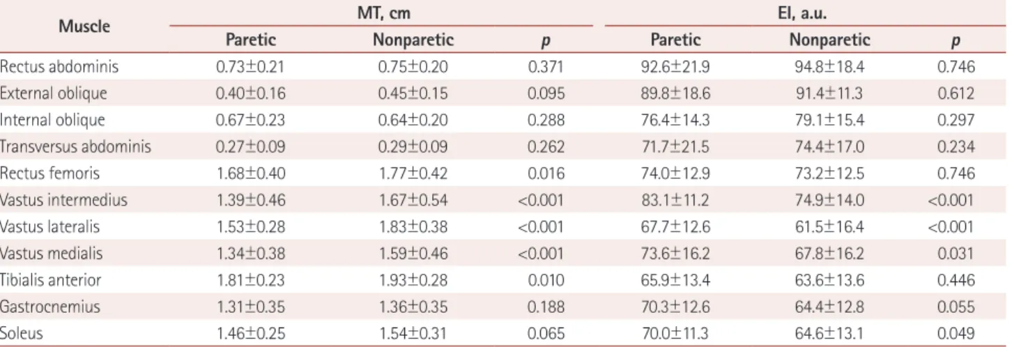
Table 1 lists the MT and EI values of the abdominal and lower limb muscles on the paretic and nonparetic sides. Re-
Monjo H et al.
JCN
garding MT, the rectus femoris, vastus intermedius, vastus lateralis, vastus medialis, and tibialis anterior were signifi- cantly thinner on the paretic side (p<0.05). The gastrocnemius,
soleus, and all abdominal muscles showed no significant dif- ferences between the paretic and nonparetic sides. The EI values of the vastus intermedius, vastus lateralis, vastus me-
A
E
B
F
C
G
D
Fig. 1. Muscle ultrasound images and measurement sites and positions. A: Rectus abdominis, at 3 cm lateral to the umbilicus, with the patient supine. B: External oblique, internal oblique, and transverse abdominis, at 2.5 cm anterior to the midaxillary line at the midpoint between the infe- rior rib and the iliac crest, with the patient supine. C: Rectus femoris and vastus intermedius, at midway between the anterior superior iliac supine and the proximal end of the patella, with the patient supine. D: Vastus lateralis, at midway between the great trochanter and the lateral condyle of the tibia, with the patient supine. E: Vastus medialis, at 30% proximal between the great trochanter and the lateral condyle of the tibia, with the pa- tient supine. F: Tibialis anterior, at 30% proximal between the lateral malleolus of the fibula and the lateral condyle of the tibia, with the patient su- pine. G: Gastrocnemius, at the medial head of the gastrocnemius at 30% proximal between the lateral malleolus of the fibula and the lateral condyle of the tibia, with the patient sitting. Soleus, at 30% proximal between the lateral malleolus of the fibula and the lateral condyle of the tibia, with the patient sitting.
Table 1. MT and EI values of abdominal and lower limb muscles on the paretic and nonparetic sides in stroke patients
Muscle MT, cm EI, a.u.
Paretic Nonparetic p Paretic Nonparetic p
Rectus abdominis 0.73±0.21 0.75±0.20 0.371 92.6±21.9 94.8±18.4 0.746
External oblique 0.40±0.16 0.45±0.15 0.095 89.8±18.6 91.4±11.3 0.612
Internal oblique 0.67±0.23 0.64±0.20 0.288 76.4±14.3 79.1±15.4 0.297
Transversus abdominis 0.27±0.09 0.29±0.09 0.262 71.7±21.5 74.4±17.0 0.234
Rectus femoris 1.68±0.40 1.77±0.42 0.016 74.0±12.9 73.2±12.5 0.746
Vastus intermedius 1.39±0.46 1.67±0.54 <0.001 83.1±11.2 74.9±14.0 <0.001
Vastus lateralis 1.53±0.28 1.83±0.38 <0.001 67.7±12.6 61.5±16.4 <0.001
Vastus medialis 1.34±0.38 1.59±0.46 <0.001 73.6±16.2 67.8±16.2 0.031
Tibialis anterior 1.81±0.23 1.93±0.28 0.010 65.9±13.4 63.6±13.6 0.446
Gastrocnemius 1.31±0.35 1.36±0.35 0.188 70.3±12.6 64.4±12.8 0.055
Soleus 1.46±0.25 1.54±0.31 0.065 70.0±11.3 64.6±13.1 0.049
EI: echo intensity, MT: muscle thickness.
Muscle Thickness and Echo Intensity in Stroke
JCN
dialis, and soleus were significantly higher on the paretic side (p<0.05). The EI values of the rectus femoris, tibialis anterior, gastrocnemius, and all abdominal muscles did not differ sig- nificantly between the paretic and nonparetic sides.
Correlation analysis revealed that the frequency of going out was significantly correlated with the MT values of the paretic rectus femoris (r=0.421, p=0.026), vastus intermedius (r=0.582, p=0.001), and vastus lateralis (r=0.439, p=0.019) af- ter adjusting for age, sex, BMI, and latency from stroke onset.
Latency from stroke onset was significantly correlated with the MT of the nonparetic vastus lateralis (r=0.487, p=0.009) after adjusting for age, sex, BMI, and frequency of going out.
The other muscles were not significantly correlated with the frequency of going out or the latency from stroke onset.
DISCUSSION
To the best of our knowledge, the present study is the first to investigate changes in muscle quantity and quality in the ab- dominal, thigh, and lower leg regions using ultrasound in chronic stroke patients. It was found that the MT values of the quadriceps femoris and tibialis anterior were lower and the EI values of the vastus intermedius, vastus lateralis, vastus medialis, and soleus were higher in the paretic limb than the nonparetic limb. Conversely, none of the abdominal muscles showed any significant changes in either MT or EI. Addi- tionally, we observed significant associations between the MT values of the paretic rectus femoris, vastus intermedius, and vastus lateralis and the frequency of going out, as well as between the MT of the nonparetic vastus lateralis and the la- tency from stroke onset.
Previous studies have found lower muscle mass and high- er intramuscular fat or noncontractile tissue in the lower ex- tremities in stroke survivors.4,8,9,11 The present study found significant differences between the sides of the lower leg mus- cles only for the MT of the tibialis anterior and the EI of the soleus. In contrast, there were substantial differences for the quadriceps, particularly in the vastus muscles, for which the MT was 13.3–14.9% lower and EI was 11.3–13.9% higher on the paretic side.
Atrophy of the quadriceps muscles in stroke survivors might be associated with a reduction in physical activity. The quad- riceps are the lower limb muscles that are particularly suscep- tible to age-related loss of muscle mass.20-22 Abe et al.21,22 spec- ulated that a reduction in physical activity with advancing age may contribute to the loss of muscle mass in the quadriceps.
Physical activity is significantly lower in stroke survivors than in healthy older individuals23 and is associated with physical functioning,24 muscle cross-sectional area,25 con- centric strength,26 and the prevention of secondary complica-
tions.27 Hachisuka et al.25 found a strong correlation between physical activity—quantified as the number of steps per day—
and muscle fiber diameter of the vastus lateralis in stroke sur- vivors. Therefore, the thinner quadriceps femoris and its high- er EI observed in the present study might have been caused by reduced physical activity. The frequency of going out per week was positively correlated with the MT values of the rec- tus femoris, vastus intermedius, and vastus lateralis on the pa- retic side after adjusting for age, sex, BMI, latency from stroke onset, and LE-FM score. In healthy adults, the weekly fre- quency of performing activities including walking, exercise, and sport has been used as a measure of physical activity.28-30 Although the present study did not ask the purpose of going out, it is possible that the frequency of going out represents a simple alternative parameter for measuring the extent of physical activity in stroke survivors. However, the EI values of the quadriceps and other muscles were not found to be relat- ed to the frequency of going out. Clarifying the association between physical activity with EI may require the use of a measurement device for actual physical activity (e.g., an ac- celerometer) in future studies. It was particularly interesting that a positive association was also observed between the MT of the vastus lateralis on the nonparetic side and the latency from stroke onset. It is possible that the long-term gait pat- tern associated with lower limb asymmetry31,32 induces hy- pertrophy of the nonparetic vastus lateralis; this possibility should also be examined in future studies.
One strength of this study was related to it measuring the EI of the abdominal muscles. Kim et al.33 reported that the MT did not differ significantly between the paretic and non- paretic sides, which is consistent with the present study find- ing that the MT and EI of the abdominal muscles did not differ significantly between the two sides. The absence of a significant difference in the abdominal muscles could be at- tributed to them being innervated bilaterally. Previous stud- ies have found the abdominal muscles to be spared following a stroke due to them being innervated bilaterally from the motor cortex.34,35 Therefore, if a cerebral lesion occurs on one side only, the abdominal muscles might be not affected. How- ever, Bohannon et al.13 reported that the strength of trunk muscles is impaired in stroke patients compared to healthy controls, which indicates the need to compare the MT and EI values of abdominal muscles between stroke survivors and healthy controls.
Our study was subject to some limitations. First, the num- ber of subjects was small, and so type 2 errors might have been present. Second, our cross-sectional data did not allow us to identify any causal relationship between the frequency of go- ing out and the MT or EI in the paretic leg. Third, it is not clear whether the frequency of going out reflects physical ac-
Monjo H et al.
JCN
tivity. Longitudinal studies are needed to identify whether objectively measured physical activity contributes to future changes in MT and EI in a large number of stroke survivors.
In conclusion, a thinner quadriceps and tibialis anterior and a higher EI of the vastus intermedius, vastus lateralis, vastus medialis, and soleus were observed in the paretic limb, with no significant changes in MT or EI of the abdominal muscles in stroke survivors. In addition, the MT values of the rectus femoris, vastus intermedius, and vastus lateralis on the paretic side were associated with the frequency of going out, while that of the vastus lateralis on the nonparetic side was associated with the latency from stroke onset. Although fur- ther investigation is required to confirm our findings, the pres- ent results provide valuable information for assessing and maintaining the abdominal and lower extremity muscles in stroke survivors.
Conflicts of Interest
The authors have no financial conflicts of interest.
Acknowledgements
The authors thank Hiroki Kubo, Akihiko Kasuya, Kensuke Oshima, Hi- rotsugu Tajitsu, and Shota Koyama (Graduate School of Medical Rehabili- tation, Kobe Gakuin University) for their practical and technical assis- tance. The authors also acknowledge the superb support provided to staff members at the ‘ARUKU Studio REHA-REHA’, the ‘RE=VAL studio’ Day Service Center, and the Miyabinosato Rehabilitation Center.
REFERENCES
1. Scherbakov N, von Haehling S, Anker SD, Dirnagl U, Doehner W.
Stroke induced sarcopenia: muscle wasting and disability after stroke.
Int J Cardiol 2013;170:89-94.
2. Hafer-Macko CE, Ryan AS, Ivey FM, Macko RF. Skeletal muscle changes after hemiparetic stroke and potential beneficial effects of ex- ercise intervention strategies. J Rehabil Res Dev 2008;45:261-272.
3. English C, McLennan H, Thoirs K, Coates A, Bernhardt J. Loss of skel- etal muscle mass after stroke: a systematic review. Int J Stroke 2010;5:
395-402.
4. Ryan AS, Buscemi A, Forrester L, Hafer-Macko CE, Ivey FM. Atrophy and intramuscular fat in specific muscles of the thigh: associated weak- ness and hyperinsulinemia in stroke survivors. Neurorehabil Neural Re- pair 2011;25:865-872.
5. Prado-Medeiros CL, Silva MP, Lessi GC, Alves MZ, Tannus A, Lindquist AR, et al. Muscle atrophy and functional deficits of knee ex- tensors and flexors in people with chronic stroke. Phys Ther 2012;92:
429-439.
6. Goodpaster BH, Carlson CL, Visser M, Kelley DE, Scherzinger A, Harris TB, et al. Attenuation of skeletal muscle and strength in the el- derly: the Health ABC Study. J Appl Physiol 2001;90:2157-2165.
7. Fukumoto Y, Ikezoe T, Yamada Y, Tsukagoshi R, Nakamura M, Mori N, et al. Skeletal muscle quality assessed from echo intensity is asso- ciated with muscle strength of middle-aged and elderly persons. Eur J Appl Physiol 2012;112:1519-1525.
8. Ryan AS, Dobrovolny CL, Smith GV, Silver KH, Macko RF. Hemiparet- ic muscle atrophy and increased intramuscular fat in stroke patients.
Arch Phys Med Rehabil 2002;83:1703-1707.
9. Ramsay JW, Barrance PJ, Buchanan TS, Higginson JS. Paretic muscle atrophy and non-contractile tissue content in individual muscles of the post-stroke lower extremity. J Biomech 2011;44:2741-2746.
10. Lee SS, Spear S, Rymer WZ. Quantifying changes in material proper- ties of stroke-impaired muscle. Clin Biomech (Bristol, Avon) 2015;30:
269-275.
11. Berenpas F, Martens AM, Weerdesteyn V, Geurts AC, van Alfen N. Bi- lateral changes in muscle architecture of physically active people with chronic stroke: a quantitative muscle ultrasound study. Clin Neuro- physiol 2017;128:115-122.
12. Pillen S, Tak RO, Zwarts MJ, Lammens MM, Verrijp KN, Arts IM, et al. Skeletal muscle ultrasound: correlation between fibrous tissue and echo intensity. Ultrasound Med Biol 2009;35:443-446.
13. Bohannon RW, Cassidy D, Walsh S. Trunk muscle strength is impaired multidirectionally after stroke. Clin Rehabil 1995;9:47-51.
14. Hsieh CL, Sheu CF, Hsueh IP, Wang CH. Trunk control as an early predictor of comprehensive activities of daily living function in stroke patients. Stroke 2002;33:2626-2630.
15. Dettmann MA, Linder MT, Sepic SB. Relationships among walking performance, postural stability, and functional assessments of the hemiplegic patient. Am J Phys Med 1987;66:77-90.
16. Duncan PW, Propst M, Nelson SG. Reliability of the Fugl-Meyer as- sessment of sensorimotor recovery following cerebrovascular accident.
Phys Ther 1983;63:1606-1610.
17. Hebert JJ, Koppenhaver SL, Parent EC, Fritz JM. A systematic review of the reliability of rehabilitative ultrasound imaging for the quanti- tative assessment of the abdominal and lumbar trunk muscles. Spine 2009;34:E848-E856.
18. Koppenhaver SL, Hebert JJ, Fritz JM, Parent EC, Teyhen DS, Magel JS. Reliability of rehabilitative ultrasound imaging of the transversus abdominis and lumbar multifidus muscles. Arch Phys Med Rehabil 2009;90:87-94.
19. Ikezoe T, Nakamura M, Shima H, Asakawa Y, Ichihashi N. Associa- tion between walking ability and trunk and lower-limb muscle atrophy in institutionalized elderly women: a longitudinal pilot study. J Physiol Anthropol 2005;28:34-31.
20. Miyatani M, Kanehisa H, Azuma K, Kuno S, Fukunaga T. Site-related differences in muscle loss with aging. Int J Sport Health Sci 2003;1: 34- 21. Abe T, Sakamaki M, Yasuda T, Bemben MG, Kondo M, Kawakami Y, 40.
et al. Age-related, site-specific muscle loss in 1507 Japanese men and women aged 20 to 95 years. J Sports Sci Med 2011;10:145-150.
22. Abe T, Loenneke JP, Thiebaud RS, Fukunaga T. Age-related site-spe- cific muscle wasting of upper and lower extremities and trunk in Jap- anese men and women. Age (Dordr) 2014;36:813-821.
23. English C, Healy GN, Coates A, Lewis L, Olds T, Bernhardt J. Sitting and activity time in people with stroke. Phys Ther 2016;96:193-201.
24. Verschuren O, Mead G, Visser-Meily A. Sedentary behaviour and stroke: foundational knowledge is crucial. Transl Stroke Res 2015;6:9- 25. Hachisuka K, Umezu Y, Ogata H. Disuse muscle atrophy of lower 12.
limbs in hemiplegic patients. Arch Phys Med Rehabil 1997;78:13-18.
26. Eng JJ, Lomaglio MJ, Macintyre DL. Muscle torque preservation and physical activity in individuals with stroke. Med Sci Sports Exerc 2009;
41:1353-1360.
27. Billinger SA, Arena R, Bernhardt J, Eng JJ, Franklin BA, Johnson CM, et al. Physical activity and exercise recommendations for stroke survi- vors: a statement for healthcare professionals from the American Heart Association/American Stroke Association. Stroke 2014;45:2532- 2553.
28. Katano H, Ohno M, Yamada K. Protection by physical activity against deleterious effect of smoking on carotid intima-media thickness in young Japanese. J Stroke Cerebrovasc Dis 2013;22:176-183.
29. Laurin D, Verreault R, Lindsay J, MacPherson K, Rockwood K. Physi- cal activity and risk of cognitive impairment and dementia in elderly persons. Arch Neurol 2001;58:498-504.
30. Lytle ME, Vander Bilt J, Pandav RS, Dodge HH, Ganguli M. Exercise level and cognitive decline: the MoVIES project. Alzheimer Dis Assoc
Muscle Thickness and Echo Intensity in Stroke
JCN
Disord 2004;18:57-64.
31. Laufer Y, Sivan D, Schwarzmann R, Sprecher E. Standing balance and functional recovery of patients with right and left hemiparesis in the early stages of rehabilitation. Neurorehabil Neural Repair 2003;17:207- 32. Patterson KK, Gage WH, Brooks D, Black SE, McIlroy WE. Changes 213.
in gait symmetry and velocity after stroke: a cross-sectional study from weeks to years after stroke. Neurorehabil Neural Repair 2010;24:783- 790.
33. Kim HD, You JM, Han N, Eom MJ, Kim JG. Ultrasonographic mea- surement of transverse abdominis in stroke patients. Ann Rehabil Med 2014;38:317-326.
34. Adams RW, Gandevia SC, Skuse NF. The distribution of muscle weakness in upper motoneuron lesions affecting the lower limb. Brain 1990;113:1459-1476.
35. Carr LJ, Harrison LM, Stephens JA. Evidence for bilateral innervation of certain homologous motoneurone pools in man. J Physiol 1994;475:
217-227.