201 Introduction
Atrial septal defect (ASD) is one of the most common con- genital heart defect found in adults. Percutaneous device clo- sure using Amplatzer ASD occluder (AGA Medical Corp., Golden Valley, MN, USA) is widely used for treatment of ASD and proven to be effective and safe as traditional surgical repair.1) However, procedure or device related complications can occur, which can be potentially fatal.2) We report a rare case of subacute and silent embolization of Amplatzer device to the pulmonary artery, on the next day of successful percuta- neous ASD closure.
Case
A 45-year-old female was admitted due to symptom of dys- pnea developed several months before admission. Transthorac- ic echocardiography (TTE) showed a secundum ASD measur- ing 28 mm anterior-posteriorly at apical 4 chamber view (Fig.
1A). Cardiac catheterization revealed pulmonary hypertension with pulmonary artery systolic pressure 46 mmHg and a large left to right shunt with a Qp/Qs 3.1. Transesophageal echo- cardiography (TEE) was performed and revealed a large secun- dum ASD measuring 27 mm, with sufficient superior vena cava (11 mm), inferior vena cava (14 mm) rim (Fig. 1B) and relative small atrioventricular rim (5 mm) (Fig. 1C). Posterior rim was sufficient in length (13 mm) but relatively thin in na-
ture, and aortic rim was nearly absent (Fig. 1D). Despite rela- tive large size of ASD with insufficient aortic rim, percutane- ous device closure with Amplatzer was planned because she refused surgical treatment.
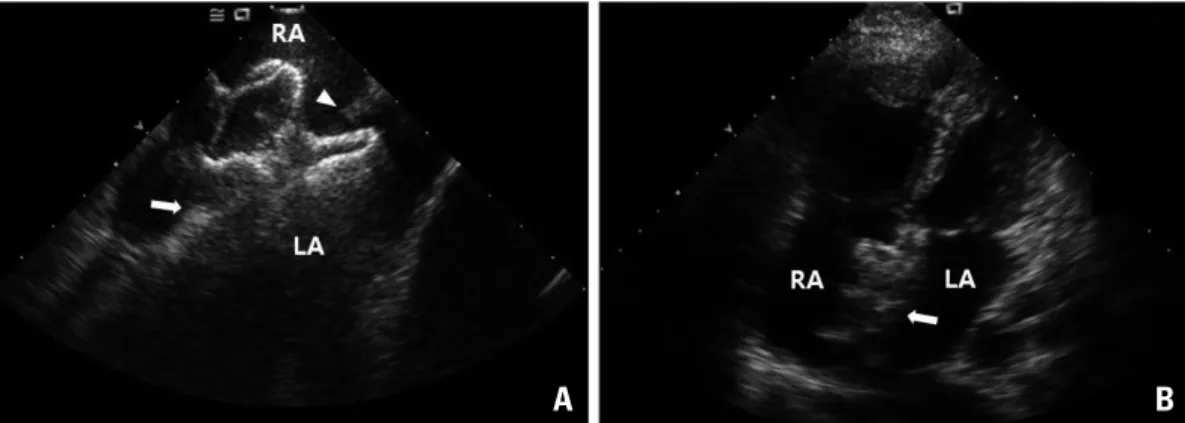
During procedure, intracardiac echocardiography (ICE) was used for guiding intervention instead of TEE as our routine practice. ASD size measured by sizing balloon under fluorosco- py was 26 mm. An oversized 32 mm Amplatzer ASD device was selected because of insufficient aortic rim and deployed successfully after several failure of capturing atrioventricular rim. Prior to final release of the device, a secure and stable posi- tion of the device within the defect was checked by a push-pull maneuver and cessation of flow across the inter-atrial septum was confirmed by ICE and TTE (Fig. 2).
On the day following device closure, she was asymptomatic and routine follow up chest X-ray and TTE was performed.
On chest X-ray, Amplatzer device shadow was found at the main pulmonary trunk area (Fig. 3A) and TTE revealed reap- pearance of the large ASD with embolized Amplatzer device in the ostium of right pulmonary artery (Fig. 3B). The right ventricular systolic pressure was increased to 63 mmHg, but she still remained asymptomatic and hemodynamically stable.
We started intravenous infusion of heparin and asked for emergency surgery to remove the Amplatzer device and repair of the ASD.
pISSN 1975-4612/ eISSN 2005-9655 Copyright © 2012 Korean Society of Echocardiography www.kse-jcu.org http://dx.doi.org/10.4250/jcu.2012.20.4.201
CASE REPORT J Cardiovasc Ultrasound 2012;20(4):201-204
Subacute, Silent Embolization
of Amplatzer Atrial Septal Defect
Closure Device to the Pulmonary Artery
Jang-Won Son, MD1 and Jong-Seon Park, MD, PhD2
1Division of Cardiology, Severance Cardiovascular Hospital, Seoul, Korea
2Division of Cardiology, Department of Internal Medicine, Yeungnam University Medical Center, Daegu, Korea
Embolization of the closure device is a rare but potentially fatal complication of percutaneous atrial septal defect (ASD) closure.
We report a case of 45-year-old woman who underwent ASD device closure with 32 mm Amplatzer device, which was embolized to the pulmonary artery without symptom one day after successful device implantation.
KEY WORDS: Atrial septal defect · Septal occluder device · Embolization.
• Received: July 28, 2012 • Revised: August 20, 2012 • Accepted: November 21, 2012
• Address for Correspondence: Jong-Seon Park, Division of Cardiology, Department of Internal Medicine, Yeungnam University Medical Center, 170 Hyeonchung-ro, Nam-gu, Daegu 705-717, Korea Tel: +82-53-620-3847, Fax: +82-53-654-8386, E-mail: [email protected]
• This is an Open Access article distributed under the terms of the Creative Commons Attribution Non-Commercial License (http://creativecommons.org/licenses/by-nc/3.0) which permits unrestricted non-commercial use, distribution, and reproduction in any medium, provided the original work is properly cited.
Journal of Cardiovascular Ultrasound 20 | December 2012
202
The operation was carried out with median sternotomy. Af- ter cardiopulmonary bypass, pulmonary artery was opened and the Amplatzer occlude was identified in the bifurcation site of pulmonary artery trunk. Some of the marginal tissue in inferoposterial portion of the atrial septum was composed with friable membranous tissue. After removing the thin and friable tissue, the size of ASD was measured up to 30 × 40 mm. The ASD was closed using pericardial patch and she dis-
charged from the hospital on the 5th postoperative day with- out other complications.
Discussion
Since the introduction in 1974, device closure of secundum type ASD is increasing and became an alternative to surgical treatment.3) Although immediate procedural success rate of Amplatzer septal occluder is 95-98%, adverse events includ-
Fig. 1. Large secundum atrial septal defect measuring 28 mm on transthoracic echocardiography (A). Transesophageal echocardiographic findings of sufficient superior and inferior vena caval rim (B) and preserved atrioventricular rim (C).
Posterior rim was sufficient in length but relatively thin in nature, and aortic rim was nearly absent (D). RA: right atrium, LA:
left atrium, IVC: inferior vena caval rim, SVC: superior vena caval rim, RV: right ventricle, AV: atrioventricular rim, SP:
superoposterior rim, P: posterior rim, Ao: aortic rim.
Fig. 2. Intracardiac echocardiographic still image during procedure (A). The Amplatzer device was successfully deployed and its secure and stable position was confirmed by push-pull maneuver. Inferior vena caval rim (white arrow) and superior vena caval rim (white arrowhead) were captured and well positioned between left and right discs. Still frame of apical 4 chamber view by transthoracic echocardiography after procedure (B). Amplatzer occluder (white arrow) is well positioned in interatrial septum without residual shunt. RA: right atrium, LA: left atrium.
C
A A
D
B B
Embolization of Amplatzer Closure Device | Jang-Won Son and Jong-Seon Park
203 ing arrhythmia, cerebral embolism, cardiac tamponade and
device embolization requiring immediate surgical removal can occur.4) Among them, device embolization is a potential life threatening complication requiring immediate removal via percutaneous or surgical intervention. Although the re- ported incidence is 0.01-0.55%, it would be higher in less ex- perienced operators.4-6) The common reasons for the device embolization are undersized ASD device, small left atrium to accommodate the device, inadequate or floppy rim and opera- tor-related technical issues.6) Most of the device embolization occurs during or several days after the procedures.4) Immediate embolization occurs in the procedural field and thought to be caused by malposition or incorrect device size. Undersizing of the device is the most common reason for the embolization in such case.6) However, subacute embolization within several days of the procedure is thought to be associated in large part with aortic rim erosion or floppy septum.7)
In present case, nearly absent aortic rim and large defect size (28 mm) could be one reason for device migration. However, large ASD size and the small or deficient aortic rim itself is not a contraindication and often considered as suitable for de- vice closure with Amplatzer occluder.8)9) Another important reason for the device migration is thin and floppy membra- nous nature of posterial portion of the atrial septum which was confirmed in operation field. Combination of small aortic rim and floppy membranous nature of counterpart rim (infer- oposterior rim) increased the instability of oversized Amp- latzer device and may lead to migration and embolization of device in our patient.
In case of complicated ASD, as our present case, it is often difficult to reconstruct the spatial structure with the use of two dimensional (2D) images. In this occasion, 2D TEE or balloon sizing may not be sufficient and additional imaging
modalities such as cardiac multidetector computed tomogra- phy or three dimensional TEE can be useful complementary option to 2D TEE for morphologic evaluation of ASD and guidance of transcatheter closure.10)11)
About 50% of cases with Amplatzer occluder embolization, percutaneous retrieval is possible by using the devices includ- ing large sheaths, gooseneck snares, or endomyocardial biopsy forcep.12) However, surgical removal and repair of the ASD is more preferable in the situation of inappropriate ASD rims for the second procedure as present case.
In conclusion, application of the strict criteria for selecting the device closure by comprehensive evaluation of ASD, and careful monitoring for the possible delayed embolization of device are mandatory in the case of complicated ASD.
References
1. Jo SS, Han SJ, Jung MJ, Lee SJ, Seol KH, Kim GH, Lee HS, Shin EK, Park IS, Kim SH. Transcatheter closure of atrial septal defect using amplatzer septal occluder. Korean Circ J 2002;32:17-24.
2. Chessa M, Carminati M, Butera G, Bini RM, Drago M, Rosti L, Giamberti A, Pomè G, Bossone E, Frigiola A. Early and late compli- cations associated with transcatheter occlusion of secundum atrial septal de- fect. J Am Coll Cardiol 2002;39:1061-5.
3. King TD, Mills NL. Nonoperative closure of atrial septal defects. Surgery 1974;75:383-8.
4. Majunke N, Bialkowski J, Wilson N, Szkutnik M, Kusa J, Ba- ranowski A, Heinisch C, Ostermayer S, Wunderlich N, Sievert H.
Closure of atrial septal defect with the Amplatzer septal occluder in adults.
Am J Cardiol 2009;103:550-4.
5. Du ZD, Hijazi ZM, Kleinman CS, Silverman NH, Larntz K; Amp- latzer Investigators. Comparison between transcatheter and surgical closure of secundum atrial septal defect in children and adults: results of a multi- center nonrandomized trial. J Am Coll Cardiol 2002;39:1836-44.
6. Levi DS, Moore JW. Embolization and retrieval of the Amplatzer septal occluder. Catheter Cardiovasc Interv 2004;61:543-7.
7. Misra M, Sadiq A, Namboodiri N, Karunakaran J. The ‘aortic rim’
Fig. 3. One day after the procedure, the embolized Amplatzer device was seen in pulmonary artery (white arrow) on chest X-ray (A), which was lodged on right pulmonary artery ostium (white arrow) on transthoracic echocardiography (B). MPA:
main pulmonary artery, RPA: right pulmonary artery, Ao: aorta.
A B
Journal of Cardiovascular Ultrasound 20 | December 2012
204
recount: embolization of interatrial septal occluder into the main pulmonary artery bifurcation after atrial septal defect closure. Interact Cardiovasc Tho- rac Surg 2007;6:384-6.
8. Braga SL, Sousa AG, Pedra CA, Esteves CA, Pedra SR, Fontes VF.
[Clinical efficacy and safety of the percutaneous treatment of secundum atrial septal defect with the Amplatzer occluder]. Arq Bras Cardiol 2004;83 Spec No:7-13.
9. Lin SM, Tsai SK, Wang JK, Han YY, Jean WH, Yeh YC. Supple- menting transesophageal echocardiography with transthoracic echocardiogra- phy for monitoring transcatheter closure of atrial septal defects with attenu- ated anterior rim: a case series. Anesth Analg 2003;96:1584-8.
10. Kijima Y, Taniguchi M, Akagi T, Nakagawa K, Kusano K, Ito H, Sano S. Torn atrial septum during transcatheter closure of atrial septal de- fect visualized by real-time three-dimensional transesophageal echocardiogra- phy. J Am Soc Echocardiogr 2010;23:1222.e5-8.
11. Ko SF, Liang CD, Yip HK, Huang CC, Ng SH, Huang CF, Chen MC. Amplatzer septal occluder closure of atrial septal defect: evaluation of transthoracic echocardiography, cardiac CT, and transesophageal echocar- diography. AJR Am J Roentgenol 2009;193:1522-9.
12. Balbi M, Pongiglione G, Bezante GP. Percutaneous rescue of left ven- tricular embolized amplatzer septal occluder device. Catheter Cardiovasc In- terv 2008;72:559-62.