26
Introduction
Fabry disease (FD) is an X-linked lysosomal storage disor- der caused by α-galactosidase A (α-Gal A) deficiency. Because the disease is X-linked, males are predominantly affected.
This enzyme deficiency leads to widespread deposition of neu- tral glycosphingolipids (mainly globotriaosylceramide and, to a lesser extent, galabiosylceramide) on blood vessel walls throughout the body, resulting in a multiple-system disorder with a wide spectrum of physical signs and symptoms that pre- dominantly affect the central and peripheral nervous systems, skin, heart, kidneys, and eyes.1) In the heart, glycosphingolipids deposition causes progressive left ventricular hypertrophy (LVH) that mimics the morphological and clinical characteris- tics of hypertrophic cardiomyopathy (HCM).2)3)
Enzyme replacement therapy is effective in reversing the microvascular changes in FD by catabolizing the lipid depos- its and improving cardiac function in patients with cardiac in- volvement.4)5) We report a case of FD with end-stage renal dis- ease (ESRD) which was suspected based upon two-dimensional transthoracic echocardiographic finding.
Case
A 44-year-old man was admitted to evaluation of aggravat- ed exertional dyspnea with orthopnea for two weeks in 2010.
He had been diagnosed with ESRD of unknown etiology at age 41 followed by renal transplantation in 2007. He had been admitted for azotemia three times after renal transplantation.
Percutaneous biopsy of the transplanted kidney was performed three times in 2008, 2009, 2010. The findings on renal biopsy were acute rejection, chronic renal calcineurin inhibitor toxicity, antibody mediated rejection and no evidence of FD in the graft kidney. He had been treated with oral immunosuppres- sive agents, including prednisolone (10 mg daily), tacrolimus (4 mg, bid) and mizoribine (50 mg, qd). Vital signs on arrival included a blood pressure of 162/98 mmHg and a regular pulse rate of 73 bpm, and body temperature of 36.5°C. The initial electrocardiogram showed LVH with a strain pattern, ST-T changes in leads II, III, aVF, V3-V6 and short PR inter- val (Fig. 1). Chest radiography demonstrated cardiomegaly (cardiothoracic ratio = 70%) and blunting of both costophren- ic angle (Fig. 2). Laboratory studies revealed that hemoglobin
pISSN 1975-4612/ eISSN 2005-9655 Copyright © 2013 Korean Society of Echocardiography www.kse-jcu.org http://dx.doi.org/10.4250/jcu.2013.21.1.26
CASE REPORT J Cardiovasc Ultrasound 2013;21(1):26-29
Fabry Cardiomyopathy
Jae Yong Yoon, MD, Joon Hyuk Song, MD, Sang Soo Cheon, MD, Hyun Jun Cho, MD, Myung Hwan Bae, MD, Jang Hoon Lee, MD, Dong Heon Yang, MD, Hun Sik Park, MD, Yongkeun Cho, MD and Shung Chull Chae, MD
Department of Internal Medicine, Kyungpook National University Hospital, Daegu, Korea
Fabry disease is a progressive X-linked disorder of glycosphingolipid metabolism caused by a deficiency of the α-galactosidase lysosomal enzyme. The partial or complete deficiency of the lysosomal enzyme leads to an accumulation of neutral glycosphingolipids in the vascular endothelium and visceral tissues throughout the body. In the heart, glycosphingolipids deposition causes progressive left ventricular hypertrophy (LVH). We report a case of Fabry disease which was suspected based upon two-dimensional echocardiographic finding of LVH. A 44-year-old man was admitted to evaluation of aggravated exertional dyspnea of two weeks duration. He had been diagnosed with end-stage renal disease of unknown etiology at age 41 followed by renal transplantation that year. He had been treated with oral immunosuppressive agents. On hospital day two, transthoracic echocardiography revealed concentric LVH. Left ventricular systolic function was preserved but diastolic dysfunction was present. Fabry disease was confirmed by demonstration of a low plasma α-galactosidase A (α-Gal A) activity. Analysis of genomic DNA showed α-Gal A gene mutation.
The patient was diagnosed with Fabry disease.
KEY WORDS: Fabry disease · Alpha-galactosidase A · Cardiomyopathies.
• Received: September 21, 2012 • Revised: November 19, 2012 • Accepted: February 13, 2013
• Address for Correspondence: Dong Heon Yang, Department of Internal Medicine, Kyungpook National University Hospital, 130 Dongdeok-ro, Jung-gu, Daegu 700-721, Korea Tel: +82-53-420-6587, Fax: +82-53-426-2046, E-mail: [email protected]
• This is an Open Access article distributed under the terms of the Creative Commons Attribution Non-Commercial License (http://creativecommons.org/licenses/by-nc/3.0) which permits unrestricted non-commercial use, distribution, and reproduction in any medium, provided the original work is properly cited.
Fabry Cardiomyopathy | Jae Yong Yoon, et al.
27 was 6.2 g/dL, BUN 64.2 mg/dL, creatinine 6 mg/dL, sodium
134 mEq/L, potassium 6.1 mEq/L, and serum N-terminal pro-B type natriuretic peptide level 126043 pg/mL. On hos- pital day two, two-dimensional transthoracic echocardiogra- phy revealed concentric LVH (interventricular septal dimension 23 mm, LV posterior wall dimension 22.8 mm), mimicking non-obstructive HCM (Fig. 3). The interventricular septal di-
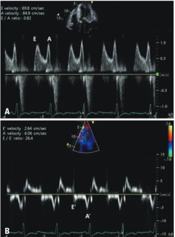
mension and posterior wall dimension was thicker than 3 years ago (interventricular septal dimension 17 mm, LV poste- rior wall dimension 17 mm). And left atrial enlargement was seen (4.5 cm). Left ventricular systolic function was preserved (ejection fraction = 59%), but diastolic dysfunction was pres- ent. Pulsed-wave Doppler recording of mitral inflow revealed a phase resembling an abnormal relaxation diastolic filling pattern, with an ratio between early (E) and late (A) mitral in- flow velocity (E/A) of 0.82 (Fig. 4A). The mitral annulus early diastolic tissue Doppler velocity (E’) and the E/E’ index were 2.64 cm/s and 26.4, respectively, indicating increased LV filling pressure and a pseudonormal pattern (Fig. 4B). The patient was prescribed diuretics for dyspnea and epokine for anemia. And the patient’s condition improved. The patient’s history of early onset ESRD and echocardiographic findings were suggestive of Fabry cardiomyopathy as well as idiopathic HCM. Alpha- galactosidase activity assay was performed. The assay was per- formed by fluorescence assay with 4-methylumbelliferyl and sequencing. The patient was confirmed FD by demonstration of a low plasma α-Gal A activity of 3.8 nmoles/hr/mg (normal mean, 7.5-12.5 nmol/hr/mg). Sequent analysis of genomic DNA showed c.639 + 5G > A [IVS4 (+5)G > A] mutation
Fig. 1. The initial electrocardiogram showed left ventricular hypertrophy with a strain pattern, ST-T changes in leads II, III, aVF, V3-V6.
Fig. 2. Chest radiography. Chest radiography demonstrated cardiome- galy (cardiothoracic ratio = 70%) and blunting of both costophrenic angle.
Fig. 3. Two dimensional echocardiography. Severe concentric left ventricular hypertrophy is shown in a parasternal long-axis view. The interventricular septal dimension (A) was 23 mm and the left ventricular posterior wall dimension (B) was 22.8 mm in thickness.
Fig. 4. Pulse-waved Doppler echocardiography (A) and tissue Doppler echocardiography (B). Decreased mitral annulus velocities (E’) and increased mitral peak Doppler E-wave (E) to peak mitral annulus velocity ratio (E/E’) are seen, suggesting a pseudonormal pattern.
A
B
Journal of Cardiovascular Ultrasound 21 | March 2013
28
in the α-Gal A gene leading to a low plasma α-Gal A activity.
Family screening was done, and his brother was also con- firmed FD by α-Gal A enzyme activity test and renal biopsy.
Enzyme replacement therapy with recombinant α-Gal A was started on an out-patient basis.
Discussion
FD is a progressive X-linked disorder of glycosphingolipid metabolism caused by a deficiency of the α-galactosidase lyso- somal enzyme.1) Overall, the prevalence of FD has been estimat- ed to be 1 in 40000 to 117000 male individuals.6-8) However, several recent studies suggested that the prevalence may be higher in the hemodialysis population, in which values up to 1.2% have been reported.6)9-13) The progressive accumulation of neutral glycosphingolipids in many tissues throughout the body, particularly the vascular endothelium, heart, and kid- ney.1) The manifestation of FD varies and may include angio- keratoma, corneal opacity, acroparesthesia, cerebrovascular dis- ease, ischemic heart disease, and chronic kidney disease. Most men and some women with FD exhibit deterioration of renal function, and many eventually develop ESRD.14)15) Typically the diagnosis of FD is made in male adolescents, but it may be missed or delayed. The “variant” phenotypes usually have a low level of residual α-galactosidase activity resulting in a lack of the classic phenotype. The heart can be the only organ in- volved in male patients with specific gene mutations and in fe- male carriers provided by low enzymatic activity, the so called
“cardiac Fabry variant”. The cardiac variant of FD has primarily cardiac manifestations, including LVH, valvular involvement, arrhythmia, and diastolic dysfunction, but no other classical symptoms of FD.16)17) Because effective enzyme replacement therapy is now available for FD, it is important to diagnosis the disease earlier, when it is potentially treatable.4)5)
We present a case of FD with cardiac involvement and early onset ESRD of unknown etiology. In our patient, transthorac- ic echocardiography revealed concentric LVH and grade 2 dia- stolic dysfunction. Recently, both enzyme activity enhance- ment and enzyme-replacement therapy have been revealed effective in reducing glycosphingolipid accumulation and in clearing existing deposits with improvement and even regres- sion of the cardiomyopathy.9) Patients with ESRD can be pro- tected from cardiovascular and cerebrovascular complications by treatment with enzyme replacement therapy.16-19)
Therefore, early diagnosis of Fabry cardiomyopathy has be- come important to allow prompt institution of the treatment and prevent cardiac complications as LVH with diastolic heart failure, in addition to cardiac arrhythmias, ischemic heart dis- ease, and systemic thromboembolic events.
Because the prevalence may be higher in the hemodialysis population than other patients without ESRD in several stud- ies, screening test for FD is needed for early diagnosis and treatment in the patient with ESRD who has LVH and/or dia- stolic dysfunction in transthoracic echocardiography.
References
1. Desnick RJ, Ioannou YA, Eng CM. α-galactosidase A deficiency: Fabry disease. In: Scriver CR, Beaudet AL, Sly WS, Valle D, editors. The meta- bolic and molecular bases of inherited disease. 8th ed. New York: McGraw- Hill;2001. p.3733-74.
2. Tanaka H, Adachi K, Yamashita Y, Toshima H, Koga Y. [Four cases of Fabry’s disease mimicking hypertrophic cardiomyopathy]. J Cardiol 1988;18:705-18.
3. Jeong JW. Hypertrophic cardiomyopathy. Korean Circ J 2002;32:7-14.
4. Eng CM, Guffon N, Wilcox WR, Germain DP, Lee P, Waldek S, Caplan L, Linthorst GE, Desnick RJ; International Collaborative Fabry Disease Study Group. Safety and efficacy of recombinant human alpha-galactosidase A--replacement therapy in Fabry’s disease. N Engl J Med 2001;345:9-16.
5. Weidemann F, Breunig F, Beer M, Sandstede J, Turschner O, Voelker W, Ertl G, Knoll A, Wanner C, Strotmann JM. Improvement of cardiac function during enzyme replacement therapy in patients with Fab- ry disease: a prospective strain rate imaging study. Circulation 2003;108:
1299-301.
6. Mehta A, Ricci R, Widmer U, Dehout F, Garcia de Lorenzo A, Kampmann C, Linhart A, Sunder-Plassmann G, Ries M, Beck M.
Fabry disease defined: baseline clinical manifestations of 366 patients in the Fabry Outcome Survey. Eur J Clin Invest 2004;34:236-42.
7. Desnick RJ, Brady R, Barranger J, Collins AJ, Germain DP, Gold- man M, Grabowski G, Packman S, Wilcox WR. Fabry disease, an under-recognized multisystemic disorder: expert recommendations for diagno- sis, management, and enzyme replacement therapy. Ann Intern Med 2003;
138:338-46.
8. Meikle PJ, Hopwood JJ, Clague AE, Carey WF. Prevalence of lyso- somal storage disorders. JAMA 1999;281:249-54.
9. Linthorst GE, Hollak CE, Korevaar JC, Van Manen JG, Aerts JM, Boeschoten EW. alpha-Galactosidase A deficiency in Dutch patients on dialysis: a critical appraisal of screening for Fabry disease. Nephrol Dial Transplant 2003;18:1581-4.
10. Kotanko P, Kramar R, Devrnja D, Paschke E, Voigtländer T, Au- inger M, Pagliardini S, Spada M, Demmelbauer K, Lorenz M, Hauser AC, Kofler HJ, Lhotta K, Neyer U, Pronai W, Wallner M, Wieser C, Wiesholzer M, Zodl H, Födinger M, Sunder-Plassmann G. Results of a nationwide screening for Anderson-Fabry disease among di- alysis patients. J Am Soc Nephrol 2004;15:1323-9.
11. Tsakiris D, Simpson HK, Jones EH, Briggs JD, Elinder CG, Men- del S, Piccoli G, dos Santos JP, Tognoni G, Vanrenterghem Y, Valderrabano F. Report on management of renale failure in Europe, XXVI, 1995. Rare diseases in renal replacement therapy in the ERA- EDTA Registry. Nephrol Dial Transplant 1996;11 Suppl 7:4-20.
12. Thadhani R, Wolf M, West ML, Tonelli M, Ruthazer R, Pastores GM, Obrador GT. Patients with Fabry disease on dialysis in the United States. Kidney Int 2002;61:249-55.
13. Nakao S, Kodama C, Takenaka T, Tanaka A, Yasumoto Y, Yoshida A, Kanzaki T, Enriquez AL, Eng CM, Tanaka H, Tei C, Desnick RJ. Fabry disease: detection of undiagnosed hemodialysis patients and iden- tification of a “renal variant” phenotype. Kidney Int 2003;64:801-7.
14. Ortiz A, Cianciaruso B, Cizmarik M, Germain DP, Mignani R, Oliveira JP, Villalobos J, Vujkovac B, Waldek S, Wanner C, War- nock DG. End-stage renal disease in patients with Fabry disease: natural history data from the Fabry Registry. Nephrol Dial Transplant 2010;25:
769-75.
15. Schiffmann R, Warnock DG, Banikazemi M, Bultas J, Linthorst GE, Packman S, Sorensen SA, Wilcox WR, Desnick RJ. Fabry dis- ease: progression of nephropathy, and prevalence of cardiac and cerebrovascu- lar events before enzyme replacement therapy. Nephrol Dial Transplant 2009;24:2102-11.
Fabry Cardiomyopathy | Jae Yong Yoon, et al.
29 16. von Scheidt W, Eng CM, Fitzmaurice TF, Erdmann E, Hübner G,
Olsen EG, Christomanou H, Kandolf R, Bishop DF, Desnick RJ.
An atypical variant of Fabry’s disease with manifestations confined to the myocardium. N Engl J Med 1991;324:395-9.
17. Whybra C, Kampmann C, Willers I, Davies J, Winchester B, Kriegsmann J, Brühl K, Gal A, Bunge S, Beck M. Anderson-Fabry disease: clinical manifestations of disease in female heterozygotes. J Inherit
Metab Dis 2001;24:715-24.
18. Brenner BM, Grünfeld JP. Renoprotection by enzyme replacement therapy.
Curr Opin Nephrol Hypertens 2004;13:231-41.
19. Choi JH, Cho YM, Suh KS, Yoon HR, Kim GH, Kim SS, Ko JM, Lee JH, Park YS, Yoo HW. Short-term efficacy of enzyme replacement therapy in Korean patients with Fabry disease. J Korean Med Sci 2008;
23:243-50.