ISSN 2234-3806 • eISSN 2234-3814
http://dx.doi.org/10.3343/alm.2012.32.2.162
A Case of B-cell Lymphoma, Unclassifiable, with Features Intermediate between Diffuse Large B-cell Lymphoma and Burkitt Lymphoma in a Korean Child
Jeong Yeal Ahn, M.D., Yiel Hea Seo, M.D., Pil Whan Park, M.D., Kyung Hee Kim, M.D., Mi Jung Park, M.D., Ji Hoon Jeong, M.D., Soon Ho Park, M.D., and Young Hee Song, M.D.
Department of Laboratory Medicine, Gachon University Gil Hospital, Incheon, Korea
B-cell lymphoma, unclassifiable, with features intermediate between diffuse large B-cell lymphoma (DLBCL) and Burkitt lymphoma (BL) (intermediate DLBCL/BL), is a heteroge- neous group with some features resembling DLBCL and others resembling BL. Here, we report a case of intermediate DLBCL/BL in a Korean child. A 2-yr-old male was admitted for evaluation and management of left hip pain. Immunohistochemistry of a biopsy of the femur neck revealed tumor cells positive for CD20, CD10, BCL2, BCL6, and Ki67. A bone marrow (BM) aspirate smear revealed that 49.3% of all nucleated cells were abnormal lymphoid cells, composed of large- and medium-sized cells. Immunophenotyping of the neoplastic cells revealed positivity for CD19, CD10, CD20, and sIg lambda and negativity for CD34, Tdt, and myeloperoxidase (MPO). Cytogenetic and FISH analyses showed a complex karyotype, including t(8;14)(q24.1;q32) and IGH-MYC fusion. Intensive chemo- therapy was initiated, including prednisone, vincristine, L-asparaginase, daunorubicin, and central nervous system prophylaxis with intrathecal methotrexate (MTX) and cytarabine.
One month after the initial diagnosis, BM examination revealed the persistent of abnormal lymphoid cells; cerebrospinal fluid cytology, including cytospin, showed atypical lymphoid cells. The patient was treated again with cyclophosphamide, vincristine, prednisone, adri- amycin, MTX, and intrathecal MTX and cytarabine. The patient died of sepsis 5 months after the second round of chemotherapy.
Key Words: Diffuse large B-cell lymphoma, Burkitt lymphoma, Gray zone lymphoma
Received: June 14, 2011
Revision received: November 4, 2011 Accepted: December 28, 2011 Corresponding author: Jeong Yeal Ahn Department of Laboratory Medicine, Gachon University Gil Hospital, 1198 Guwol-dong, Namdong-gu, Incheon 405-760, Korea
Tel: +82-32-460-3831 Fax: +82-32-460-3415 E-mail: [email protected]
© The Korean Society for Laboratory Medicine.
This is an Open Access article distributed under the terms of the Creative Commons Attribution Non-Commercial License (http://creativecom- mons.org/licenses/by-nc/3.0) which permits un- restricted non-commercial use, distribution, and reproduction in any medium, provided the origi- nal work is properly cited.
INTRODUCTION
In most cases, a better understanding of the molecular patho- genesis underlying lymphoid malignancies has allowed for a pathogenesis-based approach to lymphoma classification [1, 2].
Improved immunophenotyping and genetic studies have in- creased the recognition of lymphoma cases that lie on a biologic continuum between 2 entities [3]. Specifically, these borderline categories, known as “gray zone lymphoma,” “atypical Burkitt lymphoma (BL),” or “Burkitt-like lymphoma (BLL),” have over-
lapping clinical, morphological, and/or immunophenotypic fea- tures belonging to diffuse large B-cell lymphoma (DLBCL) and BL. The 2008 WHO classification includes provisional borderline categories for cases that are not clearly DLBCL or BL. This new category is called as B-cell lymphoma, unclassifiable, with fea- tures intermediate between DLBCL and BL (intermediate DLBCL/
BL) [4]. Many cases of intermediate DLBCL/BL that morphologi- cally resemble BL occur in adults and are rare in patients under the age of 18 [5]. In general, these cases have a poor prognosis, with the exception of children. In children, intermediate DLBCL/
ISSN 2234-3806 • eISSN 2234-3814
BL does not have an adverse prognosis, based on gene expres- sion profiling (GEP), although the number of patients studied is low. The prognosis for young adults is still unclear [6]. Herein, we report a case of intermediate DLBCL/BL in a Korean child and review the relevant literature.
CASE REPORT
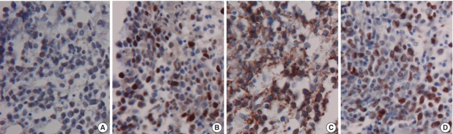
A 2-yr-old male was admitted for evaluation and management of left hip pain. On admission, physical examination was significant for several palpable lymph nodes in the left inguinal area and an ill-defined heterogeneous mass-like lesion in the left anterome- dial muscle. Abdominopelvic computed tomography and mag- netic resonance imaging of both hips were performed on suspi- cion of myositis. Radiologic studies suggested osteomyelitis of the left proximal femur with subperiosteal abscess, myositis, and a small-cell tumor, such as lymphoma, leukemia, or Ewing’s sarcoma. Bone biopsy of the femur neck, bone marrow (BM) aspiration, and biopsy of both posterior superior iliac crests were performed. Immunohistochemistry on the biopsy specimen of the femur neck revealed tumor cells positive for CD20, CD10, BCL2, BCL6, CD99, and Ki67 (-90%) and negative for myeloper- oxidase (MPO), Tdt, CD3, and cyclin D1 (Fig. 1). In situ hybrid- ization for Epstein-Barr virus-encoded RNAs (EBER) was nega- tive in the tumor cells. The final diagnosis from the bone biopsy was high-grade B-cell lymphoma, suggestive of B lymphoblastic lymphoma. Peripheral blood examination revealed the following:
hemoglobin, 9.6 g/dL; white blood cell count, 6.83×10⁹/L; plate- let count, 182×109/L, and 5 atypical lymphocytes per 100 white blood cells. Based on the laboratory findings and bone biopsy results, we suspected lymphoma or lymphoblastic leukemia;
therefore, we performed BM examination, cytogenetic analysis,
and immunophenotyping. The BM biopsy was insufficient for evaluating cellularity; 49.3% of all nucleated cells were abnor- mal lymphoid cells, consisting of large- and medium-sized cells.
Large neoplastic cells had irregular nuclei with 1-2 distinct nu- cleoli and abundant deeply basophilic cytoplasm. Medium-sized cells had round nuclei with 1-4 prominent nucleoli and scantly to moderately basophilic cytoplasm with some vacuoles (Fig. 2).
Immunophenotyping of the neoplastic cells revealed positivity for CD45 (99.0%), CD19 (94.84%), CD10 (27.64%), CD20 (94.24%), HLA-DR (95.70%), sIg lambda (96.45%), CD13 (22.46%), and CD117 (22.22%) and negativity for CD34 (0.01%), Tdt (0.64%), MPO (0.01%), CD33 (1.58%), CD14 (1.18%), CD41 (3.08%), CD2 (3.00%), sCD3 (7.78%), CD5 (1.42%), CD7 (1.34%), and CD56 (5.82%). Cytogenetic analysis of the cells in the BM aspi- rates revealed that the cells had the following karyotypes: 46,XY, t(8;14)(q24.1;q32),del(11)(q13),dup(11)(q22q13),der(17)del(17) (p12)t(1;17)(q21;q25)[29]/46,idem,t(12;19)(q13;p13.2)[4]/46, idem,add(19))(p11)[4]/46,idem,add(13)(q34)[3] (Fig. 3). FISH analysis of BM aspirate cells was performed using Vysis LSI IGH/MYC, CEP 8 tri-color, dual fusion translocation probes (Ab- bott Molecular, Des Plaines, IL, USA). We detected an IGH-MYC rearrangement in 81.2% of the nuclei examined with typical 2 fusions, 1 orange, 1 green, and 2 aqua signals, which was de- scribed as nuc ish (D8Z2x2,MYCx3,IGHx3)(MYC con IGHx2) [325/400] (Fig. 4). Neither BCL2 nor BCL6 rearrangements were observed by FISH analysis on BM aspirates cells using Vysis LSI BCL2 and BCL6 dual color, break apart rearrangement probes (Abbott Molecular). The patient was diagnosed with intermedi- ate DLBCL/BL. Intensive chemotherapy with prednisone, vin- cristine, L-asparaginase, daunorubicin, and central nervous sys- tem prophylaxis with intrathecal methotrexate (MTX) and cytara- bine were initiated. One month after the initial diagnosis, follow-
Fig. 1. Immunohistochemistry shows positivity for BCL2 (A), BCL6 (B), CD10 (C), and Ki67 (D) of tumor cells (Femur neck, immunohisto- chemical stains, 400× magnification).
A B C D
up BM examination demonstrated persistence of abnormal lym- phoid cells. Cerebrospinal fluid (CSF) analysis was performed after induction of chemotherapy; CSF cytology, including cyto- spin, showed atypical lymphoid cells consistent with malignant lymphoma. The patient was treated again with cyclophospha- mide, vincristine, prednisone, adriamycin, MTX, and intrathecal MTX and cytarabine. The patient died of sepsis 5 months after initiation of the second round of chemotherapy.
DISCUSSION
Gray zone B-cell lymphoma, such as intermediate DLBCL/BL, cannot be classified into a single distinct disease entity. This new category of lymphoma has morphologic, immunopheno- typic, and genetic features that include aspects of both DLBCL and BL, but differ with respect to one or more findings [7]. The 2008 WHO classification affirms that the following lymphoma cases should not be diagnosed as intermediate DLBCL/BL: those Fig. 2. Bone marrow smear (A, Wright–Giemsa stain, 1,000× magnification) and biopsy (B, H&E stain, 1,000× magnification) reveal ab- normal lymphoid cells composed of large and medium sized cells. Large neoplastic cells showed irregular nuclei with 1-2 distinct nucleoli and abundant deeply basophilic cytoplasm. Medium-sized cells showed round nuclei with 1-4 prominent nucleoli and had scantly to mod- erately basophilic cytoplasm with some vacuoles.
A B
1
6 7 8 9 10 11 12
13 14 15
19 20
16 17 18
2 3 4 5 1
6 7 8 9 10 11 12
13 14 15
Fig. 3. Giemsa–trypsin banding showed the following karyotypes: 46,XY,t(8;14)(q24.1;q32),del(11)(q13),dup(11)(q22q13),der(17)del(17) (p12)t(1;17)(q21;q25)[29]/46,idem,t(12;19)(q13;p13.2)[4]/46,idem,add(19))(p11)[4]/46,idem,add(13)(q34)[3].
21 22 X Y 19 20 21 22 X Y
16 17 18
2 3 4 5
A B
with a typical DLBCL morphology and a very high proliferation index, typical DLBCL with a MYC translocation, typical BL with- out a MYC rearrangement, and those with IG-MYC rearrange- ment as the only abnormality [4].
Intermediate DLBCL/BL most often occurs in adults, some with a history of follicular lymphoma; it is extremely rare in pedi- atric patients. The majority of patients present with generalized lymphadenopathy or mass lesions in extranodal sites and fre- quent involvement of the BM. Some patients have a leukemic presentation [5, 8]. Liang et al. [8] reported the clinicopathologic features of 2 pediatric patients with gray zone lymphoma, who presented with common features, such as male gender, older than 10 yr of age at the time of diagnosis, and presentation with a mediastinal mass. In children, high cure rates are achieved with treatment strategies similar or identical to those for BL and DL- BCL [9]. The gray zone between BL and DLBCL currently does not impact therapy decision or outcome in childhood lympho- mas [7].
Morphologic feature are useful in the differential diagnosis of intermediate DLBCL/BL. Such morphologic features include variable cellular forms, such as those smaller than typical DL- BCL, those resembling BL cells, those larger than typical BL, and those resembling DLBCL cells. The immunophenotype is similar to BL, showing positivity for CD19, CD20, CD22, CD79a, and germinal center-associated molecules, CD10 and BCL6. BCL2 expression may be absent, weak, or strong, and the Ki67 labeling index shows varying positivity [3]. Genetically, 35-50% of
cases of intermediate DLBCL/BL show 8q24/MYC translocations but often with atypical features, including one or more of the fol- lowing: (1) rearrangement with a non-IG partner, (2) part of a complex karyotype, and (3) concurrent rearrangements of the BCL2 and/or BCL6 genes, suggesting a “double-hit” or “triple- hit” lymphoma. Double-hit lymphomas are characterized by a second translocation in addition to t(8;14), t(8;22), or t(2;8). In the majority of the double-hit cases, an 18q21/BCL2 breakpoint can be found, mostly as a t(8;14) plus t(14;18) karyotype [10-13].
Double-hit lymphomas are almost always absent in children, consistent with the almost complete lack of t(14;18) found among lymphomas in those younger than 18 yr [6].
More recently, GEP analysis using microarrays can establish molecular categories within the gray zone between DLBCL and BL [1]. The bioinformatic core group extension method applied by the Molecular Mechanisms in Malignant Lymphomas group identified a set of 53 mature aggressive B-cell lymphomas with a molecular BL (mBL) index between that of mBL (mBL index
>0.95) and that of non-mBL (mBL index <0.05). These lym- phomas could not be classified as mBL or as non-mBL and were called “molecular intermediate lymphomas” [1, 5, 6]. Molecular intermediate DLBCL/BL in children has a significantly higher Burkitt index by GEP than in adult patients, frequent IG-MYC positivity, and a good outcome. Interestingly, significant numbers (31%) of morphologic DLBCL in children show mBL by GEP, with more than half of them having IG-MYC. These findings sug- gest that, in children, biologic BL might be hidden among DL- BCL [6]. Salaverria et al. [6] suggested that GEP should be con- sidered a single diagnostic criterion, like MYC status or CD10 positivity.
In the present case, the patient was diagnosed with interme- diate DLBCL/BL based on the intermediate morphological fea- tures of both BL and DLBCL, the expression of CD10, BCL6, BCL2, and Ki67 labeling index, and a complex karyotype with 8q34/MYC.
Based on existing literature and the findings of this case, eval- uation for intermediate DLBCL/BL should include an immuno- phenotypic panel with CD10, BCL6, BCL2, and Ki67. In addition, evaluation should include conventional cytogenetic analysis for detection of simple or complex karyotypic abnormalities and molecular cytogenetic evaluation (FISH) with MYC-IgH fusion probe for t(8;14), BCL2-IgH fusion probe for t(14;18), BCL2 break-apart probe, BCL6 break-apart probe, MYC break-apart probe, IGH break-apart probe, and IGL break-apart probe for t(2;8) and t(8;22) [12].
However, several questions remain unanswered. A major con- Fig. 4. FISH analysis using Vysis LSI IGH/MYC, CEP 8 tri-color, dual
fusion translocation probe reveals IGH-MYC rearrangement with 2 fusions (IGH-MYC fusions on der(8)t(8;14) and der(14)t(8;14)), 1 or- ange (native MYC), 1 green (native IGH), and 2 aqua (D8Z2) signals.
cern is that cases of intermediate DLBCL/BL only slightly differ from BL and DLBCL. Therefore, it is important that the diagnos- tic border of DLBCL and BL is clearly defined in order to identify the specific morphology, immunophenotyping, genetics, and molecular lesions based on a gene expression. Another issue is that the current treatment of intermediate DLBCL/BL may com- promise the validity of clinical trials evaluating the efficacy and safety of therapies on well-established diagnostic entities and may obscure the pathologic predictors of their outcome and treat- ment response.
Authors’ Disclosures of Potential Conflicts of Interest
No potential conflict of interest relevant to this article was re- ported.
REFERENCES
1. Hummel M, Bentink S, Berger H, Klapper W, Wessendorf S, Barth TF, et al. A biologic definition of Burkitt’s lymphoma from transcriptional and genomic profiling. N Engl J Med 2006;354:2419-30.
2. Saglio G, Bosa M, Gino M, Ulisciani S, Parvis G. Molecular pathogene- sis of diffuse large B-cell lymphoma. Hematologica reports 2006;2:68-9. 3. Carbone A, Gloghini A, Aiello A, Testi A, Cabras A. B-cell lymphomas
with features intermediate between distinct pathologic entities. From pathogenesis to pathology. Hum Pathol 2010;41:621-31.
4. Kluin PM, Harris NL, Stein H, Leoncini L, Raphael M, Campo E, et al.
B-cell lymphoma, unclassifiable, with features intermediate between diffuse large B-cell lymphoma and Burkitt lymphoma. In: Swerdlow SH,
Campo E, et al., eds. WHO classification of tumours of haematopoietic and lymphoid tissues. 4th ed. Lyon: IARC Press, 2008:265-6.
5. Klapper W, Szczepanowski M, Burkhardt B, Berger H, Rosolowski M, Bentink S, et al. Molecular profiling of pediatric mature B-cell lympho- ma treated in population-based prospective clinical trials. Blood 2008; 112:1374-81.
6. Salaverria I and Siebert R. The gray zone between Burkitt’s lymphoma and diffuse large B-cell lymphoma from a genetics perspective. J Clin Oncol 2011;29:1835-43.
7. HasserJian RP, Ott G, Elenitoba-Johnson KS, Balague-Ponz O, de Jong D, de Leval L. Commentary on the WHO classification of tumors of lym- phoid tissues (2008): “Gray zone” lymphomas overlapping with Burkitt lymphoma or classical Hodgkin lymphoma. J Hematop 2009;2:89-95. 8. Liang X, Greffe B, Cook B, Giller R, Graham DK, McGranahan AN, et al.
Gray zone lymphomas in pediatric patients. Pediatr Dev Pathol 2011;14: 57-63.
9. Patte C, Auperin A, Gerrard M, Michon J, Pinkerton R, Sposto R, et al.
Results of the randomized international FAB/LMB96 trial for intermedi- ate risk B-cell non-Hodgkin lymphoma in children and adolescents: it is possible to reduce treatment for the early responding patients. Blood 2007;109:2773-80.
10. Quintanilla-Martinez L, de Jong D, de Mascarel A, Hsi ED, Kluin P, Nat- kunam Y, et al. Gray zones around diffuse large B cell lymphoma. Con- clusions based on the workshop of the XIV meeting of the European As- sociation for Hematopathology and the Society of Hematopathology in Bordeaux, France. J Hematop 2009;2:211-36.
11. Le Gouill S, Talmant P, Touzeau C, Moreau A, Garand R, Juge-Morineau N, et al. The clinical presentation and prognosis of diffuse large B-cell lymphoma with t(14;18) and 8q24/c-MYC rearrangement. Haematologi- ca 2007;92:1335-42.
12. Bellan C, Stefano L, Giulia de F, Rogena EA, Lorenzo L. Burkitt lympho- ma versus diffuse large B-cell lymphoma: a practical approach. Hema- tol Oncol 2009;27:182-5.
13. Aukema SM, Siebert R, Schuuring E, van Imhoff GW, Kluin-Nelemans HC, Boerma EJ, et al. Double-hit B-cell lymphomas. Blood 2011;117: 2319-31.
![Fig. 3. Giemsa–trypsin banding showed the following karyotypes: 46,XY,t(8;14)(q24.1;q32),del(11)(q13),dup(11)(q22q13),der(17)del(17) (p12)t(1;17)(q21;q25)[29]/46,idem,t(12;19)(q13;p13.2)[4]/46,idem,add(19))(p11)[4]/46,idem,add(13)(q34)[3].](https://thumb-ap.123doks.com/thumbv2/123dokinfo/5190499.114198/3.918.79.842.146.453/fig-giemsa-trypsin-banding-showed-following-karyotypes-idem.webp)
