Management of intractable pancreatic leak from iatrogenic pancreatic duct injury following resection of choledochal cyst in
an adult patient
Jin Uk Choi, Shin Hwang, and Yong-Kyu Chung
Department of Surgery, Asan Medical Center, University of Ulsan College of Medicine, Seoul, Korea
Iatrogenic pancreatic duct injury can occur during resection of the choledochal cyst (CC). We herein present a case of postoperative pancreatic fistula (POPF) developed after resection of the CC in an adult patient with variant anom- alous union of pancreatobiliary duct. The 55-year-old female patient underwent surgery after the diagnosis of CC-asso- ciated gallbladder cancer. During surgery, the CC mass was accidentally pulled out, by which the intrapancreatic CC portion was torn out from the main pancreatic duct. Since the pancreatic duct stump was not identified due to its small size, repair was not possible. The excavated defect at the pancreas head was closed securely combined with insertion of multiple drains. Postoperative POPF and peripancreatic fluid collection developed and the patient had to be fasted for 4 weeks. She was first discharged at 6 weeks after surgery. At 10 weeks, she was readmitted due to progression of peripancreatic fluid collection, which was controlled by percutaneous drain insertion. At 6 months, she was readmitted again due to repeated progression of peripancreatic fluid collection, which were controlled by endo- scopic transmural duodenocystostomy. It took 8 months to resolve the pancreatic duct injury-associated pancreatitis.
The experience in this case suggests that iatrogenic pancreatic duct injury during resection of CC can induce cata- strophic complications, thus special attention should be paid to prevent pancreatic duct injury. (Ann Hepatobiliary Pancreat Surg 2020;24:228-233)
Key Words: Pancreatic leak; Iatrogenic injury; Resection; Anomalous union of pancreatobiliary duct
Received: March 9, 2020; Revised: March 13, 2020; Accepted: March 15, 2020 Corresponding author: Shin Hwang
Department of Surgery, Asan Medical Center, University of Ulsan College of Medicine, 88 Olympic-ro 43-gil, Songpa-gu, Seoul 05505, Korea Tel: +82-2-3010-3930, Fax: +82-2-3010-6701, E-mail: [email protected]
Copyright Ⓒ 2020 by The Korean Association of Hepato-Biliary-Pancreatic Surgery
This is an Open Access article distributed under the terms of the Creative Commons Attribution Non-Commercial License (http://creativecommons.org/
licenses/by-nc/4.0) which permits unrestricted non-commercial use, distribution, and reproduction in any medium, provided the original work is properly cited.
Annals of Hepato-Biliary-Pancreatic Surgery ∙ pISSN: 2508-5778ㆍeISSN: 2508-5859
INTRODUCTION
The choledochal cyst (CC) is often associated with anomalous union of the pancreatobiliary duct (AUPBD).
Since the CC has malignant potential as well as develop- ment of de novo malignancy after incomplete resection, complete resection of CC is highly recommended in adult patients.1,2 However, the patterns of AUPBD are variable, thus there is a potential risk of iatrogenic pancreatic duct injury when surgeons attempt to remove the deeply-seated CC portion within the pancreas head. Such an iatrogenic pancreatic duct injury can lead to a major pancreatic leak, which is different from the usual postoperative pancreatic fistula (POPF) following pancreatoduodenectomy. We herein report our experience in the management of major POPF developed after resection of CC in an adult patient
with variant AUPBD.
CASE
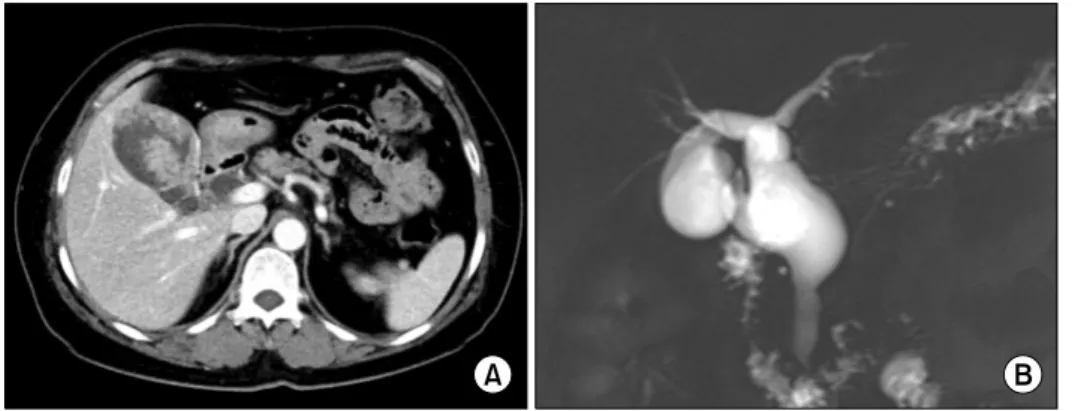
A 55-year-old female patient was admitted under the initial diagnosis of CC-associated gallbladder cancer. The computed tomography (CT) scan showed a highly sus- pected gallbladder cancer arising from the intracystic pap- illary neoplasm with the underlying CC type I (Fig. 1A).
Magnetic resonance cholangiopancreatography (MRCP) showed intracystic papillary neoplasm of the gallbladder and CC combined with AUPBD type I (Fig. 1B). Fludeo- xyglucose-positron emission tomography scan showed a definite hypermetabolic uptake at the gallbladder, suggest- ing malignant changes.
The extent of surgical resection was planned to be com-
Fig. 1. Preoperative radiologic findings. The computed tomogra- phy scan showed highly suspected gallbladder cancer arising from the intracystic papillary neoplasm with underlying choledochal cyst (A). Magnetic resonance cholan- giopancreatography showed cho- ledochal cyst combined with ano- malous union of pancreatobiliary duct (B).
Fig. 2. The extent of resection overlapped at the magnetic res- onance cholangiopancreatography image. A green bidirectional arrow was the preplanned transection line. The red thick dot- ted line indicates the main pancreatic duct and the blue thin dotted line indicates the accessory pancreatic duct. The yel- low dotted area indicates the actual extent of resection.
patible with the extended cholecystectomy and CC re- section with Roux-en-Y hepaticojejunostomy. After per- forming transection of the hepatic duct and extended chol- ecystectomy, dissection continued to remove the intra- pancreatic cystic portion. At this time, the second assistant forcefully pulled out the CC mass, by which the intra- pancreatic bile duct portion was accidentally torn out from the main pancreatic duct (Fig. 2). Since the pancreatic duct stump was not identified probably due to its small size, primary repair or internal stenting was not possible.
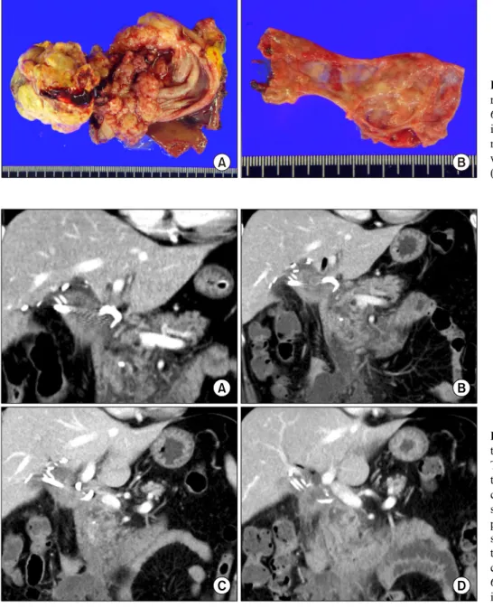
After primary closure of the distal bile duct stump, the excavated defect at the pancreas head was closed securely with multiple sutures. Other parts of this CC surgery were similar to the standard procedures. Multiple Jackson-Pratt type abdominal drains and a 14-Fr pigtail catheter were inserted to cope with potential development of major POPF. The pathology report revealed that there was a 6 cm-sized moderately differentiated adenocarcinoma aris- ing in the intracystic papillary neoplasm (Fig. 3). The depth of the gallbladder wall invasion was extension to the perimuscular connective tissue. There was no lympho- vascular or perineural invasion. There was no malignant change at the CC specimen. One of the 3 regional lymph nodes were tumor-positive, making the tumor stage T2N1M0.
A water-clear pancreatic enzyme-rich fluid was drained up to 500 ml/day through the pigtail catheter, which was located at the pancreatic repair site. The dynamic CT scan taken after 5 days showed swelling of the pancreas with peripancreatic and right anterior pararenal space fluid col- lection, indicating development of acute pancreatitis (Fig.
4A). Acute pancreatitis was exacerbated and peripancre- atic fluid collection increased on the 10-day CT scan (Fig.
4B). At the 4-week CT scan, pancreatitis and peripancre- atic fluid collection began to resolve (Fig. 4C). Until this
time point, the patient was permitted to have only sips of water under total parenteral nutrition. Pancreatitis was slowly improved with a marked decrease in pigtail drain- age until 6 weeks (Fig. 4D). During these sequences, her general condition was good without febrile episodes. Thus, she was discharged after removal of the abdominal drains.
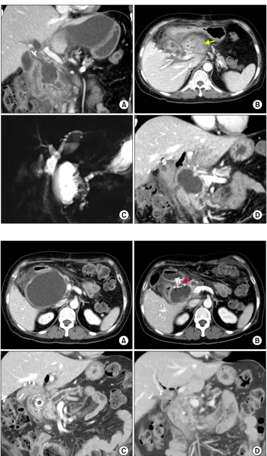
At the 10-week CT scan, there was noticeable pro- gression of acute necrotic collection around the pancreas head, the small bowel mesentery, and along the right ante- rior pararenal space (Fig. 5A). She was readmitted and a new pigtail catheter was inserted percutaneously. At the 14-week CT scan, a liver abscess developed in the left liver (Fig. 5B). MRCP showed no abnormality at the in- trahepatic bile duct and the remnant pancreatic duct was dilated with peripancreatic fluid collection (Fig. 5C). At the 17-week CT scan, the liver abscess improved and the
Fig. 3. Gross photographs of the resected specimen. There was a 6 cm-sized adenocarcinoma aris- ing in the intracystic papillary neoplasm (A). The choledocal cyst was free of malignant changes (B).
Fig. 4. Postoperative computed tomography (CT) findings. (A) The 5-day CT showed pancrea- titis with peripancreatic fluid collection. (B) The 10-day CT showed exacerbation of acute pancreatitis. (C) The 4-week CT scan showed progressive resolu- tion of pancreatitis and peripan- creatic fluid collection. (D) The 6-week CT scan showed further improvement of pancreatitis.
walled-off necrosis around the pancreas gradually de- creased (Fig. 5D). After removal of the pigtail catheter, she was discharged again.
At the 6-month CT scan, there was a marked increase in complicated fluid collection posterior to the pancreatic head and the proximal duodenum (Fig. 6A). She was re- admitted and endoscopic ultrasonography-guided trans- mural duodenocystostomy was performed to drain the per- ipancreatic fluid (Fig. 6B). She was discharged again for observation. After 2 months, she was readmitted to re- move the wall stent. At the 8-month CT scan, there was a marked decrease in pancreatic pseudocyst, with further dilation of the pancreatic duct and atrophy of the pancre-
atic parenchyma (Fig. 6C). At the 12-month CT scan, there was no abnormal fluid collection and pancreatitis was completely controlled (Fig. 6D).
Adjuvant chemotherapy had been initially planned after surgery, but it was not performed due to delayed recovery from postoperative pancreatitis. Currently, she is doing well without any physical discomfort 13 months after the operation. She was diagnosed with new-onset type II dia- betes during follow-up, thus administration of antidiabetic medication was necessary.
Fig. 5. Postoperative computed tomography (CT) and magnetic resonance imaging findings. (A) The 10-week CT showed progres- sion of peripancreatic necrotic collection. (B) The 14-week CT scan showed liver abscess at the left liver (arrow). (C) Magnetic resonance cholangiopancreato- graphy image showed no abnor- mality at the intrahepatic bile duct and dilation of the remnant accessory pancreatic duct. (D) The 17-week CT showed resolu- tion of liver abscess and impro- vement of peripancreatic necrosis.
Fig. 6. Postoperative computed tomography (CT) findings. (A) The 6-month CT scan showed increased complicated fluid collec- tion. (B) Endoscopic ultrasono- graphy-guided transmural duode- nocystostomy was performed to drain the peripancreatic fluid col- lection (arrow). (C) The 8-mon- th CT scan showed marked decrease of pancreatic pseudocyst with further dilation of the pan- creatic duct and atrophy of the pancreatic parenchyma. (D) The 12-month CT scan showed com- plete resolution of pancreatitis.
DISCUSSION
Complete resection of the CC has been the mainstay
of the management of CC. However, some problems re- main with intrahepatic and distal end parts of the CC.
Although radical cyst excision is well known to be a treat-
tions exist such as morbidity of the porta hepatis dis- section and postoperative complications such as POPF and pancreatitis.3 As a result, surgeons occasionally tend to be reluctant to perform aggressive complete excision, espe- cially in patients with unusual anatomical variations.
POPF is one of the most serious complication following surgical resection of the CC. Since CC is often buried deep within the pancreatic parenchyma, extensive dis- section into the pancreatic parenchyma is an essential part of surgical procedure. It is important to protect the junc- tion portion of the distal bile duct and pancreatic duct.
If this portion is not definitely delineated at the pre- operative CT or MRCP, special attention should be paid during pancreatic excavation. Meticulous CC dissection combined with intraluminal probing within the distal bile duct is a useful and intuitive surgical technique to prevent iatrogenic injury of the pancreatic duct.
In this case, we applied the abovementioned technique during dissection of the distal CC part. However, our sec- ond assistant accidentally pulled out the CC mass force- fully, by which the intrapancreatic cystic portion was torn out from the main pancreatic duct. We identified the in- jured junction portion of the distal bile duct and pancre- atic duct because there was a small perforation at the torn-out site of the distal bile duct wall. However, we failed to identify the remnant pancreatic duct stump due to its small size. Since primary repair with internal stent- ing was not possible, the only method we could use was secure closure of the excavated pancreatic defect and in- sertion of multiple drains.
As anticipated, a major POPF and pancreatitis devel- oped soon after surgery. We had to wait with supportive care and serial follow CT scan were repeated until the res- olution of POPF. She fasted for 4 weeks under total pa- renteral nutrition and was discharged at 6 weeks after surgery. However, acute pancreatitis with marked fluid collection developed and gradually controlled with a pig- tail drainage. At 6 months after the surgery, acute pan- creatitis with peripancreatic fluid collection developed again. We supposed that initial acute pancreatitis from ia- trogenic pancreatic duct injury was not completely con- trolled, thus pancreatitis was relapsed until marked atro- phy of the pancreatic parenchyma. Since percutaneous drainage did not appear safe, endoscopic transmural duo-
atic fluid collection, which was maintained for 2 months.
It took at least 8 months to resolve the pancreatic duct injury-associated pancreatitis.
The present case was the only case where a patient suf- fered from pancreatic duct injury in the personal (SH) ex- perience of one hundred cases of CC surgery. After re- viewing this case retrospectively, we presumed that there was an unusual pattern of AUPBD, illustrated at Fig. 2.
We had to transect the distal bile duct at the junction of the CC portion, but accidental torn-out of the pancreatic duct junction led to inevitable closure of the main pancre- atic duct. On follow-up MRCP, we identified the existence of an accessory pancreatic duct inserted at the end of the distal bile duct. If such an accessory pancreatic duct was absent, we presume that pancreatitis might continue until complete atrophy of the pancreatic parenchyma.
Afraid of POPF, some surgeons were reluctant to per- form aggressive complete resection of the intrapancreatic CC. We previously presented a case of cancer develop- ment at the remnant CC within the pancreatic parenchyma after 16 years, which was treated by pylorus-preserving pancreatoduodenectomy.4 In a Chinese study including 78 patients with partial resection of the CC, carcinogenesis in the residual intrapancreatic biliary duct developed in 14.1%.1 Development of cholangiocarcinoma more than 10 years after excision of CC is rare, with a median time of recurrence being 6 years.5-12 The risk of interval malig- nancy after excision of CC is known well, but there are no practical guidelines for long-term follow-up. Most pa- tients diagnosed with cholangiocarcinoma long period af- ter resection of the CC were not followed up routinely with radiological imaging and tumor markers.5
For diagnosis and treatment of CC, MRCP is the most important imaging study. Preoperative MRCP examination is essential to avoid damaging the pancreatic duct during surgery because it can visualize a specific morphology of the pancreatobiliary duct junction.13,14
The experience of our present case suggests that iatro- genic pancreatic duct injury during resection of CC can induce catastrophic complications, thus special attention should be paid to prevent pancreatic duct injury.
CONFLICT OF INTEREST
None of the authors has any conflict of interest.
ORCID
Jin Uk Choi: https://orcid.org/0000-0001-8078-0593 Shin Hwang: https://orcid.org/0000-0002-9045-2531 Yong-Kyu Chung: https://orcid.org/0000-0002-2132-2450
AUTHOR CONTRIBUTIONS
Conceptualization: SH. Data curation: JUC, YKC.
Visualization: SH. Writing - original draft: SH. Writing - review & editing: SH.
REFERENCES
1. Fan F, Xu DP, Xiong ZX, Li HJ, Xin HB, Zhao H, et al. Clinical significance of intrapancreatic choledochal cyst excision in surgi- cal management of type I choledochal cyst. J Int Med Res 2018;46:1221-1229.
2. Cho MJ, Hwang S, Lee YJ, Kim KH, Ahn CS, Moon DB, et al. Surgical experience of 204 cases of adult choledochal cyst disease over 14 years. World J Surg 2011;35:1094-1102.
3. Ando H, Kaneko K, Ito T, Watanabe Y, Seo T, Harada T, et al. Complete excision of the intrapancreatic portion of chol- edochal cysts. J Am Coll Surg 1996;183:317-321.
4. Oh SY, Kwon JH, Hwang S. Development of adenocarcinoma at the remnant intrapancreatic cyst 16 years after resection of the choledochal cyst. Ann Hepatobiliary Pancreat Surg 2019;
23:192-196.
5. Ng DW, Chiow AK, Poh WT, Tan SS. Metachronous chol- angiocarcinoma 13 years post resection of choledochal cyst-is long-term follow-up useful?: a case study and review of the literature. Surg Case Rep 2016;2:60.
6. Shimamura K, Kurosaki I, Sato D, Takano K, Yokoyama N, Sato Y, et al. Intrahepatic cholangiocarcinoma arising 34 years after excision of a type IV-A congenital choledochal cyst: report of a case. Surg Today 2009;39:247-251.
7. Kumamoto T, Tanaka K, Takeda K, Nojiri K, Mori R, Taniguchi K, et al. Intrahepatic cholangiocarcinoma arising 28 years after excision of a type IV-A congenital choledochal cyst: report of a case. Surg Today 2014;44:354-358.
8. Nishiyama R, Shinoda M, Tanabe M, Masugi Y, Ueno A, Hibi T, et al. Intrahepatic cholangiocarcinoma arising 33 years after excision of a choledochal cyst: report of a case. Int Surg 2011;96:320-325.
9. Ohashi T, Wakai T, Kubota M, Matsuda Y, Arai Y, Ohyama T, et al. Risk of subsequent biliary malignancy in patients under- going cyst excision for congenital choledochal cysts. J Gastroenterol Hepatol 2013;28:243-247.
10. Koike M, Yasui K, Shimizu Y, Kodera Y, Hirai T, Morimoto T, et al. Carcinoma of the hepatic hilus developing 21 years after biliary diversion for choledochal cyst: a case report. Hepatogastro- enterology 2002;49:1216-1220.
11. Suzuki S, Amao K, Harada N, Tanaka S, Hayashi T, Suzuki M, et al. A case of intrahepatic cholangiocarcinoma arising 26 years after excision of congenital biliary dilatation. Jpn J Gastroenterol Surg 2004;37:416-421.
12. Ono S, Sakai K, Kimura O, Iwai N. Development of bile duct cancer in a 26-year-old man after resection of infantile choledochal cyst. J Pediatr Surg 2008;43:E17-E19.
13. Yoon JH. Magnetic resonance cholangiopancreatography diag- nosis of choledochal cyst involving the cystic duct: report of three cases. Br J Radiol 2011;84:e18-e22.
14. Shimizu T, Suzuki R, Yamashiro Y, Segawa O, Yamataka A, Kuwatsuru R. Magnetic resonance cholangiopancreatography in assessing the cause of acute pancreatitis in children. Pancreas 2001;22:196-199.