서 론
비결핵항산균(nontuberculous mycobacteria)은 토양 및 물 등 의 자연환경에 흔히 존재하고 정상 건강인에서도 검출되어 비병원
성으로 알려져 왔다[1]. 그러나 후천성면역결핍증 환자에서 기회 감염균으로 확인되면서 그 중요성이 부각되기 시작하였고 면역기 능이 정상인 환자에서도 감염을 일으킬 수 있음이 알려졌다[2].
전국적인 실태조사에 의하면 국내의 결핵유병률은 감소하고 있는 반면에 비결핵항산균증은 1990년대 이후 증가하고 있다[3]. 비결 핵항산균은 임상양상이 결핵과 감별하기 힘들고, 기존 항결핵제에 높은 내성을 보이며, 치료반응이 좋지 않아 장기간 약제병용요법 으로 치료하여야 한다. 또한 동정된 균과 감염부위에 따라서 치료 약제 선택과 치료방법이 달라지므로[4] 비결핵항산균을 결핵균과 구별하고 균종 수준까지 동정할 필요가 있다.
40 40
비결핵항산균 동정에서 다상-실시간-중합효소연쇄반응과 융해곡선 분석의 유용성
Usefulness of Multiplex Real-Time PCR and Melting Curve Analysis in Identification of Nontuberculous Mycobacteria
Seong-Ho Kang, M.D.
1, Kwang Cheol Yoo
2, Kyoung Un Park, M.D.
1,2, Junghan Song, M.D.
1,2, and Eui Chong Kim, M.D.
1Departments of Laboratory Medicine, Seoul National University College of Medicine1, Seoul; and Seoul National University Bundang Hospital2, Seongnam, Korea
강성호1∙유광철2∙박경운1,2∙송정한1,2∙김의종1
서울대학교 의과대학 검사의학교실1, 분당서울대학교병원 진단검사의학과2
40 40
Background : Nontuberculous mycobacteria (NTM) should be correctly identified to the species level, because of different treatment plans among NTM species. This study was performed to assess the usefulness of real-time PCR and melting curve analysis in the identification of NTM.
Methods :One hundred fifty-two clinical NTM isolates were identified to the species level by PCR- restriction fragment length polymorphism analysis (PRA). Those strains were then identified by mul- tiplex real-time PCR and melting curve analysis on the 16S rRNA gene and hsp65 gene.
Results :In the 16S rRNA gene fragment analysis, M. abscessus-M. chelonae group showed melt- ing point at temperatures above 65°C and M. avium complex (MAC; M. avium and M. intracelluare) below 48°C, which differentiated M. abscessus-M. chelonae group and MAC from other NTM. In the hsp65 gene fragment analysis, M. abscessus-M. chelonae group was clearly divided into M. absces- sus type I, M. abscessus type II, and M. chelonae according to the melting points at 61.25°C, 66.06°C, and 57.58°C, respectively.
Conclusions :With the multiplex real-time PCR and melting curve analysis of 16S rRNA and hsp65 genes, M. abscessus and M. chelonae were readily identified and MAC were differentiated from other NTM. Especially, M. abscessus and M. chelonae, which were not differentiated from each other with the 16S rRNA gene fragment analysis, were identified with hsp65 gene fragment analysis. (Korean J Lab Med 2007;27:40-5)
Key Words : Nontuberculous mycobacteria, Species identification, Real-time PCR, Melting curve analysis
접 수: 2006년 9월 17일 접수번호:KJLM1989 수정본접수: 2006년 12월 21일
게재승인일: 2006년 12월 22일 교 신저 자: 박 경 운
우 463-707 경기도 성남시 분당구 구미동 300 분당서울대학교병원 진단검사의학과 전화: 031-787-7692, Fax: 031-787-4015 E-mail: [email protected]
전통적으로 비결핵항산균은 균 집락의 색, 모양, 성장속도 등으 로 구분하였고 니아신(niacin) 생성, 질산염(nitrate) 환원, Twe- en-80 가수분해와 같은 생화학적 방법을 이용하여 동정하였다. 이 생화학적인 방법은 정확한 균종 동정이 힘들고 시간이 많이 걸리 며 숙달된 인력이 필요하다. 최근에는 고작위액체크로마토그래피 (high performance liquid chromatography, HPLC)를 이용하여 미콜산(mycolic acid)을 분석하는 방법[5], 핵산탐색자법[6], 중 합효소연쇄반응-제한절편길이다형성 분석(PCR-restriction frag- ment length polymorphism analysis, PRA)[7, 8] 등이 비결핵 항산균을 동정하는데 사용하고 있다.
실시간-중합효소연쇄반응(real-time PCR)은 증폭된 유전자 산물에 결합된 올리고핵산(oligonucleotide) 탐색자의 형광물질로 부터 방출되는 형광량을 실시간으로 측정한다. 이후 융해곡선 분 석(melting curve analysis)에서는 온도를 서서히 올리면서 형광 의 변화가 가장 심한 온도를 융해온도로 정하고 이를 통해 유전자 의 염기서열차이를 검출한다.
근래 실시간 중합효소연쇄반응과 융해곡선 분석을 결핵균의 동 정과 정량에 이용하며 항산균을 비결핵항산균과 분리하고 비결핵 항산균의 동정에 사용하고 있다[9, 10]. 실시간 중합효소연쇄반응 과 융해곡선 분석은 결핵균 정량에 민감도가 높으며 신속한 동정 이 가능한 것으로 알려져 있다[9]. 본 연구에서는 중합효소연쇄반 응-제한절편길이다형성 분석으로 균종 수준까지 동정된 비결핵항 산균을 대상으로 다상-실시간-중합효소연쇄반응(multiplex real- time PCR)과 융해곡선 분석을 시행하여 비결핵항산균 동정에서 그 유용성을 알아보고자 하였다.
대상 및 방법
1. 대상분당서울대학교병원 결핵균검사실에서 3% Ogawa 배지(Shin- yang chemical, Seoul, Korea)에 배양된 임상균주 중 AccuProbe Mycobacterium complex culture identification kit (Gen-Probe, San Diego, CA, USA)를 이용하여 비결핵항산균을 분리하였다.
그 후rpoB 유전자에 대한 중합효소연쇄반응-제한절편길이다형성 분석으로 균종을 구분하는 Myco-ID kit (M&D, Seoul, Korea) 를 사용하여 비결핵항산균의 균종 수준까지 동정한 152 임상 균주 (M. abscessus21균주, M. asiaticum, 3균주, M. avium 29균주, M. chelonae 3균주, M. fortuitum 14균주, M. gordonae 3균주, M. intracellulare 17균주, M. kansasii 10균주, M. margeritense 8균주, M. mucogenicum 6균주, M. nonchromogenicum 9균주, M. peregrinum 5균주, M. pulveris 2균주, M. scrofulaceum 4 균주, M. septicum 10균주, M. shimoidei 5균주, M. smegmatis 3균주)를 대상으로 하였다. 대조군으로는 AccuProbe MTBC kit에서 양성으로 확인된Mycobacterium tuberculosis complex
5균주를 사용하였다.
2. 다상-실시간-중합효소연쇄반응과 융해곡선 분석
LightCycler 2.0 (Roche, Penzerg, Germany)을 이용하여 비 결핵항산균의 16S rRNA 유전자와hsp65유전자를 대상으로 다 상-실시간-중합효소연쇄반응 후 융해곡선 분석을 시행하였다. 시 발체로는 16S rRNA 유전자의 1,000 bp 절편의 중합효소연쇄반 응을 시행하는 시발체[9] 및hsp65 유전자의 439 bp 절편의 중 합효소연쇄반응을 시행하는 시발체[11]를 이용하였다(Table 1).
16S rRNA 시발체는M. tuberculosis16S rRNA 유전자 염기서 열(Genbank no. NC_000962)의 12번째에서 1,037번째의 염기를 증폭하는 것으로 항산성 염색에서 양성 반응을 보일 수 있는No- cardia와Rhodococcus는 증폭이 일어나지 않는 것이다[9]. hsp65 시발체는 M. tuberculosis의hsp65 유전자 염기서열[12]의 396 번째에서 836번째 염기를 증폭한다. 비결핵항산균의 균종을 감별 하기 위해 쓰인 16S rRNA 탐색자[13] 및M. abscessus와M.
chelonae를 감별하기 위한hsp65 탐색자[13]는 Table 1과 같다.
(16S rRNA 유전자는 비결핵항산균 중 완속성장항산균(slowly growing mycobacteria)에서는 유전자 다형성이 상당 부분 있으 나 신속성장항산균(rapidly growing mycobacteria)에서는 유전 자 변이가 거의 없는 것으로 알려져 있다[14]. 또한hsp65 유전자 는 항산균에서 16S rRNA 유전자 보다 변이가 더 많으며 신속성 장항산균의 동정에서 유용한 것으로 알려져 있다[15]. 그리하여 비결핵항산균의 균종 감별에 16S rRNA 탐색자를M. abscessus 와M. chelonae 감별에는hsp65 탐색자를 사용하였다.) 이용된 16S rRNA 탐색자는M. abscessus 유전자 염기서열과는 완전히 부합하나M. tuberculosis 유전자 염기서열과는 상당 부분 부합하 지 않는다. 다른 비결핵항산균은 탐색자 염기서열과의 부합 정도 에 따라서 탐색자 형광의 증폭이 일어나지 않거나 융해온도가 달 라지게 되고 이 융해온도의 차이를 비결핵항산균의 동정에 이용하 였다. hsp65 탐색자는M. abscessus와M. chelonae를 감별하는 부분과M. abscesus type I과M. abscessustype II를 구분하는 hsp65 유전자의 542번째 염기[15]를 포함한 것이다.
16S rRNA 유전자와hsp65 유전자를 동시에 증폭하는 다상- 실시간-중합효소연쇄반응을 기술하면 다음과 같다. 배지에서 자
Genes Primers and probes (5′→ 3′)
16S rRNA Forward primer: GAG TTT GAT CCT GGC TCA GGA Reverse primer: TGC ACA CAG GCC ACA AGG GA LightCycler Red 705-CAA AAG CTT TGC ACC ACT CAC GGC CGC GGG CCC ATC CCA CAC-fluorescein hsp65 Forward primer: ACC AAC GAT GGT GTG GCC AT
Reverse primer: CTT GTC GAA CCG CAT ACC CT LightCycler Red 640-GGT GGT GGT GCC GTC ACC GAG CCT GGG CAA GCA CGG TGG-fluorescein Table 1.Primers and probes used in real-time polymerase chain reaction and melting curve analysis
란 균 집락을 루프를 이용하여 150 L Tris-EDTA 용액에 풀고 100℃에서 10분간 가열 후, 15,000 g에서 5분간 원심분리하여 상
층액 100 L를 취하였다. 반응혼합용액(20 L)에는 DNA가 포 함된 상층액 3 L, LightCycler FastStart DNA Master Hyb- ridization Probe (Roche, Penzerg, Germany) 2.0 L, 각 시발 체 0.25 M, 각 탐색자 0.1 M, MgCl2 2.0 mM이 포함되었다.
LightCycler 2.0을 사용하여 94℃에서 10분간 초기변성을 시행 하였고 94℃ 3초, 56℃ 2초, 72℃ 40초의 증폭반응을 50회 시행 한 후, 95℃에서 30초, 38℃에서 30초, 38℃에서 80℃까지 초당 0.2℃ 상승시켜 융해과정을 시행하였다. 16S rRNA 유전자 절편 의 융해곡선 분석은 705 nm에서 시행하였고, hsp65 유전자 절편 의 융해곡선 분석은 640 nm에서 시행하였다.
결 과
1. 16S rRNA 융해곡선 분석152균주의 비결핵항산균 모두에서 16S rRNA 융해곡선 분석 이 가능하였다. Mycobacterium tuberculosis (MTB) complex 는 형광의 증폭이 일어나지 않았다. 평균 융해온도는 최소 46.11℃
에서 최대 66.22℃이었다. M. abscessus와M. chelonae의 평균 융해온도는 각각 65.62℃과 65.30℃로 65℃ 이상인 반면에 두 균 종을 제외한 나머지 균종의 융해온도는 57℃ 이하로써 M. ab- scessus-M. chelonae군을 다른 균주와 명확히 구분할 수 있었다 (Table 2). M. abscessus와M. chelonae를 제외한 나머지 균종 의 평균 융해온도는 46.00-56.71℃이었는데, M. avium complex Abbreviations: SD, standard deviation; MTB complex, Mycobacterium
tuberculosis complex.
Abbreviations: See Table 2.
N of isolates Mycobacteria
species
Mean (°C)
SD Range (°C) (°C) Tested Amplified
M. abscessus 21 21 65.62 65.28-66.22 0.24
M. chelonae 3 3 65.30 65.21-65.36 0.08
M. septicum 10 10 53.97 53.29-56.71 1.05 M. peregrinum 5 5 53.85 53.42-54.64 0.48 M. fortuitum 14 14 53.49 53.17-53.84 0.17 M. margeritense 8 8 53.34 53.11-53.47 0.13 M. mucogenicum 6 6 53.14 53.07-53.22 0.05
M. pulveris 2 2 53.03 53.02-53.03 0.01
M. nonchromoge- 9 9 51.68 48.81-53.81 1.85 nicum
M. shimoidei 5 5 49.86 48.72-53.52 2.06
M. gordonae 3 3 48.98 48.79-49.14 0.18
M. scrofulaceum 4 4 48.93 48.67-49.29 0.26 M. kansasii 10 10 48.57 46.99-49.36 0.65 M. asiaticum 3 3 47.67 46.82-48.69 0.95 M. avium complex 46 46 46.70 46.05-47.51 0.34
(MAC)
M. avium 29 29 46.89 46.60-47.51 0.22
M. intracelluare 17 17 46.39 46.05-47.14 0.28
M. smegmatis 3 3 46.41 46.11-46.63 0.27
MTB complex 5 0 No
amplification
Table 2.Melting point temperatures of the 16S rRNA gene frag- ment in the 705 nm channel
N of isolates
Mycobacteria species Mean (°C) Range (°C) SD (°C)
Tested Amplified
M. abscessus type I 6 6 61.25 60.95-62.01 0.39
M. abscessus type II 15 15 66.06 65.31-66.59 0.41
M. chelonae 3 3 57.58 57.54-57.62 0.04
M. septicum 10 10 53.39 50.45-56.43 2.39
M. peregrinum 5 5 55.73 52.54-56.95 1.81
M. fortuitum 14 14 53.84 52.18-56.64 1.88
M. margeritense 8 8 52.52 52.06-52.96 0.24
M. mucogenicum 6 6 60.13 59.97-60.29 0.23
M. pulveris 2 1 59.87
M. nonchromogenicum 9 7 62.41 54.68-66.91 4.13
M. shimoidei 5 1 60.07
M. gordonae 3 0 No amplification
M. scrofulaceum 4 0 No amplification
M. kansasii 10 0 No amplification
M. asiaticum 3 0 No amplification
M. avium complex (MAC) 46 0 No amplification
M. avium 29 0 No amplification
M. intracelluare 17 0 No amplification
M. smegmatis 3 0 No amplification
MTB complex 5 0 No amplification
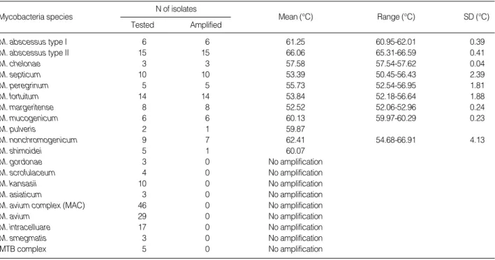
Table 3.Melting point temperatures of the hsp65 gene fragment in the 640 nm channel
(MAC; M. avium 및M. intracellulare)의 융해온도는 48℃ 이 하였고, M. fortuitum의 평균 융해온도는 53.49℃이었다. MAC 의 평균 융해온도가 46.70℃로 다른 비결핵항산균과 구분이 가능 하였다.
2.
hsp65
융해곡선 분석152균주의 비결핵항산균 중 M. gordonae, M. scrofulaceum, M. kansasii, M. asiaticum, M. avium, M. intracelluare, M.
smegmatis는 모든 균주에서 형광의 증폭이 일어나지 않았고M.
pulveris 2균주 중 1균주, M. nonchromogenicum 9균주 중 2균 주, M. schimoidei 5균주 중 4균주에서 형광의 증폭이 일어나지 않았다. 대조군으로 사용된 MTB complex에서도 형광의 증폭이 일어나지 않았다. 융해온도는 최소 50.45℃에서 최대 66.91℃이었 다. M. abscessus type I, M. abscessus type Ⅱ, M. chelonae 의 평균 융해온도는 각각 61.25℃, 66.06℃, 57.58℃로 16S rRNA 융해곡선 분석에서M. abscessus-M chelonae군으로 구분된 균 주를M. abscessus type I, M. abscessus type Ⅱ, M. chelon- ae로 구분할 수 있었다(Table 3).
고 찰
국내의 비결핵항산균의 분리율은 과거에 비해서 증가하고 있으 며[3, 16], 균종별 분리율은M. avium, M. intracelluare가 속하 는M. avium complex (MAC)가 42.1%에서 65.2%로 가장 높 고, 그 다음으로 분리율이 높은 균종은 보고마다 차이가 있으나 M. abscessus, M. fortuitum complex, M. kansasii, M. gordon- ae 등이다[3, 5, 17, 18]. 비결핵항산균은 균종에 따라 치료방법 이 다르고 항균제 감수성이 달라서 정확한 균 동정이 필요하다.
현재 비결핵항산균을 동정하는데는 중합효소연쇄반응-제한절편 길이다형성 분석이 가장 흔히 사용되고 있다. 중합효소연쇄반응- 제한절편길이다형성 분석은 고가의 장비가 불필요한 중합효소연 쇄반응을 시행하므로 신속하고 저렴하게 검사하는 장점이 있다.
그러나 증폭된 DNA 산물의 절편 양상으로 균종을 감별하는 과 정에서 절편양상이 때로는 해석하기 힘들고 명확하지 않을 수 있 어 여러 제한효소를 사용하여야 하므로 다소 번거롭고 시간이 더 소요될 수 있다.
본 연구에서 항산균 동정의 기준으로 사용된 Myco-ID는Hae
Ⅲ 제한효소반응에 의한 절편의 크기를 분석하여 M. abscessus 와M. chelonae를 감별한다. 이때 절편의 크기는M. abscessus 가 125/105/95/42 bp이고M. chelonae가 125/105/95 bp로 마 지막 42 bp의 유무로 감별하는데, 그 크기가 너무 작아 발견되지 못할 수 있고 시발체의 비 특이적인 반응이 나타날 수 있으므로 M. abscessus와M. chelonae를 정확히 감별하기 위해서는Bst E Ⅱ 제한효소를 처리하여 재 확인을 필요로 한다. 그러나 본 연
구의 융해온도분석은 두 비결핵항산균을 빠르고 간편하고 정확하 게 감별할 수 있었다.
본 연구의 16S rRNA 융해곡선 분석과hsp65 융해곡선 분석 을 종합하면M. abscessus type I, M. abscessus type Ⅱ, M.
chelonae를 명확히 구분할 수 있다. M. abscessus type I과M.
abscessus type Ⅱ는hsp65 유전자의 542번째 유전자의 염기가 시토신(C), 티민(T) 여부에 따라서 구분되는 것으로[15] 임상적 인 의의는 알려져 있지 않으며 본 연구에서 항산균 동정의 기준 으로 사용된 중합효소연쇄반응-제한절편길이다형성 분석(Myco- ID)으로는 이루어지지 않는 것이다. M. abscessus와M. chelon- ae가 속하는 신속성장항산균은 림프절염, 창상감염, 폐감염 등을 일으킨다. 폐질환을 주로 일으키는M. abscessus는M. cheloneii subspecies abscessus, M. chelonae subspecies abscessus로 불 리다가 1992년에서야M. abscessus로 분리되었다[14]. 신속성장 항산균은 모든 항결핵제에 내성이므로 항결핵제 대신 amikacin, cefoxitin, imipenem, clarithromycin 등의 항생제(antibacterial agent)로 치료한다[4, 19]. 신속성장항산균 중M. abscessus는 만성 폐 감염에서 분리되는 신속성장항산균의 82%를 차지하는 균이고[20] 창상감염을 일으키며M. abscessus에 의한 폐질환은 외과적 절제가 유일한 치료법이다. M. chelonae는 주로 면역억제 환자에서 발견되며 전신적인 피부병변이나 정맥관 감염의 원인균 으로 항균제로 치료한다. M. abscessus와M. chelonae는 항균제 감수성에 있어서 신속성장항산균의 치료제인 clarithromycin에 대한 감수성 정도가 차이가 있는 균주이다[21]. 본 연구에서M.
abscessus와M. chelonae 융해온도의 표준편차는 16S rRNA와
hsp65 융해곡선 분석 모두에서 다른 균종에 비해 낮은 편으로
(Table 2, 3), M. abscessus와M. chelonae를 감별하는 정확도 가 상대적으로 높을 것으로 생각된다.
이번 연구에서 이용한 다상-실시간-중합효소연쇄반응과 융해 곡선 분석은 가장 흔히 발견되는 비결핵항산균인 MAC을 구분할 수 있었다. 실제로 MAC은 본 검사실에서 분리된 비결핵항산균의 62.2%를 차지하는 균이다[17]. MAC의 치료에는 macrolide와 항 결핵제인 rifampin, ethambutol을 사용하며M. avium과M.
intracelluare 두 균종은 임상적으로 증상, 방사선학적 소견, 치료 에 대한 반응의 차이를 구별할 수 없는 것으로 알려져 있다[22].
본 연구에서 MAC을 평균 융해온도로 다른 비결핵항산균과 구분 할 수 있었으나 MAC을M. avium과M. intracelluare로 분리할 수 없었으며 드물게 발견되는M. smegmatis와M, asiaticum의 융해온도 범위가 중복되는 점이 제한점으로 생각된다. 또한 M.
abscessus, M. chelonae, MAC을 제외한 여러 비결핵항산균의 융해온도가 균종별로 구분되지 않는 것도 본 연구의 제한점으로 생각된다. 다른 실시간 중합효소연쇄반응과 융해곡선 분석을 이용 한 항산균의 동정 연구는 주로 16S rRNA 유전자를 이용하였다.
3개의 16S rRNA 탐색자를 이용한 연구[9]에서는 MTB com- plex와M. avium을 동정할 수 있었다. 16S rRNA 유전자의 과 변이 부위 A (hypervariable region A)의 탐색자를 이용한 연구
[10]에서는M. tuberculosis, M. avium, M. intracellulare, M.
kansasii 등을 동정할 수 있었다. 위의 연구들은 MTB complex 와M. avium, M. intracelluare를 동정할 수 있으나M. absces- sus와M. chelonae를 감별하지는 못하였다.
본 연구에서 주목해야 할 것은 MTB complex가 형광의 증폭이 일어나지 않았다는 것이다. 이는M. tuberculosis에서 16S rRNA 유전자의 1,000 bp 절편과hsp65 유전자의 439 bp 절편의 중합 효소연쇄반응은 일어나지만 M. tuberculosis의 염기서열과 16S rRNA 탐색자, hsp65 탐색자의 염기서열이 부합하지 않아 형광 의 증폭이 일어나지 않은 것이다. 형광의 증폭이 일어나지 않은 비결핵항산균주 또한 염기서열이 탐색자와 부합하지 않기 때문일 것이다. MTB complex는 형광의 증폭이 일어나지 않았고 비결 핵항산균만 증폭이 관찰되어 다상-실시간-중합효소연쇄반응과 융 해곡선 분석이 MTB complex를 구별하는 데에도 유용하게 사용 될 수 있을 것이다.
저자들은 비결핵항산균의 동정에 있어서 다상-실시간-중합효소 연쇄반응과 융해곡선 분석의 유용성을 확인하였다. 다상-실시간- 중합효소연쇄반응과 융해곡선 분석에 의한 비결핵항산균의 동정 은M. abscessus와M. chelonae의 신속한 동정과 MAC 분리에 유용하게 사용될 수 있을 것이다.
요 약
배경 : 비결핵항산균은 균종에 따라 항균제 감수성과 치료방법 에 차이가 있어서 정확한 균종 동정이 필요하다. 본 연구에서는 비결핵항산균 동정에서 실시간 중합효소연쇄반응-융해곡선 분석 의 유용성을 알아보고자 하였다.
방법 : 중합효소연쇄반응-제한절편다형성분석으로 균종 수준까 지 동정된 152개의 임상 균주를 대상으로 하였다. 이 균주에서 16S rRNA 융해곡선 분석과 hsp65 융해곡선 분석을 시행하여 균종을 동정하였다.
결과 : 16S rRNA 융해곡선 분석에서M. abscessus-M. che- lonae군의 융해온도는 65℃ 이상이었고 M. avium complex (MAC; M. avium 및 M. intracellulare)의 융해온도는 48℃
이하로써M. abscessus-M. chelonae군과 MAC을 다른 비결핵 항산균으로부터 구분할 수 있었다. hsp65 융해곡선 분석으로M.
abscessus-M. chelonae군을 융해온도 61.25℃, 66.06℃, 57.58℃
에 따라서 각각M. abscessus type I, M. abscessus type Ⅱ, M. chelonae로 동정할 수 있었다.
결론 : 16S rRNA와hsp65 유전자에 대한 다상-실시간-중합 효소연쇄반응과 융해곡선 분석을 통하여M. abscessus와M. che- lonae를 동정하고 MAC을 분리할 수 있었다. 특히M. abscessus 와M. chelonae는 16S rRNA 융해곡선 분석으로 감별되지 못하 였으나hsp65융해곡선 분석으로 감별할 수 있었다.
참고문헌
1. Portaels F. Epidemiology of mycobacterial diseases. Clin Dermatol 1995;13:207-22.
2. Prince DS, Peterson DD, Steiner RM, Gottlieb JE, Scott R, Israel HL, et al. Infection with Mycobacterium avium complex in patients with- out predisposing conditions. N Engl J Med 1989;321:863-8.
3. Scientific Committee in Korean Academy of Tuberculosis and Res- piratory Dieseases. National survey of mycobacterial diseases other than tuberculosis in Korea. Tuberc Respir Dis 1995;42:277-94. (대한 결핵및호흡기학회학술위원회. 비결핵항산균증전국실태조사. 결핵및 호흡기질환 1995;42:277-94.)
4. Koh WJ, Kwon OJ, Lee KS. Diagnosis and treatment of nontuber- culous mycobacterial pulmonary diseases: a Korean perspective. J Korean Med Sci 2005;20:913-25.
5. Jeong J, Lee SH, Jeong US, Chang CH, Kim SR. Identification of my- cobacteria using high performance liquid chromatography in clini- cal specimens. Korean J Clin Micorbiol 2004;7:148-55. (정윤성, 이선호, 정의석, 장철훈, 김성률. 임상검체에서 high performance liquid chro- matography법을이용한 mycobacteria의동정. 대한임상미생물학회지 2004;7:148-55.)
6. Kim HK, Kim YR, Park JP, Kim NH, OK CH, Jung MH, et al. Isola- tion of nontuberculous mycobacteria by DNA probe and clincal char- acteristics of patients with NTM pulmonary disease. Tuberc Respir Dis 2005;58:248-56. (김희규, 김유리, 박정필, 김낭희, 옥철호, 정만홍 등.
DNA probe를이용한비결핵항산균의분리및폐질환자들의임상적특 징. 결핵및호흡기질환 2005;58:248-56.)
7. Lee H, Park HJ, Cho SN, Bai GH, Kim SJ. Species identification of mycobacteria by PCR-restriction fragment length polymorphism of the rpoB gene. J Clin Microbiol 2000;38:2966-71.
8. Kim H, Kim SH, Shim TS, Kim MN, Bai GH, Park YG, et al. PCR restriction fragment length polymorphism analysis (PRA)-algorithm targeting 644 bp Heat Shock Protein 65 (hsp65) gene for differentia- tion of Mycobacterium spp. J Microbiol Methods 2005;62:199-209.
9. Lachnik J, Ackermann B, Bohrssen A, Maass S, Diephaus C, Punck- en A, et al. Rapid-cycle PCR and fluorimetry for detection of myco- bacteria. J Clin Microbiol 2002;40:3364-73.
10. Shrestha NK, Tuohy MJ, Hall GS, Reischl U, Gordon SM, Procop GW. Detection and differentiation of Mycobacterium tuberculosis and nontuberculous mycobacterial isolates by real-time PCR. J Clin Microbiol 2003;41:5121-6.
11. Telenti A, Marchesi F, Balz M, Bally F, Bottger EC, Bodmer T. Rapid identification of mycobacteria to the species level by polymerase chain reaction and restriction enzyme analysis. J Clin Microbiol 1993;
31:175-8.
12. Shinnick TM. The 65-kilodalton antigen of Mycobacterium tuber-
culosis. J Bacteriol 1987;169:1080-8.
13. Sedlacek L, Rifai M, Feldmann K, Bange FC. LightCycler-based dif- ferentiation of Mycobacterium abscessus and Mycobacterium che- lonae. J Clin Microbiol 2004;42:3284-7.
14. Kusunoki S and Ezaki T. Proposal of Mycobacterium peregrinum sp. nov., nom. rev., and elevation of Mycobacterium chelonae subsp.
abscessus (Kubica et al.) to species status: Mycobacterium abscessus comb. nov. Int J Syst Bacteriol 1992;42:240-5.
15. Ringuet H, Akoua-Koffi C, Honore S, Varnerot A, Vincent V, Berche P, et al. hsp65 sequencing for identification of rapidly growing my- cobacteria. J Clin Microbiol 1999;37:852-7.
16. Koh WJ, Kwon OJ, Yu CM, Jeon K, Suh GY, Chung MP, et al. Recov- ery rate of nontuberculous mycobacteria from acid-fast-bacilli smear- positive sputum speciment. Tuberc Respir Dis 2003;54:22-32. (고원 중, 권오정, 유창민, 전경만, 서지영, 정만표등. 항산균도말양성객담에 서 비결핵성 마이코박테리아의 분리 비율. 결핵 및 호흡기질환 2003;54:
22-32.)
17. Park CM, Heo SR, Park KU, Song JH, Lee JH, Lee CT, et al. Isolation of nontuberculous mycobacteria using polymerase chain reaction- restriction fragment length polymorphism. Korean J Lab Med 2006;
26:161-7. (박철민, 허세란, 박경운, 송정한, 이재호, 이춘택등. 중합효소
연쇄반응-제한절편길이다형성을 이용한 비결핵항상균의 분리. 대한진 단검사의학회지 2006;26:161-7.)
18. Lee JY, Choi HJ, Lee HY, Joung EY, Huh JW, Oh YM, et al. Recovery rate and characteristics of nontuberculous mycobacterial isolates in a university hospital in Korea. Tuberc Respir Dis 2005;58:385-91. (이 정연, 최희진, 이혜영, 정은영, 허진원, 오연목등. 한대학병원에서비결핵 항산균의분리및동정실태. 결핵및호흡기질환 2005;58:385-91.) 19. Wagner D and Young LS. Nontuberculous mycobacterial infections:
a clinical review. Infection 2004;32:257-70.
20. Griffith DE, Girard WM, Wallace RJ Jr. Clinical features of pulmo- nary disease caused by rapidly growing mycobacteria. An analysis of 154 patients. Am Rev Respir Dis 1993;147:1271-8.
21. Yang SC, Hsueh PR, Lai HC, Teng LJ, Huang LM, Chen JM, et al.
High prevalence of antimicrobial resistance in rapidly growing my- cobacteria in Taiwan. Antimicrob Agents Chemother 2003;47:1958- 62.
22. British Thoracic Society. Management of opportunist mycobacterial infections: Joint Tuberculosis Committee Guidelines 1999. Subcom- mittee of the Joint Tuberculosis Committee of the British Thoracic Society. Thorax 2000;55:210-8.