Prognostic factors after hepatic resection for the single hepatocellular carcinoma larger than 5 cm
Ji Hyun Noh, Tae-Seok Kim, Keun Soo Ahn, Yong Hoon Kim, Koo Jeong Kang
Department of Surgery, Dongsan Medical Center, Keimyung Univsersity School of Medicine, Daegu, Korea
INTRODUCTION
Liver resection has been accepted as the best treatment modality to achieve curative goals of hepatocellular carcinoma (HCC), particularly in patients with a single tumor, although nonsurgical treatments such as transarterial chemoembolization (TACE), radiofrequency ablation (RFA), percutaneous ethanol injection (PEIT), and radiation treatment have been performed widely for treatment of HCC in cases of small tumors, multiple bilateral tumors, and anatomically or functionally unresectable tumors. Single HCC has generally manifested good prognosis after resection and accepted as a good candidate. However, tumor size has been considered as an important prognostic factor and adopted in the recent staging system, the 7th edition
of American joint Committee on Cancer (AJCC) cancer staging with a cutoff value 5 cm in size [1,2].
Despite recent advances in diagnostic imaging, HCC fre
quently presents in large size and advanced stage as a result of the absence of early symptoms and poorly performed screening. Consequently tumor sizes were over 10 cm in 7% to 14% of the patients with HCC who underwent surgical resection [36]. Because of increased risk of morbidity and mortality after resection, nonsurgical treatments such as TACE and ra
diation treatment have been often performed for huge HCC [3]. However, surgical indications in liver surgery have been ex panded to include advanced cases in response to technical ad vances [6], and the feasibility of hepatic resection for a large HCC has been established already with statistically similar peri
Purpose: This study aimed to determine which factors affect the prognosis of hepatectomy for hepatocellular carcinoma (HCC) larger than 5 cm, including the prognostic difference between tumor sizes from 5–10 cm and larger than 10 cm.
Methods: The medical records of 114 patients who underwent hepatectomy for single HCC larger than 5 cm were reviewed and analyzed retrospectively.
Results: In the analysis of the entire cohort of 114 patients, the 5-year overall and diseases-free survival rates were 50%
and 29%, respectively. In a comparison of survival rates between groups, tumor sizes of 5 to 10 cm and larger than 10 cm, the overall and disease-free survival rates were not significantly different, respectively (54% vs. 41%, P = 0.433 and 33% vs. 23%, P = 0.083). On multivariate analysis, positive hepatitis B, high prothrombin induced by vitamin K absence or antagonist-II levels over 200 mIU/mL, and vascular invasion (micro- and macrovascular invasion) were independent prognostic factors for recurrence after hepatic resection. However, tumor size larger than 10 cm was not significant for recurrence after resection.
Conclusion: This study shows that surgical resection of solitary HCC larger than 5 cm showed favorable overall survival.
And there is no survival difference with tumors between 5–10 cm and larger than 10 cm.
[Ann Surg Treat Res 2016;91(3):104-111]
Key Words: Hepatocellular carcinoma, Hepatectomy, Prognosis
Reviewed January February March April May June July August September October November December
http://dx.doi.org/10.4174/astr.2016.91.3.104 Annals of Surgical Treatment and Research
Received March 16, 2016, Revised April 27, 2016, Accepted May 16, 2016 Corresponding Author: Koo Jeong Kang
Division of Hepatobiliary & Pancreatic Surgery, Department of Surgery, Dongsan Medical Center, Keimyung University School of Medicine, 56 Dalseong-ro, Jung-gu, Daegu 41931, Korea
Tel: +82-53-250-7655, Fax: +82-53-250-7322 E-mail: kjkang@dsmc.or.kr
Copyright ⓒ 2016, the Korean Surgical Society
cc Annals of Surgical Treatment and Research is an Open Access Journal. All articles are distributed under the terms of the Creative Commons Attribution Non- Commercial License (http://creativecommons.org/licenses/by-nc/4.0/) which permits unrestricted non-commercial use, distribution, and reproduction in any medium, provided the original work is properly cited.
operative morbidity and mortality rates [3,5].
Tumor size has been considered a significant factor for intrahepatic and extrahepatic recurrence. However, many studies reported favorable survival with 5year survival rates exceeding 30% after resection, even in tumor sizes larger than 10 cm; and tumor size is not a significant prognostic factor after resection in cases of tumor size larger than 5 cm [2,3,58].
On the other hand, another study reported better survival outcome after resection in patients with tumor sizes below 10 cm than over 10 cm [3]. Therefore, it is still necessary to validate the influence of tumor size on prognosis after resection in large HCC using a larger volume of patients or different settings.
Regarding the prognosis after curative resection of HCC, various pathologic factors such as vascular invasion [5,9], multi
plicity [9,10], EdmonsonSteiner grade and tumor markers [9,11]
are known to predict the outcome after resection. In addition to pathologic factors, other factors including tumor size, viral status, margin status, and underlying liver disease should be elucidated in large HCC. Therefore, the aim of this study is to report longterm outcomes and to identify prognostic factors after surgical resection in patients with single HCC larger than 5 cm in diameter and to assess the influence of tumor size on prognosis after resection in large HCC.
METHODS
A total of 421 patients underwent hepatic resection for HCC at Keimyung Univsersity Dongsan Medical Center in Daegu between January 2001 and November 2013. Among these patients, 114 patients (27.1%) had single HCC larger than 5 cm and were included in this study. In this study, single HCC was defined based on the preoperative imaging studies regardless of post operative pathologic results including satellite nodules and/
or vascular invasion. The medical records of these patients were reviewed retrospectively and the following data were collected for each patient: demographics; laboratory data including tumor marker and hepatitis serologic test; tumor pathology; operative outcomes; date of last followup, recurrence, and death. Hepatic reserve was assessed using ChildPugh classification and preoperative Indocyanine green retention at 15 minutes (ICG R15) was routinely performed to assess liver function. Tumor size was defined as the largest diameter of the tumor in the specimen. Anatomical resection was defined as the systematic resection of hepatic segment according to the segmental and sectional anatomy described at the International Hepato
Pancreato Biliary Association Brisbane meeting in 2000.
Vascular invasion is classified as macrovascular invasion, which is grossly recognizable mostly in large to medium vessels, and microvascular invasion, which can be defined as the presence of tumor emboli mainly in small vessels such as portal vein branches in portal tracts, central veins in noncancerous
liver tissue and venous vessels in the tumor capsule [2,12].
Tumor grade was assessed using the nuclear grading scheme outlined by Edmondson and Steiner. Grades 1 and 2 were considered lowgrade HCC, and grades 3 and 4 were considered high grade.
The routine followup program consisted of physical examina
tion, CT and laboratory tests including αFP and prothrombin induced by vitamin K absence or antagonistII (PIVKAII) level every 3 month for the first year, and then every 6 month for next 5 years, thereafter annually for patients who have neither recurrence nor metastasis. Recurrence was defined as the ap
pearance of a new lesion compatible with HCC in radiologic examination during followup period.
Statistical analysis was performed with IBM SPSS ver. 18.0 (IBM Co., Armonk, NY, USA). Cumulative survival curves were analyzed by using the KaplanMeier method, and significance was determined by logrank test. To investigate the prognostic factors predicting tumor recurrence, univariate and stepwise multivariate regression analysis was performed using a Cox proportional hazard model with P < 0.05 considered statistically significant.
RESULTS
Demography and clinicopathologic features
The demographics and clinical features of the 114 patients are summarized in Table 1. Among these patients 89 patients were male (78.1%) and 25 patients were female (21.9%). The mean age of the patients was 56.2 years. All of these patients were classified as ChildPugh A in preoperative liver function assessment. The median preoperative αFP and PIVKAII were 69.1 ng/mL and 61.8 mIU/mL, and the mean preoperative ICG R15 level was 11.2%. The mean tumor size was 9.1 cm.
Among these patients, 73 patients (64%) had tumors measuring between 5 and 10 cm and 41 patients (36%) had tumors larger than 10 cm. The location of tumor was dominant in right liver (57%) and HBsAg was detected in 86 patients (75.4%). Anato
mical resection was performed in 91 patients (79.8 %) and nona
natomical tumorectomy was performed in 23 patients (20.2%).
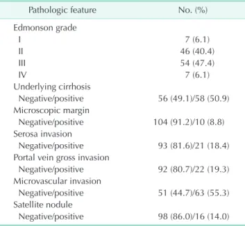
Pathological features of the patients in this study are summa
rized in Table 2. Portal vein gross invasion was identified in 22 patients (19.3%) and microscopic vascular invasion in 63 patients (55.3%), and satellite nodule was detected in 16 patients (14%).
Underlying liver cirrhosis was identified in 58 patients (50.9%).
Resection margin was grossly free from tumor in all patients.
However, microscopic margin status of 10 patients (8.8%) was positive.
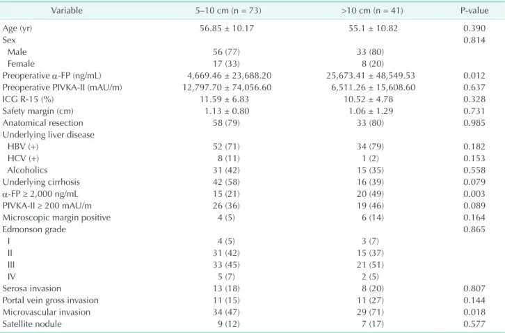
Clinicopathologic features and survival according to tumor size
In comparison of the clinicopathologic characteristics bet
ween groups stratified on the basis of tumor size (5–10 cm and over 10 cm), there were significant differences in preoperative αFP level (P = 0.01) and microvascular invasion of the tumor (P
= 0.02). However, other demographics and pathologic findings were not significantly different between groups and anatomical resection was performed similarly in both groups (Table 3).
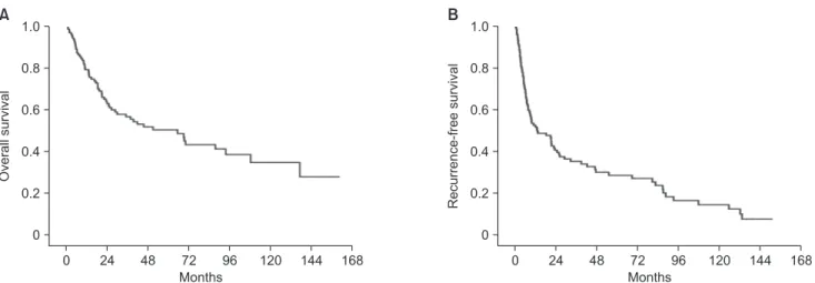
The 1, 3 and 5year overall survival rates were 84%, 62%, and 54% in patients with tumors 5–10 cm, and 72%, 46%, and 41%
in patients with tumors over 10 cm, respectively (Fig. 1A). The 1, 3 and 5year recurrencefree survival rates were 63%, 41%, and 33% in patients with tumors 5–10 cm, and 35%, 26%, and 23% in patients with tumors over 10 cm, respectively (Fig. 1B).
Although there were significant differences in preoperative αFP and the rate of microvascular invasion that are known as prognostic factors after resection, there was no significant
difference in overall and recurrencefree survival between patients with tumors 5–10 cm and those with tumors over 10 cm (P = 0.433 and P = 0.083. respectively).
Survival & recurrence of the entire cohort
The median followup period was 26.4 months (range, 0.8–159.7 months). During a followup period, tumor recurrence occurred in 85 patients (74.6%) and median time to recurrence was 11.2 months after surgery, respectively. At the time of last followup, 59 patients (51.8%) had died of recurrent disease progression. Inhospital mortality occurred in only 1 pa tient (0.9%). For the entire cohort of 114 patients, 1, 3 and 5year overall survival rates were 79%, 57%, and 50%, (Fig. 2A) and recurrencefree survival rates were 53%, 36%, and 29%, respec
tively (Fig. 2B).
Prognostic factor analysis for tumor recurrence
The outcome of univariate and multivariate analysis of risk factors for tumor recurrence is summarized in Table 4. In univariate analysis, positive HBsAg, high level of α-FP (≥2,000 ng/mL), high level of PIVKA-II (≥200 mIU/mL), Edmonson- Steiner grade III/IV, the presence of portal vein gross invasion and microvascular invasion, and the presence of satellite nodule were significant factors to predict tumor recurrence after re
sec tion. However, tumor size over 10 cm, positive surgical margin, and anatomical resection were not associated with tumor recurrence. Multivariate analysis revealed that positive HBsAg (hazard ratio [HR], 1.94; P = 0.043), PIVKA ≥200 mIU/
mL (HR, 3.07; P < 0.001), portal vein gross invasion (HR, 2.30; P
= 0.011) and microvascular invasion (HR, 2.15; P = 0.004) were Table 1. Clinical features of 114 patients who underwent
resection for single hepatocellular carcinoma larger than 5 cm
Variable Value
Age (yr) 56.2 ± 10.40
Sex
Male:female 89 (78.1):25 (21.9)
Etiology of liver disease
Hepatitis B 86 (75.4)
Hepatitis C 9 (7.9)
Hepatitis B & C 3 (2.6)
Alcoholics 46 (40.4)
Nonviral, nonalcoholic 10 (8.8) Preoperative ICG R15 11.2 ± 6.17
αFP (ng/mL) 69.05 (1–200,000)
PIVKAII (mAU/m) 61.82 (3–589,500)
Tumor size (cm) 9.1 ± 3.58
5–10 73 (64.0)
>10 41(36.0)
Tumor location
Right 65 (57.0)
Left 35 (30.7)
Central 12 (10.5)
Caudate 2 (1.8)
Surgical resection
Anatomical 91 (79.8)
Nonanatomical 23 (20.2)
Inhospital mortality 1 (0.9)
Recurrence time (mo)
Mean 29.0 ± 37.48
Median (range) 11.2 (0.3–150.9)
Followup period (mo)
Mean 42.3 ± 39.86
Median (range) 26.4 (0.3–159.7)
Values are presented as mean ± standard deviation, number (%), or median (range).
ICG R15, Indocyanine green retention at 15 minutes; PIVKAII, prothrombin induced by vitamin K absence or antagonistII.
Table 2. Pathologic features of 114 patients who underwent resection for single hepatocellular carcinoma larger than 5 cm
Pathologic feature No. (%)
Edmonson grade
I 7 (6.1)
II 46 (40.4)
III 54 (47.4)
IV 7 (6.1)
Underlying cirrhosis
Negative/positive 56 (49.1)/58 (50.9) Microscopic margin
Negative/positive 104 (91.2)/10 (8.8) Serosa invasion
Negative/positive 93 (81.6)/21 (18.4) Portal vein gross invasion
Negative/positive 92 (80.7)/22 (19.3) Microvascular invasion
Negative/positive 51 (44.7)/63 (55.3) Satellite nodule
Negative/positive 98 (86.0)/16 (14.0)
independent risk factors for HCC recurrence after resection in patients with single HCC larger than 5 cm. The differences of recurrentfree survival according to independent prognostic fac
tors revealed in this study were shown in Fig. 3.
DISCUSSION
In patients with large HCC, longterm prognosis is generally considered to be poor [13,14]. In previous studies, the size of Table 3. Comparison of clinicopathologic features of patients underwent resection for single hepatocellular carcinoma larger than 5 cm
Variable 5–10 cm (n = 73) >10 cm (n = 41) Pvalue
Age (yr) 56.85 ± 10.17 55.1 ± 10.82 0.390
Sex 0.814
Male 56 (77) 33 (80)
Female 17 (33) 8 (20)
Preoperative αFP (ng/mL) 4,669.46 ± 23,688.20 25,673.41 ± 48,549.53 0.012
Preoperative PIVKAII (mAU/m) 12,797.70 ± 74,056.60 6,511.26 ± 15,608.60 0.637
ICG R15 (%) 11.59 ± 6.83 10.52 ± 4.78 0.328
Safety margin (cm) 1.13 ± 0.80 1.06 ± 1.29 0.731
Anatomical resection 58 (79) 33 (80) 0.985
Underlying liver disease
HBV (+) 52 (71) 34 (79) 0.182
HCV (+) 8 (11) 1 (2) 0.153
Alcoholics 31 (42) 15 (35) 0.558
Underlying cirrhosis 42 (58) 16 (39) 0.079
αFP ≥ 2,000 ng/mL 15 (21) 20 (49) 0.003
PIVKAII ≥ 200 mAU/m 26 (36) 19 (46) 0.089
Microscopic margin positive 4 (5) 6 (14) 0.164
Edmonson grade 0.865
I 4 (5) 3 (7)
II 31 (42) 15 (37)
III 33 (45) 21 (51)
IV 5 (7) 2 (5)
Serosa invasion 13 (18) 8 (20) 0.807
Portal vein gross invasion 11 (15) 11 (27) 0.144
Microvascular invasion 34 (47) 29 (71) 0.018
Satellite nodule 9 (12) 7 (17) 0.577
Values are presented as mean±standard deviation or number (%).
PIVKAII, prothrombin induced by vitamin K absence or antagonistII; ICG R15, Indocyanine green retention at 15 minutes.
Fig. 1. Overall survival curve (A) and recurrencefree survival curve (B) according to tumor size, between 5–10 cm and over 10 cm.
5 10 cm
> 10 cm
0 24 48 72 96 120 144 168
Overallsurvival
0 1.0
0.8
0.6
0.4
0.2
A
0 24 48 72 96 120 144 168
Recurrence-freesurvival
0 1.0
0.8
0.6
0.4
0.2
B
Months Months
5 10 cm
> 10 cm
P = 0.433 P = 0.082
HCC has been considered an independent risk factor for patient survival and tumor recurrence. It may be related with higher incidence of occult vascular invasion, satellite nodules, and more advanced histologic grade in large HCC than small HCC [9].
The cutoff value that has an influence on the survival after resection for HCC was defined as 5 cm in the 7th edition of AJCC cancer staging system. In the AJCC 7th cancer staging system, T stage of HCC is classified based on vascular invasion, tumor multiplicity, and tumor size (5 cm). This is based on a report that identified independent prognostic factors after surgical resection by survival analysis of 557 patients collected from 4 centers [2]. In this report, tumor size had no effect on patient survival in patients with a single tumor without vascular
invasion, while large tumor size over 5 cm had an effect on patient survival in cases of multiple tumors or presence of vascular invasion. However, tumor size was not a prognostic factor after resection even if tumor size was larger than 10 cm in patients with HCC over 5 cm in size [2]. In addition to this study, another study by a Tokyo group reported comparable survival between patients with HCC 5–10 cm and over 5 cm, regardless of significant differences in prognostic factors such as underlying liver status including cirrhosis, tumor markers, histologic grade, microvascular invasion and satellite nodules [6]. Moreover, excellent longterm survival rates after resection in patients with single large HCC have been reported in several studies [1517].
Fig. 2. Overall survival curve (A) and recurrencefree survival curve (B) for entire cohort after surgical resection for single hepa
tocellular carcinoma larger than 5 cm. The 1, 3, and 5year overall survival rates were 79%, 57%, and 50%. The 1, 3, and 5year recurrencefree survival rates were 53%, 36%, and 29%, respectively.
0 24 48 72 96 120 144 168
Overallsurvival
0 1.0
0.8
0.6
0.4
0.2
A
0 24 48 72 96 120 144 168
Recurrence-freesurvival
0 1.0
0.8
0.6
0.4
0.2
B
Months Months
Table 4. Prognostic factors associated with hepatocellular carcinoma recurrence
Variable Univariate Multivariate
Pvalue HR (95% CI) Pvalue HR (95% CI)
HBV (+) 0.03 1.77 (1.06–2.97) 0.043 1.94 (1.02–3.69)
HCV (+) 0.21 1.60 (0.76–3.36)
Alcoholics 0.296 0.79 (0.51–1.22)
αFP > 2,000 ng/mL 0.031 1.64 (1.04–2.59)
PIVKAII > 200 mAU/m 0.001 2.26 (1.39–3.67) <0.001 3.07 (1.80–5.23)
Underlying cirrhosis (+) 0.233 1.30 (0.84–2.00)
Tumor size > 10 cm 0.084 1.47 (0.94–2.29)
Nonanatomic resection 0.884 1.04 (0.68–1.75)
Edmonson grade III/IV 0.008 1.81 (1.17–2.79)
Margin < 0.5 0.79 1.06 (0.67–1.70)
Microscopic margin positive 0.087 1.78 (0.92–3.46)
Serosa invasion (+) 0.281 1.34 (0.78–2.29)
Portal vein gross invasion 0.004 2.09 (1.26–3.47) 0.011 2.30 (1.21–4.38)
Microvascular invasion 0.002 1.98 (1.28–3.06) 0.004 2.15 (1.28–3.61)
Satellite nodule positive 0.016 2.06 (1.14–3.70)
HR, hazard ratio; CI, confidence interval; PIVKAII, prothrombin induced by vitamin K absence or antagonistII.
In our study, our group analyzed the survival rate after resection in patients with HCC larger than 5 cm, and the 3, 5year overall and recurrentfree survival rates were 57%, 50% and 36%, 29%, respectively. The overall and recurrent
free survival rates after surgical resection in patients with HCC over 10 cm were not significantly different from those in patients with HCC 5–10 cm even though there were significant differences in αFP level and microvascular invasion, and these results are comparable with the data from other reports [3,5
7]. Therefore, our data suggest that surgical resection of large single HCC is feasible and should be considered in patients with resectable HCC regardless of tumor size.
Recent studies showed that macrovascular invasion [5,9], microvascular invasion [9,10], elevated αFP level [10,11], pre
sence of liver cirrhosis [9,10], presence of satellite nodules [6]
and multiple tumor nodules [911] influence prognosis after hepatic resection in patients with single large HCC. In our study, 4 prognostic factors predicting tumor recurrence after resection for single large HCC were identified; hepatitis B, PIVKAII level over 200 mIU/mL, portal vein gross invasion and microvascular invasion. However, αFP, presence of satellite nodule, presence
of liver cirrhosis, and histologic grade, which have been known as poor prognostic factors, were not associated with tumor recurrence in our study. The presence of satellite nodules was significantly associated with tumor recurrence in univariate analysis; however, not significant in multivariate analysis.
This may have been caused by the small number of patients in whom satellite nodules were identified.
One of the interesting results in our study is that microscopic positive resection margin status shows no adverse effect on recurrence and survival. This result is consistent with other reports [6,18]. In our study, capsule exposure on resection margin was interpreted as microscopic positive margin. How
ever, tumor capsule was preserved intact in most cases. It is likely that tumor capsule exposure on resection margin would have less impact on prognosis after resection in encapsulated large HCC. Therefore, if tumor capsule is grossly intact and has no evidence of tumor invasion, capsule exposure on resection margin could be considered as negative margin. Practically, in cases where the tumor is very close to major vessels, paren
chymal dissection near vessels should be performed using careful techniques, which can preserve capsule and prevent Fig. 3. Recurrencefree survival curves according to HBV (A), prothrombin induced by vitamin K absence or antagonistII (B), the presence of portal vein gross invasion (C), and the presence of microvascular invasion (D).
0 24 48 72 96 120 144 168
0 1.0
0.8
0.6
0.4
0.2
A
Months
Recurrence-freesurvival
0 24 48 72 96 120 144 168
0 1.0
0.8
0.6
0.4
0.2
C
Months
Recurrence-freesurvival
HBV ( ) HBV (+)
P = 0.028
0 24 48 72 96 120 144 168
Recurrence-freesurvival
0 1.0
0.8
0.6
0.4
0.2
B
Months
PIVKA < 200 PIVKA > 200
P = 0.001
No invasion PV invasion
P = 0.003
0 24 48 72 96 120 144 168
Recurrence-freesurvival
0 1.0
0.8
0.6
0.4
0.2
D
Months
No invasion MV invasion
P = 0.002
cancer spread, such as Kelly clamping technique rather than using Cavitron Ultrasonic Surgical Aspirator technique.
Our study showed high recurrence rate (74.6%, 11.2 median recurrence months) after resection of single HCC larger than 5 cm. Because of the high recurrence rate, postoperative restric
tive surveillance by checking tumor markers and imaging studies every 3–6 months are needed and appropriate treat
ments by multimodal approach such as repeat resection, TACE, RFA, and PEIT should be performed if recurrence is detected.
There are some limitations in this study. This study is a retrospective, singlecenter study and thus the results cannot be generalized. Therefore, a highpowered multicenter study with large cohort should be performed to validate our results. And the presence of comorbidity such as diabetes, cardiopulmonary disease, and cerebrovascular disease, which can have influence on the survival, was not assessed exactly in this study due to the limitations of a retrospective study. Another limitation of this study is that the different types and strengths of treatment after HCC recurrence in each patient were not assessed in this study. However, the survival status of all patients was com
pletely identified through the assistance of the Korean National Health Insurance Service, and the most important variables for predicting prognosis are included in this study.
In conclusion, this study showed that tumor recurrence after surgical resection for single HCC larger than 5 cm is signi
ficantly influenced by the presence of portal vein gross invasion and microscopic vascular invasion, HBV, and elevated PIVKA
II. Hepatic resection for single HCC larger than 5 cm showed favorable survival outcome and there was no significant differ
ence in survival after hepatic resection for single HCC larger than 10 cm compared to tumor sizes of 5–10 cm. Therefore, tumor size alone is not a poor prognostic factor after resection and surgical resection could be considered in patients with resectable single large HCC regardless of size.
CONFLICTS OF INTEREST
No potential conflict of interest relevant to this article was reported.
1. Huang WJ, Jeng YM, Lai HS, Sheu FY, Lai PL, Yuan RH. Tumor size is a major deter
minant of prognosis of resected stage I hepatocellular carcinoma. Langenbecks Arch Surg 2015;400:72534.
2. Vauthey JN, Lauwers GY, Esnaola NF, Do KA, Belghiti J, Mirza N, et al. Simplified staging for hepatocellular carcinoma. J Clin Oncol 2002;20:152736.
3. Chen JH, Wei CK, Lee CH, Chang CM, Hsu TW, Yin WY. The safety and adequacy of resection on hepatocellular carcinoma lar
ger than 10 cm: a retrospective study over 10 years. Ann Med Surg (Lond) 2015;4:193
9.
4. Kee KM, Wang JH, Lee CM, Chen CL, Changchien CS, Hu TH, et al. Validation of clinical AJCC/UICC TNM staging sys
tem for hepatocellular carcinoma: analy
sis of 5,613 cases from a medical cen ter in southern Taiwan. Int J Cancer 2007;120:
26505.
5. Lee SG, Hwang S, Jung JP, Lee YJ, Kim KH, Ahn CS. Outcome of patients with huge hepa tocellular carcinoma after primary
resec tion and treatment of recurrent le
sions. Br J Surg 2007;94:3206.
6. Lim C, Mise Y, Sakamoto Y, Yamamoto S, Shindoh J, Ishizawa T, et al. Above 5 cm, size does not matter anymore in patients with hepatocellular carcinoma. World J Surg 2014;38:29108.
7. Shah SA, Wei AC, Cleary SP, Yang I, Mc
Gilvray ID, Gallinger S, et al. Prognosis and results after resection of very large (>or=10 cm) hepatocellular carcinoma. J Gastrointest Surg 2007;11:58995.
8. Hwang S, Lee YJ, Kim KH, Ahn CS, Moon DB, Ha TY, et al. Longterm outcome after resection of huge hepatocellular car cinoma ≥ 10 cm: single-institution ex- pe rience with 471 patients. World J Surg 2015;39:251928.
9. Pawlik TM, Delman KA, Vauthey JN, Nagorney DM, Ng IO, Ikai I, et al. Tumor size predicts vascular invasion and histo
logic grade: implications for selection of surgical treatment for hepatocellular car
cinoma. Liver Transpl 2005;11:108692.
10. Pandey D, Lee KH, Wai CT, Wagholikar
G, Tan KC. Long term outcome and prog
no stic factors for large hepatocellular car cinoma (10 cm or more) after surgical re section. Ann Surg Oncol 2007;14:2817
23.
11. Yeh CN, Lee WC, Chen MF. Hepatic re sec
tion and prognosis for patients with hepa
tocellular carcinoma larger than 10 cm:
two decades of experience at Chang Gung memorial hospital. Ann Surg Oncol 2003;
10:10706.
12. Ikai I, Yamamoto Y, Yamamoto N, Tera
jima H, Hatano E, Shimahara Y, et al.
Results of hepatic resection for hepa to
cellular carcinoma invading major portal and/or hepatic veins. Surg Oncol Clin N Am 2003;12:6575.
13. The Liver Cancer Study Group of Japan.
Predictive factors for long term prognosis after partial hepatectomy for patients with hepatocellular carcinoma in Japan.
Cancer 1994;74:277280.
14. TungPing Poon R, Fan ST, Wong J. Risk factors, prevention, and management of postoperative recurrence after resection
REFERENCES
of hepatocellular carcinoma. Ann Surg 2000;232:1024.
15. Ariizumi S, Kotera Y, Takahashi Y, Katagiri S, Yamamoto M. Impact of hepatectomy for huge solitary hepatocellular carci
noma. J Surg Oncol 2013;107:40813.
16. Pawlik TM, Poon RT, Abdalla EK, Zorzi D, Ikai I, Curley SA, et al. Critical appraisal
of the clinical and pathologic predictors of survival after resection of large hepa
tocellular carcinoma. Arch Surg 2005;140:
4507.
17. Shimada K, Sakamoto Y, Esaki M, Kosuge T. Role of a hepatectomy for the treatment of large hepatocellular carcinomas mea
suring 10 cm or larger in diameter. Lan
genbecks Arch Surg 2008;393:5216.
18. Shindoh J, Hasegawa K, Inoue Y, Ishizawa T, Nagata R, Aoki T, et al. Risk factors of postoperative recurrence and adequate surgical approach to improve longterm outcomes of hepatocellular carcinoma.
HPB (Oxford) 2013;15:319.