Received September 24, 2012, Revised March 19, 2013, Accepted for publication April 27, 2013
Corresponding author: Seong Jun Seo, Department of Dermatology, Chung-Ang University Hospital, 102 Heukseok-ro, Dongjak-gu, Seoul 156-755, Korea. Tel: 82-2-6299-1538, Fax: 82-2-6264-0129, E-mail:
This is an Open Access article distributed under the terms of the Creative Commons Attribution Non-Commercial License (http://
creativecommons.org/licenses/by-nc/3.0) which permits unrestricted non-commercial use, distribution, and reproduction in any medium, provided the original work is properly cited.
ORIGINAL ARTICLE
Calcitriol May Down-Regulate mRNA Over-Expression of Toll-Like Receptor-2 and -4, LL-37 and Proinflammatory Cytokines in Cultured Human Keratinocytes
Mi Sook Jeong, Ji-Yun Kim, He In Lee, Seong Jun Seo
Department of Dermatology, Chung-Ang University Hospital, Seoul, Korea
Background: Although vitamin D analogs have been used in the topical treatment of psoriasis, their mechanisms of action are not well understand. Calcitriol, the hormonally active vitamin D3 metabolite, has been demonstrated to exert immunomodulatory effects in the skin by down-regulating the expression of Toll-like receptors (TLRs) and proinflamm- atory cytokines. Objective: We investigated the effects of calcitriol on the expression of TLR2, TLR4, antimicrobial peptide LL-37, and proinflammatory cytokines in cultured human keratinocytes. Methods: The mRNA expression levels of TLR2, TLR4, tumor necrosis factor α (TNF-α), interleukin (IL)-1β and LL-37 in cultured human keratino- cytes were measured by real-time polymerase chain reaction (PCR) and reverse transcription (RT). Furthermore, we measured supernatant TNF-α levels by an enzyme- linked immunosorbent assay (ELISA) to confirm the effects of cal- citriol on TLR2 and TLR4. Results: As measured by RT-PCR and real-time PCR, calcitriol was found to suppress the lipopolysaccharide- and ultraviolet B radiation-mediated induction of expression of TLRs, LL-37 and proinflammatory cytokines such as TNF-α and IL-1β in normal human keratinocytes. The supernatant TNF-α levels measured by ELISA were also suppressed after treatment with calcitriol.
Conclusion: Calcitriol may down- regulate inflammatory
stated over-expression of LL-37 and proinflammatory cyto- kines. (Ann Dermatol 26(3) 296∼302, 2014)
-Keywords-
Calcitriol, TLR2, TLR4
INTRODUCTION
The hormonally active vitamin D3 metabolite calcitriol (also known as 1,25-dihydroxyvitamin D3 or 1,25(OH)2D3) has immunomodulatory effects in the skin in addition to its roles in bone metabolism, calcium homeostasis, cell differentiation, proliferation. Calcitriol also cause changes in cytokine expression, suppresses keratinocyte prolifera- tion, promotes keratinocyte differentiation, and induces the expression of antimicrobial peptides (AMPs) such as hu- man β-defensin 3 (HBD-3) and the human cathelicidin family member LL-37.
There is convincing evidence that vitamin D3 directly regulates AMP gene expression in human skin1,2. The pro- moter regions of the human cathelicidin AMPs (CAMP) and defensin 2 genes contain consensus vitamin D res- ponsive elements (VDREs) that mediate calcitriol-depen- dent gene expression3, and vitamin D has been shown to enhance the expression of LL-37 in cultured human kera- tinocytes in vivo1. Induction of CAMP expression in kera- tinocytes and monocytes is mediated by Toll-like recep- tors (TLRs)4,5. However, vitamin D3 was shown to down- regulate the expression of TLR2 and TLR4 in a human monocyte in vitro model6, to dose-dependently suppress the protein and mRNA levels of TLR2 and TLR4 in monocytes7. Vitamin D-induced LL-37 up-regulation wou- ld therefore be expected to worsen inflammation in pso- riasis; however, vitamin D analogs have long been used in
the topical treatment of psoriasis. While one study demon- strated that the vitamin D analog calcipotriol suppressed the expression of HBD-2 and LL-37 induced by lipopo- lysaccharide (LPS) or ultraviolet B (UVB) irradiation in cultured human keratinocytes8, the molecular effects of vitamin D on TLRs and AMP such as LL-37 have not been elucidated in keratinocytes.
For this reason, we sought to determine the effects of cal- citriol on the expression of TLR2, TLR4, LL-37 in cultured human keratinocytes. Furthermore, we performed cytoki- ne analysis for tumor necrosis factor α (TNF-α) and inter- leukin 1β (IL-1β) to confirm the effects of calcitriol on TLR2 and TLR4.
MATERIALS AND METHODS
Cell isolation and culture
For harvesting normal human keratinocytes (NHKs), neon- atal foreskin was obtained from neonatal circumcision specimens and primary culture was carried out. Briefly, neonatal foreskin was chopped into 1-mm pieces and trypsinized at room temperature overnight. After vortexing the sample vigorously and incubating for 5 minutes, the supernatant was plated in 25-cm2 culture flasks and in- cubated in 5% CO2 at 37oC in keratinocyte growth me- dium (KGM; Clonetics, East Rutherford, NJ, USA) con- taining growth supplements and a calcium concentration of 0.03 mM. After 4 passages, cultured keratinocytes were plated at 2×10⁵ cells/ml in a standard flat-bottomed plate.
The keratinocytes were starved overnight in keratinocyte basal medium supplemented with serum-free KGM . The cells were divided into six groups as follows: negative control group, LPS-treated group (5 μg/ml; Sigma, St. Louis, MO, USA), UVB-irradiated group (20 mJ/cm2), calcitr- iol-treated group (10 nM; Sigma), LPS plus calcitriol- treated group, and UVB plus calcitriol-treated group. The LPS plus calcitriol-treated group was treated with 10 nM calcitriol 30 minutes after the LPS treatment, and the UVB plus calcitriol-treated group was treated with 10 nM calcitriol 30 minutes after the UVB irradiation and incu- bated for 24 hours.
Ultraviolet B irradiation
The UVB irradiation of 20 mJ/cm2, which was chosen based on preliminary data, was delivered with a Philips TL 20W/12 (Philips, Eindhoven, Netherlands) fluorescent bu- lb emitting 280 to 320 nm wavelengths with a peak at 313 nm. Before UVB irradiation, the medium was removed and replaced with phosphate-buffered saline. Irradiation output was monitored with a Waldmann UV-meter (Wal- dmann, Villigen-Schwenningen, Germany).
Preparaton of primers
Polymerase chain reaction (PCR) primers were designed based on Gene Bank (www.ncbi.nlm.nih.gov) data using a DNA synthesizer (Pharmacia; Bjorkgatan, Uppsala, Swe- den). The sequences were as follows:
TLR-2 (298bp)
5’- GGC CAG CAA ATT ACC TGT GT-3’ (sense) and 5’- TTC TCC ACC CAG TAG GCA TC-3’ (anti-sense);
TLR-4 (167bp)
5’- TGA GCA GTC GTG CTG GTA TC-3’ (sense) and 5’- CAG GGC TTT TCT GAG TCG TC-3’ (anti-sense);
TNF-α (219bp)
5’- CAG AGG GCC TGT ACC TCA TCT GA-3’ (sense) and 5’- GGA AGA CCC CTC CCA GAT AG-3’ (anti-sense);
IL-1β (205bp)
5’- GGG CCT CAA GGA AAA GAA TC -3’ (sense) and 5’- TTC TGC TTG AGA GGT GCT GA-3’ (anti-sense);
LL-37 (183bp)
5’- GCT AAC CTC TAC CGC CTC CT -3’ (sense) and 5’- GGT CAC TGT CCC CAT ACA CC-3’ (anti-sense);
GAPDH (238bp)
5’- GAG TCA ACG GAT TTG GTC GT-3’ (sense) and 5’-TTG ATT TTG GAG GGA TCT CG-3’ (anti-sense).
Reverse transcription-polymerase chain reaction Total RNA was isolated from one dish of cultured kera- tinocytes using 1 ml of TRIzol reagent (Invitrogen, Carls- bad, CA, USA). After 5 minutes at room temperature, 0.2 ml of chloroform/ml of TRIzol reagent was added, tubes were shaken vigorously by hand for 15 seconds, incu- bated at 15oC to 30oC for 3 minutes. The mixtures were centrifuged at 12,000 rpm (14,000 g) at 4oC for 15 mi- nutes, the upper aqueous phase was transferred to a fresh tube, and an equal amount of 2-propanol was added. After mixtures were incubated at 4oC for 15 minutes, they were centrifuged at 12,000 rpm at 4oC for 15 minutes. The supernatant was removed, and RNA pellets were washed with 500 μl of 70% ethanol, centrifuged at 12,000 rpm at 4oC for 5 minutes, briefly dried. The purified RNA was dissolved in 30 μl of diethyl pyrocarbonate-distilled wa- ter. Three micrograms of total cellular RNA was reverse transcribed at 42oC for 30 minutes in a 20 μl volume containing 1 μl of reverse transcriptase (TaKaRa; Shiga, Japan), 2 μl of 10× buffer, 2 μl of 10 mM dNTP, 1 μl of oligo dT primer solution, 0.5 μl of RNase inhibitor and 4 μl of 25 mM MgCl2. Two microliters of each resulting cDNA sample was amplified by PCR in 25 μl containing 2.5 μl of 10× buffer, 2.5 μl of 25 mM MgCl2 and 0.75 μl of 10 pmol primer solution.
Thermal cycle profiles were conducted using the follo-
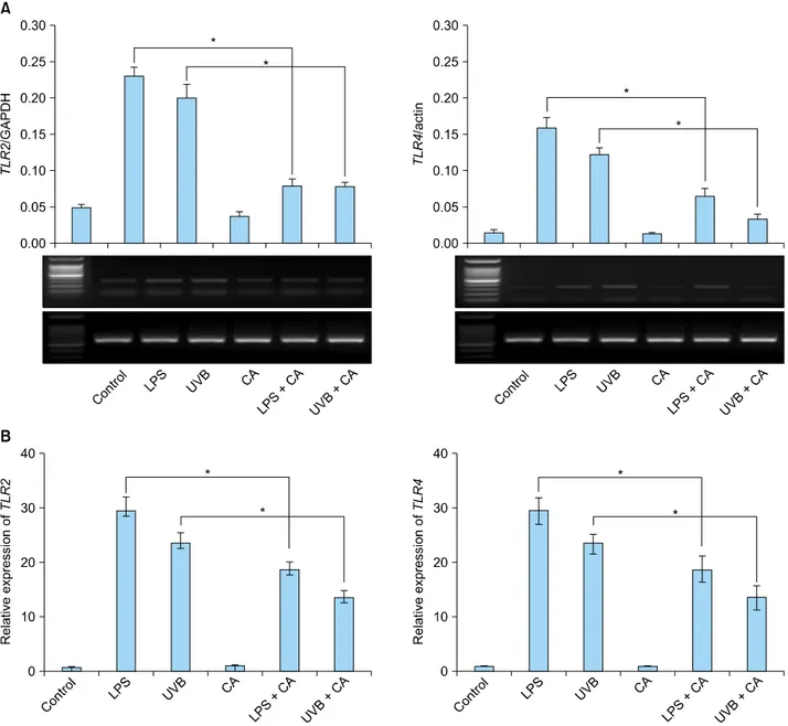
Fig. 1. The expression of TLR2 and TLR4 mRNA in keratinocytes increased in response to LPS or UVB irradiation. The increased TLR2 and TLR4 mRNA expression was down-regulated upon treatment with 10 nM calcitriol. (A) Reverse transcription-polymerase chain reaction. (B) Real-time polymerase chain reaction. TLR: Toll-like receptor, LPS: lipopolysaccharide, UVB: ultraviolet B, CA:
calcitriol. *p<0.05.
wing conditions: 94oC for 5 minutes, 35 cycles of 94oC for 1 minute, 59oC for 1 minute, 72oC for 1 minute and a final extension step of 72oC for 10 minutes.
Electrophoresis
The PCR products were run on a 1.5% agarose gel, sepa- rated by electrophoresis for 15 minutes at 100 volts, and visualized by UV transillumination.
Real-time polymerase chain reaction
RNA was isolated using TRIzol Reagent (Invitrogen), and
cDNA was synthesized using the RevertAidTM First Strand cDNA Synthesis Kit (Fermentas, California, MD, USA).
The primer sequences for GAPDH, TLR2, TLR4, TNF-α, IL-1β and LL-37 were the same as those described above.
The purity and quantity of each sample were determined by UV absorption and gel electrophoresis. Real-time PCR of target cDNA was conducted for TLR2, TLR4, TNF-α, IL-1β and LL-37, and normalized to GAPDH gene ex- pression. All SYBR Green reactions used SsoFastTM Eva- Green (BioRad, Mississauga, ON, Canada). Real-time PCR amplification was performed on a CFX96TM Real-Time
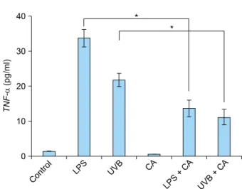
Fig. 3. The levels of secretory cytokine TNF-α in the supernatant increased in response to LPS or UVB irradiation. The increased TNF-α was down-regulated upon treatment with calcitriol. The expression level of TNF-α was quantified by enzyme-linked im- munosorbent assay. TNF: tumor necrosis factor, LPS: lipopo- lysaccharide, UVB: ultraviolet B, CA: Calcitriol. *p<0.05.
Fig. 2. The expression of TNF-α mRNA in keratinocytes increa- sed in response to LPS or UVB irradiation. The increase in TNF-α mRNA expression was suppressed upon treatment with calcitriol.
Expression levels of TNF-α mRNA was measured by real-time polymerase chain reaction. TNF: tumor necrosis factor, LPS:
lipopolysaccharide, UVB: ultraviolet B, CA: Calcitriol. *p<0.05.
System (BioRad).
Enzyme-linked immunosorbent assay
Cell culture supernatants were collected after drug treat- ment, centrifuged to remove cellular components, and stored at −80oC. To determine the amount of TNF-α in each supernatant, antibodies directed against human TNF-α were used as the capture and detection antibodies. The fluorescent substrate horseradish peroxidase-avidin (R&D Systems, Baltimore, MD, USA) was used for color de- velopment. The amount of cytokine in the test sample was determined from standard curves established with serial dilutions of recombinant human TNF-α (R&D Systems).
TNF-α concentrations were measured with the Spec- traMax 340PC384 System (Molecular Devices, Woking- ham, UK).
Statistical analysis
Data are presented as mean±standard deviation. Multiple comparisons were adjusted according to ANOVA. p-val- ues are two-sided and p<0.05 was considered statistically significant. All statical analyses were performed by using a Sigma Plot for Windows (Systat software Inc., San Jose, CA, USA).
RESULTS
Increased TLR2 and TLR4 mRNA expression is supp- ressed by calcitriol
Calcitriol may act through various pattern recognition re- ceptors9. Hence, we investigated whether various pattern
recognition receptors such as TLR2 and TLR4 could be influenced by calcitriol in NHKs. Because there was no change in the expression of TLR2 and TLR4 when NHKs were treated with calcitriol (10 nM), the keratinocytes were stimulated with LPS (5 μg/ml) or UVB irradiation (20 mJ/cm2). As expected, mRNA levels of TLR2 and TLR4 were found to be increased as much as 30 times (Fig. 1).
This effect was suppressed when the cells were treated with calcitriol (10 nM) prior to stimulation with LPS or UVB irradiation (20 mJ/cm2) (Fig. 1). These differences were statistically significant relative to the control group (p<
0.05).
Calcitriol suppresses over-expressed proinflammatory cytokines
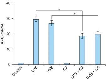
We investigated the effects of calcitriol on the immune response by stimulating keratinocytes in the presence of 10 nM calcitriol and measuring the cytokine levels in the cells and supernatant. The expression of TNF-α mRNA in NHKs was up-regulated 24 hours after stimulation with LPS or UVB irradiation. This effect was diminished upon treatment with calcitriol (Fig. 2). The levels of supernatant TNF-α were also up-regulated upon stimulation with LPS or UVB irradiation. However, the amount of secreted TNF-α was reduced after calcitriol treatment (Fig. 3). The expression of IL-1β mRNA in NHKs was also up-regu- lated upon stimulation with LPS or UVB irradiation, and this effect was diminished after treatment with calcitriol (Fig. 4).
Fig. 4. The expression of IL-1β mRNA in keratinocytes increased in response to LPS or UVB irradiation. The increase in IL-1β mRNA expression was suppressed upon treatment with calcitriol.
Expression levels of IL-1β mRNA were measured by real-time polymerase chain reaction. IL: interleukin, LPS: lipopolysac- charide, UVB: ultraviolet B, CA: Calcitriol. *p<0.05.
Fig. 5. The expression of LL-37 mRNA in keratinocytes increased in response to LPS or UVB irradiation. The increase in LL-37 mRNA expression was suppressed upon treatment with calcitriol.
Expression levels of LL-37 mRNA were measured by real-time polymerase chain reaction. LPS: lipopolysaccharide, UVB: ultra- violet B, CA: Calcitriol. *p<0.05.
Increased LL-37 mRNA expression is suppressed by calcitriol
LL-37 mRNA expression was not detected in the cultured keratinocytes of the unstimulated controls. LL-37 mRNA expression was increased in the UVB-irradiated and LPS sti- mulated groups, and decreased after treatment with cal- citriol (Fig. 5). These effects were statistically significant when compared with the results of the control group (p<0.05).
DISCUSSION
In this study, we investigated the effect of the vitamin D analog calcitriol on the expressions levels of TLR2, TLR4, and LL-37 in cultured human keratinocytes. The mRNA levels TLR2 and TLR4 were up-regulated in keratinocytes stimulated with LPS or UVB irradiation, while this effect was diminished after treatment with calcitriol. TLRs play important roles in the innate immune response to micro- bial infection. Dysregulation of TLR signaling is linked to a number of disease conditions, and possible roles for TLRs in innate immunity activation in psoriasis have been in- vestigated. Recent studies have suggested that TLR2, TLR4, and γδ T-cell receptors may recognize heat shock pro- tein 60 as a ligand and consequently activate the immune system10-12.
TLRs signal via the transcription factor nuclear factor-κB, which regulates the transcription of proinflammatory cyto- kines such as TNF-α, IL-1 and IL-6. For this reason, we performed cytokine analysis for TNF-α and IL-1β to con- firm the effects of calcitriol on TLRs. The expression levels
of TNF-α and IL-1β in keratinocytes were up-regulated upon stimulation with LPS and UVB irradiation. This effect was diminished after treatment with calcitriol.
Secretion of TNF-α was also suppressed after treatment with calcitriol. TNF-α is a pluripotent but predominantly proinflammatory cytokine, and is a major factor in the early steps of the innate immune response. TNF-α pro- duction is induced by LPS/lipoteichoic acie stimulation through TLR2- and TLR4-dependent pathways. Therefore, TNF-α production might be inhibited upon TLR inhi- bition. Calcitriol would be expected to suppress TNF-α synthesis through the down-regulation of TLRs in kera- tinocytes. Our results imply that calcitriol is able to modify the cytokine response towards an anti-inflammatory pro- file. This effect of calcitriol on the expression in TNF-α in keratinocytes could be one mechanism of its of action mechanisms in the treatment of psoriasis.
The link between TLR activation and the expression of AMPs is clearly established5. Kumar et al.13 demonstrated that expression of HBD2 is regulated by TLR2-dependent pathways. Recently, the immunomodulatory effect of calcitriol through down-regulation of TLR2 and TLR4 ex- pression was demonstrated in a human monocyte in vitro model6. Do et al.7 showed that vitamin D3 was found to dose-dependently suppress the protein and mRNA levels of TLR2 and TLR4. Based on these results, calcitriol would be expected to suppress TLRs, and in turn, TLR-mediated AMP expression in keratinocytes. The present study also showed that LL-37 induction was suppressed after treat- ment with calcitriol. Although we did not confirm the pathway by which calcitriol affected AMP expression, our
results suggest a possible mechanism in which calcitriol suppresses TLR activation and in turn decreases TLR-me- diated AMP expression in keratinocytes. These results su- ggest that calcitriol may exert its therapeutic effect on psoriasis by regulating TLR2- and TLR4-mediated AMP ex- pression in keratinocytes.
Several recent reports suggest a connection between vita- min D3 and AMP expression in keratinocytes. In the pre- sence of vitamin D, human keratinocytes upregulate LL-37 expression in response to TLR2 and IL-17 signaling4,14. Vitamin D analogs activate vitamin D receptor, which in turn would be expected to bind the VDREs in the pro- moter regions of cathelicidin genes and thus increase LL-37 expression3. Increased LL-37 would then aggravate inflam- mation by binding self-DNA and activating plasmacytoid dendritic cells in psoriasis15,16. However, in reality, oppo- site is true: indeed, vitamin D analogs are a mainstay in the topical treatment of psoriasis. To date, mechanisms that could explain this paradoxical effect of vitamin D analogs on psoriasis are not completely understood. Re- cent publications highlight the role of dysregulated AMP expression in the pathogenesis of psoriasis. HBD and LL-37 levels are greatly increased in the keratinocytes of psoriatic plaques17. It was demonstrated that LL-37 is able to suppress the induction of apoptosis in keratinocytes18. Therapeutic approaches to restore normal LL-37 expre- ssion in keratinocytes may prove beneficial for the treat- ment of psoriasis. Our previous study8 demonstrated that calcipotriol decreased the expression of HBD-2 and LL-37 induced by UVB and LPS in cultured human keratino- cytes. The present study demonstrated that calcitriol su- ppress the induction of LL-37 and proinflammatory cyto- kines in addition to TLR2 and TLR4 upon LPS and UVB radiation treatment of cultured human keratinocytes.
In conclusion, calcitriol was found to suppress the LPS- and UVB-mediated induction of TLR2 and TLR4 in human keratinocytes. This study suggests that calcitriol modulates the expression of AMPs or TNF-α in chronic inflamma- tory skin diseases associated with overexpression of these factors. Although our experimental results are in conflict with the current understanding of vitamin D effects on AMPs and proinflammatory cytokines, our results support the application of calcitriol in therapy for psoriasis and the possibility for alternative pathways of vitamin D analogs activity in psoriasis patients.
ACKNOWLEDGMENT
This study was supported by a grant from the Korea Healthcare Technology R&D Project, Ministry for Health, Welfare & Family Affairs, Republic of Korea (A091121).
REFERENCES
1. Weber G, Heilborn JD, Chamorro Jimenez CI, Hammarsjo A, Törmö H, Stahle M. Vitamin D induces the antimicrobial protein hCAP18 in human skin. J Invest Dermatol 2005;124:
1080-1082.
2. Zasloff M. Sunlight, vitamin D, and the innate immune de- fenses of the human skin. J Invest Dermatol 2005;125:
xvi-xvii.
3. Wang TT, Nestel FP, Bourdeau V, Nagai Y, Wang Q, Liao J, et al. Cutting edge: 1,25-dihydroxyvitamin D3 is a direct inducer of antimicrobial peptide gene expression. J Immunol 2004; 173:2909-2912.
4. Schauber J, Dorschner RA, Coda AB, Büchau AS, Liu PT, Ki- ken D, et al. Injury enhances TLR2 function and antimi- crobial peptide expression through a vitamin D-dependent mechanism. J Clin Invest 2007;117:803-811.
5. Liu PT, Stenger S, Li H, Wenzel L, Tan BH, Krutzik SR, et al.
Toll-like receptor triggering of a vitamin D-mediated human antimicrobial response. Science 2006;311:1770-1773.
6. Sadeghi K, Wessner B, Laggner U, Ploder M, Tamandl D, Friedl J, et al. Vitamin D3 down-regulates monocyte TLR expression and triggers hyporesponsiveness to pathogen- associated molecular patterns. Eur J Immunol 2006;36:361- 370.
7. Do JE, Kwon SY, Park S, Lee ES. Effects of vitamin D on ex- pression of Toll-like receptors of monocytes from patients with Behcet's disease. Rheumatology (Oxford) 2008;47:840- 848.
8. Kim BJ, Rho YK, Lee HI, Jeong MS, Li K, Seo SJ, et al. The effect of calcipotriol on the expression of human beta de- fensin-2 and LL-37 in cultured human keratinocytes. Clin Dev Immunol 2009;2009:645898.
9. Khoo AL, Chai LY, Koenen HJ, Oosting M, Steinmeyer A, Zuegel U, et al. Vitamin D(3) down-regulates proinflam- matory cytokine response to Mycobacterium tuberculosis through pattern recognition receptors while inducing protective cathelicidin production. Cytokine 2011;55:294- 300.
10. Ohashi K, Burkart V, Flohé S, Kolb H. Cutting edge: heat shock protein 60 is a putative endogenous ligand of the toll-like receptor-4 complex. J Immunol 2000;164:558-561.
11. Cohen-Sfady M, Nussbaum G, Pevsner-Fischer M, Mor F, Carmi P, Zanin-Zhorov A, et al. Heat shock protein 60 acti- vates B cells via the TLR4-MyD88 pathway. J Immunol 2005;175:3594-3602.
12. Wadia P, Atre N, Pradhan T, Mistry R, Chiplunkar S. Heat shock protein induced TCR gammadelta gene rearrange- ments in patients with oral cancer. Oral Oncol 2005;41:175- 182.
13. Kumar A, Zhang J, Yu FS. Toll-like receptor 2-mediated expression of beta-defensin-2 in human corneal epithelial cells. Microbes Infect 2006;8:380-389.
14. Peric M, Koglin S, Kim SM, Morizane S, Besch R, Prinz JC, et al. IL-17A enhances vitamin D3-induced expression of cathelicidin antimicrobial peptide in human keratinocytes. J Immunol 2008;181:8504-8512.
15. Peric M, Koglin S, Dombrowski Y, Gross K, Bradac E, Büchau A, et al. Vitamin D analogs differentially control antimicrobial peptide/"alarmin" expression in psoriasis. PLoS One 2009;4:e6340.
16. Lande R, Gregorio J, Facchinetti V, Chatterjee B, Wang YH, Homey B, et al. Plasmacytoid dendritic cells sense self-DNA coupled with antimicrobial peptide. Nature 2007;449:564- 569.
17. Gambichler T, Skrygan M, Tomi NS, Othlinghaus N, Brock- meyer NH, Altmeyer P, et al. Differential mRNA expression of antimicrobial peptides and proteins in atopic dermatitis as compared to psoriasis vulgaris and healthy skin. Int Arch Allergy Immunol 2008;147:17-24.
18. Chamorro CI, Weber G, Grönberg A, Pivarcsi A, Ståhle M.
The human antimicrobial peptide LL-37 suppresses apop- tosis in keratinocytes. J Invest Dermatol 2009;129:937-944.