Eruptive Anetoderma in a Patient with SLE
Vol. 26, No. 5, 2014 621
Received June 7, 2012, Revised September 18, 2012, Accepted for publication September 25, 2012
Corresponding author: Young Lee, Department of Dermatology, Chungnam National University School of Medicine, 266 Munhwa-ro, Jung-gu, Daejeon 301-747, Korea. Tel: 82-42-280-7706, Fax: 82-42-280-7706, E-mail: resina20@cnu.ac.kr
This is an Open Access article distributed under the terms of the Creative Commons Attribution Non-Commercial License (http://
creativecommons.org/licenses/by-nc/3.0) which permits unrestricted non-commercial use, distribution, and reproduction in any medium, provided the original work is properly cited.
Ann Dermatol Vol. 26, No. 5, 2014 http://dx.doi.org/10.5021/ad.2014.26.5.621
CASE REPORT
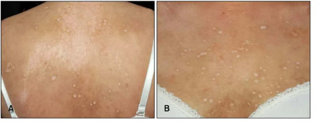
Fig. 1. Multiple widespread atro- phic whitish lesions on the upper back (A) and chest (B).
Eruptive Anetoderma in a Patient with Systemic Lupus Erythematosus
Nam-Ji Jeong, Seung-Bae Park, Myung Im, Young-Joon Seo, Jeung-Hoon Lee, Young Lee
Department of Dermatology, Chungnam National University School of Medicine, Daejeon, Korea
Anetoderma is a rare cutaneous disorder characterized by a loss of normal elastic tissue that presents clinically as atrophic patches located mainly on the upper trunk. Recent studies suggest immunological mechanisms may play a role in this process. Furthermore, a secondary form of macular atrophy occurs in the course of infectious diseases (e.g.
syphilis and tuberculosis) and autoimmune disease (e.g.
systemic lupus erythematosus [SLE]). Here, we report the case of a 20-year-old woman previously diagnosed with SLE, who presented with numerous well-circumscribed atrophic macules on the face and upper trunk. Histopathological examination showed decreased elastic tissues in the reticular dermis and mononuclear cells adhering to elastic fibers, consistent with anetoderma. Thus, the eruptive anetoderma localized widely on the face and upper trunk may have been caused by an autoimmune response of SLE. (Ann Dermatol 26(5) 621∼623, 2014)
-Keywords-
Anetoderma, Systemic lupus erythematosus
INTRODUCTION
Anetoderma is an uncommon cutaneous disease charac- terized by a decrease in the amount of normal elastic tissue. However, its precise pathophysiology remains un- clear. Clinically, anetoderma manifests as localized laxity of the skin with herniation or out-pouching resulting from abnormal dermal elastic tissue1. The associations of this condition with several immunological abnormalities such as systemic lupus erythematosus (SLE) and primary hypo- thyroidism with anti-thyroid antibodies as well as several infectious diseases such as syphilis and tuberculosis su- ggests an immunological mechanism may play a role in
NJ Jeong, et al
622 Ann Dermatol
the elastolytic process in anetoderma2,3. There are few case reports regarding anetoderma or cutis laxa-like lesions associated with SLE. Here, we report the case of a female SLE patient with eruptive anetoderma spreading gradually on the face and upper trunk. The observations in this case may help explain the immunological patho- genesis of anetoderma in SLE.
CASE REPORT
A 20-year-old woman presented with a 1-year history of gradually spreading white atrophic macules on the face and upper trunk. She had been diagnosed with SLE 3 years previously and was treated with hydroxychloroquine sulfate. Blood chemistry showed antiphospholipid, anti- cardiolipin, and lupus anticoagulant antibodies were all normal while complement levels were slightly low. The lesions were multiple 1∼2-cm shiny white atrophic ma- cules on the face and upper trunk (Fig. 1). Histopatho- logical examination of the back showed atrophic epidermis and marked decreases in the amount of elastic fibers in the upper dermis and mononuclear cells adhering
to elastic fibers. Some thrombosis of the small vessels was observed, but there were no signs of vasculitis (Fig. 2A, B).
Elastic staining revealed a loss of elastic fibers, which was more evident in the upper dermis (Fig. 2C). Biopsy con- firmed a diagnosis of anetoderma, which was hypothe- sized to be due to an autoimmune disorder. The patient is still receiving treatment for SLE with oral hydroxy- chloroquine sulfate and antihistamine, but the skin lesions have shown little improvement. As SLE progressed, the skin lesions exhibited aggravation.
DISCUSSION
Primary anetoderma is an idiopathic condition and can also be seen in association with autoimmune diseases such as SLE or primary hypothyroidism, occurring when there are no underlying skin diseases. Secondary aneto- derma implies characteristic atrophic lesions appearing at the same sites where specific dermatoses such as acne or varicella lesions have previously occurred4. An ultra- structural study investigated the changes in elastic fibers in the cutaneous lesions of 7 patients with LE; the results Fig. 2. (A) Atrophic epidermis and perivascular lymphohistiocytic infiltration in upper dermis (H&E, ×100). (B) The small vessels of the upper dermis progress toward thrombosis (arrows) without signs of vasculitis (H&E, ×200). (C) Elastic stain revealed loss of elastic fibers, which was more evident in the upper dermis (elastin, ×100).
Eruptive Anetoderma in a Patient with SLE
Vol. 26, No. 5, 2014 623 show a loss of dermal elastin matrix, resulting in a relative
increase in the predominance of elastofibrils and the formation of what are termed “dermal fibrillar bodies”5. The LE-associated loss of elastic fibers has been reported in lesions similar to anetoderma. Unlike the white firm non-follicular papules in papular elastorrhexis, depressed atrophic lesions are the main feature of anetoderma.
Moreover, it can be distinguished from extragenital lichen sclerosus because it clinically manifests as a porcelain white atrophic plaque4. Although the causative mecha- nism of anetodermic lesions is unknown, it may be due to microthrombosis in the dermal vessels, inducing the development of local ischemia and leading to elastic tissue degeneration6,7. No association between primary anetoderma and SLE has been identified, but the findings of microthrombosis in some skin biopsies and in some cases hypocomplementemia, hypergammaglobulinemia, or circulating antiphospholipid antibodies (e.g. lupus anti- coagulant antibody) can cause ischemia of dermal tissues and trigger elastic fiber degeneration8,9. On the other hand, there appears to be a correlation between increased anticardiolipin antibody levels and the development of cutaneous lesions of anetoderma in human immuno- deficiency virus type 1 (HIV-1) disease. Anetoderma may occur in patients with HIV infection, and anticardiolipin antibodies may alter the function of dermal proteinases, thereby destroying elastic fibers10. However, the present case was negative for antiphospholipid and anticardiolipin antibodies, although there were mild decreases in com- plement levels and D-dimer level was elevated. Therefore, we postulate that the spread of anetodermic lesions in the present patient was mediated by microthromboses in the dermal vessels, resulting in the development of local ischemia and degeneration of elastic tissue due to the immunological response in SLE. In addition, as the aneto- dermic lesions were aggravated following the clinical progression of SLE, we considered the anetoderma to have occurred in association with SLE. Further data in clinical cases of SLE with anetoderma must be collected to elu- cidate the pathophysiology of anetoderma in SLE patients.
Because of the possible clinical relevance of the asso- ciation between primary anetoderma and SLE, we recom- mend that patients with anetoderma be examined for
antiphospholipid antibodies or other autoimmune antibo- dies as well as for hypercoagulable states.
ACKNOWLEDGMENT
This study was supported by a grant of the Korean Health Technology R&D Project, Ministry of Health & Welfare, Republic of Korea (Grant No. HN10C0013).
REFERENCES
1. Venencie PY, Winkelmann RK. Monoclonal antibody stu- dies in the skin lesions of patients with anetoderma. Arch Dermatol 1985;121:747-749.
2. Hodak E, Shamai-Lubovitz O, David M, Hazaz B, Katzenel- son-Weissman V, Lahav M, et al. Immunologic abnormali- ties associated with primary anetoderma. Arch Dermatol 1992;128:799-803.
3. Romaní J, Pérez F, Llobet M, Planagumá M, Pujol RM.
Anetoderma associated with antiphospholipid antibodies:
case report and review of the literature. J Eur Acad Dermatol Venereol 2001;15:175-178.
4. Wolff K, Goldsmith LA, Katz SI, Gilchrest BA, Paller AS, Leffell DJ. Fitzpatrick's dermatology in general medicine. 7th ed. New York: McGraw-Hill, 2008;547,567-568.
5. Schmitt D, Thivolet J, Perrot H. Ultrastructural study of the cutaneous elastic fibres in lupus erythematosus. Br J Der- matol 1972;87:355-360.
6. Sparsa A, Piette JC, Wechsler B, Amoura Z, Francès C.
Anetoderma and its prothrombotic abnormalities. J Am Acad Dermatol 2003;49:1008-1012.
7. Hodak E, David M. Primary anetoderma and antiphosp- holipid antibodies--review of the literature. Clin Rev Allergy Immunol 2007;32:162-166.
8. Hodak E, Shamai-Lubovitz O, David M, Hazaz B, Lahav M, Sandbank M. Primary anetoderma associated with a wide spectrum of autoimmune abnormalities. J Am Acad Derma- tol 1991;25:415-418.
9. Montilla C, Alarcón-Segovia D. Anetoderma in systemic lupus erythematosus: relationship to antiphospholipid anti- bodies. Lupus 2000;9:545-547.
10. Lindstrom J, Smith KJ, Skelton HG, Redfield R, Alving BM, Wagner KF, et al. Increased anticardiolipin antibodies asso- ciated with the development of anetoderma in HIV-1 disease. Military Medical Consortium for the Advancement of Retroviral research (MMCARR). Int J Dermatol 1995;
34:408-415.