ISSN 2234-3806 • eISSN 2234-3814
http://dx.doi.org/10.3343/alm.2015.35.4.410
Characterization of Mucoid and Non-Mucoid
Streptococcus pneumoniae Isolated From Outpatients
Shinji Ogihara, B.S.1, Ryoichi Saito, Ph.D.2, Teru Akikura, B.S.3, Akiko Iwama, B.S.3, Yukari Adachi, B.S.3, Daiki Kaji, B.S.3, Kyoka Kakinuma, B.S.3, and Hiroshi Takahashi, B.S.3
Department of Clinical Laboratory1, Tokyo Medical and Dental University Hospital of Medicine, Tokyo; Department of Microbiology and Immunology2, Graduate School of Health Care Sciences, Tokyo Medical and Dental University, Tokyo; Department of Clinical Laboratory3, Kimitsu Central Hospital, Chiba, Japan
Background: Streptococcus pneumoniae causes pneumonia, sepsis, and meningitis. This study aimed to investigate the clinical characteristics of mucoid and non-mucoid isolates of S. pneumoniae, and to explore the relationship between the isolate phenotypes and their antibiotic susceptibility.
Methods: Clinical isolates from 3,453 non-repetitive S. pneumoniae (189 mucoid and 3,264 non-mucoid) infections obtained between January 2008 and December 2012 from outpatients at the Kimitsu-Central Hospital were evaluated.
Results: Compared to the non-mucoid isolates, the mucoid phenotypes were more sus- ceptible to certain antibiotics such as erythromycin, clarithromycin, and tetracycline as op- posed to clindamycin, chloramphenicol, and rifampicin. The mucoid phenotype was iso- lated more frequently from schoolchildren, adults, and elderly adults in a variety of clinical sites, including otorrhea, genitalia, pus, and eye discharge than the non-mucoid pheno- type. This suggested that mucoid isolates are more likely to be involved than non-mucoid isolates in various local infections. Systemic infection, which indicates invasiveness, was not associated with the mucoid or non-mucoid phenotype.
Conclusions: The results of this study suggest that mucoid isolates tend to have higher susceptibility than non-mucoid isolates to antibiotics. To the best of our knowledge, mucoid and non-mucoid S. pneumoniae isolates considerably differ in terms of clinical isolation site and age-specific prevalence.
Key Words: Streptococcus pneumoniae, Mucoid colony, Antimicrobial susceptibility
Received: August 4, 2014
Revision received: December 8, 2014 Accepted: March 25, 2015
Corresponding author: Shinji Ogihara Department of Clinical Laboratory, Tokyo Medical and Dental University Hospital of Medicine, 1-5-45 Yushima Bunkyo-ku, Tokyo 113-8510, Japan
Tel: +81-3-3813-6111 Fax: +81-3-5803-0110 E-mail: [email protected]
© The Korean Society for Laboratory Medicine This is an Open Access article distributed under the terms of the Creative Commons Attribution Non-Commercial License (http://creativecom- mons.org/licenses/by-nc/3.0) which permits unrestricted non-commercial use, distribution, and reproduction in any medium, provided the original work is properly cited.
INTRODUCTION
Streptococcus pneumoniae is an important pathogen that causes invasive and non-invasive pneumococcal diseases (e.g., meningitis, sepsis, pneumonia, and otitis media) in individuals of all age groups [1]. In various countries including Japan, pneu- mococcal conjugate vaccines are routinely used to protect in- fants, children, and elderly adults from pneumococcal diseases;
severe pneumococcal pneumonia and meningitis are still associ- ated with high mortality rates [2]. Furthermore, antimicrobial re-
sistance in S. pneumoniae has been observed globally since 1980s [3]. In particular, the increasing resistance of S. pneu- moniae strains to widely used anti-pneumococcal drugs, includ- ing ß-lactams and macrolides, has become a serious clinical concern in both developing and developed countries, including Japan [4, 5].
Colonies of S. pneumoniae isolates show various morphologi- cal features, such as the presence of a central depression or a mucoid appearance [6]. The mucoid phenotype has also been observed in other pathogenic bacteria, including Pseudomonas
aeruginosa [7]. A previous study showed that the minimal in- hibitory concentration (MIC) of various antibiotics in P. aerugi- nosa with the mucoid phenotype is lower than that in non-mu- coid strains [8]. However, the relationship between antibiotic susceptibility and the mucoid phenotype in S. pneumoniae re- mains unclear. Therefore, in the present study, the relationship between the mucoid phenotype and the antibiotic susceptibility patterns of S. pneumoniae isolates from outpatients with a vari- ety of infections was examined.
METHODS
1. Patients and samples
In this study, 3,453 non-repetitive S. pneumoniae (189 mucoid and 3,264 non-mucoid) clinical isolates were evaluated; they were obtained between January 2008 and December 2012 from outpatients at the Kimitsu-Central Hospital who were sus- pected to have infectious diseases. Strains isolated from hospi- talized patients within 48 hr were defined as outpatient-derived strains.
To elucidate the age-specific prevalence of different pheno- types, the patients were assigned to one of four different age groups. Patients aged 0-5 yr were assigned to the infant and preschool group; those aged 6-11 yr were assigned to the schoolchildren group; those aged 12-64 yr were assigned to the adult group, and those aged ≥65 yr were assigned to the el- derly adult group.
2. Microbiological investigations
All 3,453 strains were identified by using an optochin disk (Eiken Chemical Co. Ltd., Tokyo, Japan) and Slidex pneumo-Kit (Sismex bioMerieux, Tokyo, Japan). Mucoid strains were visually identified on the basis of colony morphology described in the Manual of Clinical Microbiology, 10th edition [9]. Susceptibility to penicillin G (meningitis and nonmeningitis), amoxicillin-clavu- lanate, cefotaxime (meningitis and nonmeningitis), ceftriaxone (meningitis and nonmeningitis), cefepime (meningitis and non- meningitis), meropenem, erythromycin, clarithromycin, clindamy- cin, tetracycline, chloramphenicol, vancomycin, rifampicin, and trimethoprim-sulfamethoxazole was tested by using MicroFAST 5J panels (Siemens Healthcare Diagnostics, Tokyo, Japan).
Only susceptibility to ceftriaxone was evaluated in 57 mucoid and 1,226 non-mucoid samples isolated after May 2011. The results were interpreted according to the Clinical Laboratory Standards Institute document M100-S24 [10]. Oral penicillin V is an unapproved antimicrobial agent in Japan; therefore, it was
not evaluated in this study. The MicroFAST 5J panels were in- oculated and read according to the manufacturer’s recommen- dations. The inoculated panels were then incubated at 35°C in ambient air for 20-24 hr prior to visual determination of the MIC.
Quality control for MIC determination was performed by using the reference strain S. pneumoniae ATCC 49619.
3. Statistical analysis
Data were analyzed by using Microsoft Excel for Mac 2011. Cat- egorical variables were compared by using either chi-squared test or Fisher’s exact test, as appropriate. Two-tailed P <0.05 was considered statistically significant. Statistical significance was assessed in comparison between the infant and preschool group and each age group for antibiotic resistance rate with ei- ther chi-squared test or Fisher’s exact test. When the infant and preschool group showed significance to all each age group, we defined this was significant.
RESULTS
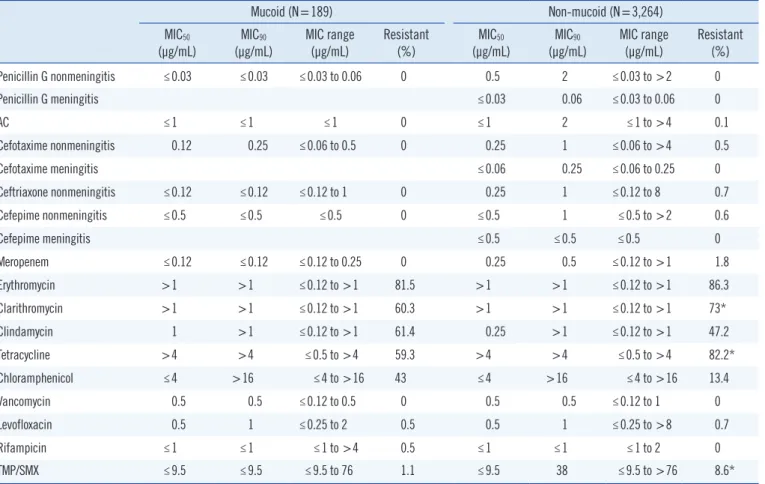
The MIC50 of penicillin G (nonmeningitis), cefotaxime (nonmen- ingitis), ceftriaxone (nonmeningitis), and meropenem in non- mucoid isolates was higher than that in mucoid isolates (Table 1). Similarly, the MIC90 of amoxicillin-clavulanate, cefepime (nonmeningitis), and trimethoprim-sulfamethoxazole in non- mucoid isolates was higher than that in mucoid isolates. Since only non-mucoid isolates were detected from patients with men- ingitis, we compared mucoid isolates and non-mucoid isolates from patients with meningitis with regard to their susceptibility to penicillin G, cefotaxime, ceftriaxone, and cefepime (Table 1).
Additionally, non-mucoid isolates from patients with meningitis before 2010 were not tested for ceftriaxone susceptibility (men- ingitis). The MIC50 and MIC90 of erythromycin, clarithromycin, tetracycline, vancomycin, levofloxacin, and rifampicin were comparable for mucoid and non-mucoid isolates, although clar- ithromycin, tetracycline, and vancomycin are not routinely used in the clinical setting. The rate of resistance and MIC of protein synthesis-inhibiting antibiotics, including erythromycin, clar- ithromycin, clindamycin, tetracycline, and chloramphenicol, were found to be high for both. Moreover, the rate of resistance to clarithromycin, tetracycline, and trimethoprim-sulfamethoxa- zole was higher in non-mucoid isolates than in mucoid isolates (P <0.01); the rate of resistance to clindamycin, chlorampheni- col, and rifampicin was higher in mucoid isolates than in non- mucoid isolates (P <0.01) (Table 1). However, the rate of resis- tance to β-lactam antibiotics, including penicillin G, amoxicillin-
clavulanate, cefotaxime, ceftriaxone, cefepime, meropenem, and vancomycin, did not differ significantly between the two phenotypes (Table 1). All mucoid and non-mucoid strains iso- lated from meningitis samples were susceptible to both penicil- lin G and cefotaxime, which have two cut-off values in meningi- tis and non-meningitis samples.
Among the schoolchildren, adult, and elderly adult groups, the frequency of mucoid isolates was significantly higher than that of non-mucoid isolates (P <0.01) (Table 2). Non-mucoid isolates were more resistant to erythromycin, clarithromycin, clindamy- cin, and tetracycline in the infant and preschool group than in the schoolchildren, adult, and elderly adult groups in all samples (P <0.05) (Table 3). The results for non-mucoid isolates were similar for all respiratory tract samples (Table 4). The invasive- ness and antibiotic resistance rate of non-mucoid isolates did not differ significantly among age groups for all antibiotics (Table 5).
The antibiotic resistance rate did not differ significantly among mucoid isolates, too. In contrast, the frequency of mucoid iso- lates obtained from otorrhea samples, genital swabs, ecthyma
Table 1. MIC50, MIC90, and range of various antibiotics for Streptococcus pneumoniae
Mucoid (N=189) Non-mucoid (N=3,264)
MIC50
(μg/mL)
MIC90
(μg/mL)
MIC range (μg/mL)
Resistant (%)
MIC50
(μg/mL)
MIC90
(μg/mL)
MIC range (μg/mL)
Resistant (%)
Penicillin G nonmeningitis ≤0.03 ≤0.03 ≤0.03 to 0.06 0 0.5 2 ≤0.03 to >2 0
Penicillin G meningitis ≤0.03 0.06 ≤0.03 to 0.06 0
AC ≤1 ≤1 ≤1 0 ≤1 2 ≤1 to >4 0.1
Cefotaxime nonmeningitis 0.12 0.25 ≤0.06 to 0.5 0 0.25 1 ≤0.06 to >4 0.5
Cefotaxime meningitis ≤0.06 0.25 ≤0.06 to 0.25 0
Ceftriaxone nonmeningitis ≤0.12 ≤0.12 ≤0.12 to 1 0 0.25 1 ≤0.12 to 8 0.7
Cefepime nonmeningitis ≤0.5 ≤0.5 ≤0.5 0 ≤0.5 1 ≤0.5 to >2 0.6
Cefepime meningitis ≤0.5 ≤0.5 ≤0.5 0
Meropenem ≤0.12 ≤0.12 ≤0.12 to 0.25 0 0.25 0.5 ≤0.12 to >1 1.8
Erythromycin >1 >1 ≤0.12 to >1 81.5 >1 >1 ≤0.12 to >1 86.3
Clarithromycin >1 >1 ≤0.12 to >1 60.3 >1 >1 ≤0.12 to >1 73*
Clindamycin 1 >1 ≤0.12 to >1 61.4 0.25 >1 ≤0.12 to >1 47.2
Tetracycline >4 >4 ≤0.5 to >4 59.3 >4 >4 ≤0.5 to >4 82.2*
Chloramphenicol ≤4 >16 ≤4 to >16 43 ≤4 >16 ≤4 to >16 13.4
Vancomycin 0.5 0.5 ≤0.12 to 0.5 0 0.5 0.5 ≤0.12 to 1 0
Levofloxacin 0.5 1 ≤0.25 to 2 0.5 0.5 1 ≤0.25 to >8 0.7
Rifampicin ≤1 ≤1 ≤1 to >4 0.5 ≤1 ≤1 ≤1 to 2 0
TMP/SMX ≤9.5 ≤9.5 ≤9.5 to 76 1.1 ≤9.5 38 ≤9.5 to >76 8.6*
*P <0.01.
Abbreviations: MIC, minimal inhibitory concentration; AC, Amoxicillin-clavulanate; TMP/SMX, Trimethoprim-sulfamethoxazole.
Table 2. Patient age at the time of detection and tissue source for Streptococcus pneumoniae isolated from 189 patients infected with mucoid strains and 3,264 patients infected with non-mucoid strains
Mucoid
(N=189) Non-mucoid (N=3,264) Age, N (%)
Infant and preschool group 63 (33.3) 2,577 (79.0)*
Schoolchildren group 27 (14.3)* 162 (4.9)
Adult group 39 (20.6)* 183 (5.6)
Elderly adult group 60 (31.8)* 342 (10.5) Source of isolates, N (%)
Respiratory tract sample 168 (88.9) 3,161 (96.4)*
Otorrhea sample 7 (3.7)* 27 (0.8)
Genital swab 3 (1.6)* 3 (0.3)
Ecthyma and abscess exudate 3 (1.6)† 10 (0.3)
Eye discharge 3 (1.6)† 15 (0.5)
Invasiveness 5 (2.6) 47 (1.4)
*P <0.01; †P <0.05.
and abscess exudate, and eye discharge was significantly higher than that of non-mucoid isolates (P <0.05) (Table 2).
DISCUSSION
Mucoid colonies of S. pneumoniae generate large amounts of capsular polysaccharides [9]. The capsule polysaccharide is the major virulence determinant of S. pneumoniae [11], particularly in hosts lacking type-specific antibodies of sufficient quantity or avidity [12]. Moreover, previous studies demonstrated that the mucoid phenotype, which is characterized by the presence of hyaluronic acid capsular polysaccharide in Streptococcus pyo- genes, is a key virulence determinant associated with severe S.
pyogenes infections [13, 14]. The mucoid phenotype of S. pneu- moniae may also show higher pathogenicity than the non-mu- coid phenotype; therefore, it is important to characterize S.
pneumoniae, especially the mucoid phenotype, with regard to
antibiotic susceptibility.
Previous studies demonstrated that P. aeruginosa isolates with mucoid phenotype are more susceptible than non-mucoid isolates to multiple antibiotics such as β-lactams and fluoroqui- nolones [8, 15]. Although the reason for antibiotic resistance was not identified in the present study, Ciofu et al. [8] provided a possible explanation for the antibiotic susceptibility of mucoid- overproducing P. aeruginosa; they proposed that non-mucoid resistant isolates co-existing with mucoid strains in biofilms may play a protective role. This explanation could be also applied to S. pneumoniae. Although similar findings have been reported in P. aeruginosa [8], to the best of our knowledge, this is the first report showing differences in antibiotic susceptibility patterns of mucoid and non-mucoid S. pneumoniae isolates.
S. pneumoniae infections are more common in infants, chil- dren, and elderly adults than in adults. In this study, it was ob- served that mucoid isolate infections were not age-dependent, whereas non-mucoid isolates were more frequent in infants and preschool children. Pastor et al. [16] reported an age-specific S.
pneumoniae prevalence of approximately 35-45% in infants and Table 3. Antibiotic resistance rate (%) of non-mucoid strains isolat-
ed from outpatients according to their age group, including those with meningitis and nonmeningitis, in all samples
Infant and preschool group (N=2,577)
Schoolchildren group (N=162)
Adult group (N=183)
Elderly adult group (N=342)
Penicillin G nonmeningitis 0 0 0 0
Penicillin G meningitis 0 0 0 0
AC 0 0 0 0
Cefotaxime nonmeningitis 0.5 1.2 0.5 0
Cefotaxime meningitis 0 0 0 0
Ceftriaxone nonmeningitis 0.9 0 0 0
Cefepime nonmeningitis 0.7 0 1.1 0
Cefepime meningitis 0 0 0 0
Meropenem 1.9 3.0 2.2 0.9
Erythromycin 88.4* 82.1 72.7 79.8
Clarithromycin 75.7* 66.7 59.0 63.5
Clindamycin 49.9† 37.7 35.0 38.3
Tetracycline 84.1† 75.3 69.9 73.1
Chloramphenicol 12.6 12.7 8.7 14.3
Vancomycin 0 0 0 0
Levofloxacin 0.1 0.6 3.3 3.5
Rifampicin 0 0 0 0
TMP/SMX 8.5 4.3 5.5 12.3
*P <0.05; †P <0.01.
Abbreviations: AC, Amoxicillin-clavulanate; TMP/SMX, Trimethoprim-sulfa- methoxazole.
Table 4. Antibiotic resistance rate (%) of non-mucoid strains isolat- ed from outpatients according to their age group, for respiratory tract samples
Infant and preschool group (N=2,525)
Schoolchildren group (N=156)
Adult group (N=163)
Elderly adult group (N=317)
Penicillin G 0 0 0 0
AC 0 0 0 0
Cefotaxime 0.4 0.6 0.6 0
Ceftriaxone 0.3 0 0 0
Cefepime 0.5 0 1.2 0
Meropenem 1.5 2.6 2.5 1.6
Erythromycin 90.1* 78.8 71.8 80.1
Clarithromycin 77.1* 62.2 60.1 61.2
Clindamycin 50.9* 34.6 35.0 37.9
Tetracycline 86.0* 73.1 69.3 72.2
Chloramphenicol 14.0 12.8 8.6 14.2
Vancomycin 0 0 0 0
Levofloxacin 0.2 0.6 3.1 4.7
Rifampicin 0 0 0 0
TMP/SMX 8.9 5.1 4.9 12.6
*P <0.01.
Abbreviations: AC, Amoxicillin-clavulanate; TMP/SMX, Trimethoprim-sulfa- methoxazole.
Table 5. Antibiotic resistance rate (%) of non-mucoid strains isolat- ed from outpatients according to their age group, including those with meningitis and nonmeningitis, with regard to invasiveness
Infant and preschool group (N=14) (%)
Schoolchildren group (N=5) (%)
Adult group (N=5) (%)
Elderly adult group (N=23) (%) Penicillin G
nonmeningitis 0 0 0 0
Penicillin G meningitis 0 0 0 0
AC 0 0 0 0
Cefotaxime nonmeningitis
0 0 0 0
Cefotaxime meningitis 0 0 0 0
Ceftriaxone
nonmeningitis 0 0 0 0
Cefepime nonmeningitis
0 0 0 0
Cefepime meningitis 0 0 0 0
Meropenem 0 0 0 0
Erythromycin 64.3 40.0 60.0 82.6
Clarithromycin 64.3 40.0 40.0 78.3
Clindamycin 28.6 0.0 20.0 39.1
Tetracycline 50.0 40.0 60.0 69.6
Chloramphenicol 7.1 40.0 40.0 13.0
Vancomycin 0 0 0 0
Levofloxacin 0 0 0 0
Rifampicin 0 0 0 0
TMP/SMX 7.1 0.0 40.0 13.0
Abbreviations: AC, Amoxicillin-clavulanate; TMP/SMX, Trimethoprim-sulfa- methoxazole.
preschool children, and 20% in elderly adults. In the present study, the prevalence of mucoid isolates in the elderly adult group was approximately 10% higher than that reported by Pas- tor et al.[16], whereas the prevalence of non-mucoid isolates was found to be approximately 35% higher in the infant and pre- school group and 10% lower in the elderly adult group. Mason et al. [17] noted that younger age is a risk factor for infection with a penicillin-resistant isolate. The present study did not evaluate penicillin-non-susceptible isolates; rather, it showed resistance to protein synthesis-inhibiting antibiotics. We observed that younger age is associated with isolation of strains not susceptible to pro- tein synthesis-inhibiting antibiotics. Regarding the similar results between respiratory tract samples and overall set of all samples, it should be noted that of the 3,264 isolates, 79% (3,161) were derived from respiratory tract samples.
Most non-mucoid isolates were obtained from patients with respiratory infections, which may suggest that mucoid isolates are more variable in terms of clinical infection site than non-mu- coid phenotypes isolated from the respiratory system. However, systemic infection, which indicates invasiveness, was not asso- ciated with either of the mucoid and non-mucoid phenotypes.
In conclusion, to the best of our knowledge, this study is that mucoid and non-mucoid S. pneumoniae isolates differ signifi- cantly in terms of clinical site of isolation and age-specific preva- lence. In particular, these results suggest that mucoid isolates tend to have greater antibiotics susceptibility than non-mucoid isolates. However, limited studies on the region-specific epidemi- ology of mucoid and non-mucoid isolates have been reported.
Furthermore, this study was not designed to collect information regarding serotype distributions. Therefore, further studies to confirm the present findings are warranted.
Authors’ Disclosures of Potential Conflicts of Interest
No potential conflicts of interest relevant to this article were re- ported.
REFERENCES
1. Fine MJ, Orloff JJ, Arisumi D, Fang GD, Arena VC, Hanusa BH, et al.
Prognosis of patients hospitalized with community-acquired pneumo- nia. Am J Med 1990;88(5N):1N-8N.
2. Chiba N, Morozumi M, Shouji M, Wajima T, Iwata S, Sunakawa K, et al.
Rapid decrease of 7-valent conjugate vaccine coverage for invasive pneumococcal diseases in pediatric patients in Japan. Microb Drug Re- sist 2013;19:308-15.
3. Adam D. Global antibiotic resistance in Streptococcus pneumoniae. J Antimicrob Chemother 2002;50(S):S1-5.
4. Song JH, Jung SI, Ko KS, Kim NY, Son JS, Chang HH, et al. High prev- alence of antimicrobial resistance among clinical Streptococcus pneu- moniae isolates in Asia (an ANSORP study). Antimicrob Agents Che- mother 2004;48:2101-7.
5. Baquero F. Pneumococcal resistance to beta-lactam antibiotics: a glob- al geographic overview. Microb Drug Resist 1995;1:115-20.
6. Richter SS, Heilmann KP, Dohrn CL, Riahi F, Beekmann SE, Doern GV.
Accuracy of phenotypic methods for identification of Streptococcus pneumoniae isolates included in surveillance programs. J Clin Microbiol 2008;46:2184-8.
7. Speert DP, Farmer SW, Campbell ME, Musser JM, Selander RK, Kuo S.
Conversion of Pseudomonas aeruginosa to the phenotype characteristic of strains from patients with cystic fibrosis. J Clin Microbiol 1990;28:
188-94.
8. Ciofu O, Fussing V, Bagge N, Koch C, Høiby N. Characterization of paired mucoid/non-mucoid Pseudomonas aeruginosa isolates from Danish cystic fibrosis patients: antibiotic resistance, beta-lactamase ac- tivity and RiboPrinting. J Antimicrob Chemother 2001;48:391-6.
9. Versalovic J, Carroll KC, et al. eds. Manual of clinical microbiology. 10th ed. Washington DC: American Society for Microbiology, 2011:331-49.
10. Clinical and Laboratory Standards Institute. Reference method for broth dilution antimicrobial susceptibility testing. Approved standard. M100- S24. Wayne, PA: Clinical and Laboratory Standards Institute, 2014.
11. Henrichsen J. Six newly recognized types of Streptococcus pneumoni- ae. J Clin Microbiol 1995;33:2759-62.
12. Kim JO and Weiser JN. Association of intrastrain phase variation in quantity of capsular polysaccharide and teichoic acid with the virulence of Streptococcus pneumoniae. J Infect Dis 1998;177:368-77.
13. Gryllos I, Tran-Winkler HJ, Cheng MF, Chung H, Bolcome R 3rd, Lu W, et al. Induction of group A Streptococcus virulence by a human antimi- cobial peptide. Proc Natl Acad Sci U S A 2008;105:16755-60.
14. Kakuta R, Yano H, Hidaka H, Miyazaki H, Irimada M, Oda K, et al. Se- vere acute otitis media caused by mucoid streptococcus pyogenes in a previously healthy adult. Tohoku J Exp Med 2014;232:301-4.
15. Shawar RM, MacLeod DL, Garber RL, Burns JL, Stapp JR, Clausen CR, et al. Activities of tobramycin and six other antibiotics against Pseudo- monas aeruginosa isolates from patients with cystic fibrosis. Antimicrob Agents Chemother 1999;43:2877-80.
16. Pastor P, Medley F, Murphy TV. Invasive pneumococcal disease in Dal- las County, Texas: results from population-based surveillance in 1995.
Clin Infect Dis 1998;26:590-5.
17. Mason EO Jr, Lamberth LB, Kershaw NL, Prosser BL, Zoe A, Ambrose PG. Streptococcus pneumoniae in the USA: in vitro susceptibility and pharmacodynamic analysis. J Antimicrob Chemother 2000;45:623-31.