ABSTRACT
Background: Rituximab (RTX), a monoclonal antibody that selectively binds to CD20+ B cells, showed favorable outcomes in patients with idiopathic inflammatory myopathies (IIM) in small case series, but the evidence is still not enough. Our goal was to determine the efficacy and safety of RTX for Korean patients with refractory IIM.
Methods: We retrospectively analyzed the medical records of 16 patients with refractory IIM treated with RTX in seven tertiary rheumatology clinics in the Korea. The efficacy of RTX was evaluated with the improvement of serum creatine phosphokinase (CPK) level and physician's global assessment (PGA), and daily corticosteroid dose reduction. A > 25%
decrease in CPK level, corticosteroid dose, or PGA was considered significant. A complete response (CR) was designated by meeting three efficacy criteria and a partial response (PR) by only two criteria.
Results: Sixteen patients with IIM were evaluated (13 female; median age, 51.8 years). All patients had received at least one conventional immunosuppressive agent (median, 3.6 [2.0–5.0]) and concomitant corticosteroids. The median CPK level and median dose of prednisolone was 421.0 units/L and 20.0 mg/day respectively. Eleven patients were treated with intravenous immunoglobulin. Seven patients received 2,000 mg of RTX and the others received lower dose. Twenty-four weeks after RTX treatment, 11 patients achieved a > 25%
reduction in corticosteroid dose and CPK levels, and nine showed improved PGA. The overall response rate was 68.8% (11 patients). At the end of follow-up (median 24 weeks), 12 (75.0%) patients responded overall: four (25.0%) and eight (50.0%) patients achieved CR and PR, respectively. Baseline muscle enzyme levels were higher in responders than non-responders, but disease duration, RTX dose, ESR and serum CRP were not significantly different between the two groups. The rate of adverse event was 25.4/1,000 person-years.
Conclusion: RTX could be an effective and relatively safe therapeutic option in patients with refractory IIM.
Original Article
Received: Mar 31, 2020 Accepted: Jul 29, 2020 Address for Correspondence:
Dae Hyun Yoo, MD, PhD
Department of Rheumatology, Hanyang University Hospital for Rheumatic Diseases, 222-1 Wangsimni-ro, Seongdong-gu, Seoul 07463, Korea.
E-mail: [email protected]
*Present address: Division of Rheumatology, Department of Internal Medicine, Korea University College of Medicine, Seoul, Korea
© 2020 The Korean Academy of Medical Sciences.
This is an Open Access article distributed under the terms of the Creative Commons Attribution Non-Commercial License (https://
creativecommons.org/licenses/by-nc/4.0/) which permits unrestricted non-commercial use, distribution, and reproduction in any medium, provided the original work is properly cited.
ORCID iDs Ga Young Ahn
https://orcid.org/0000-0003-1261-5363 Chang-Hee Suh
https://orcid.org/0000-0001-6156-393X Yong-Gil Kim
https://orcid.org/0000-0002-8029-7355 Yong-Beom Park
https://orcid.org/0000-0003-4695-8620 Seung Cheol Shim
https://orcid.org/0000-0002-3199-359X Sang-Heon Lee
https://orcid.org/0000-0002-7539-9330 Shin-Seok Lee
https://orcid.org/0000-0001-6810-7355
Ga Young Ahn ,1* Chang-Hee Suh ,2 Yong-Gil Kim ,3 Yong-Beom Park ,4 Seung Cheol Shim ,5 Sang-Heon Lee ,6 Shin-Seok Lee ,7 Sang-Cheol Bae ,1 and Dae Hyun Yoo 1
1Department of Rheumatology, Hanyang University Hospital for Rheumatic Diseases, Seoul, Korea
2Department of Rheumatology, Ajou University School of Medicine, Suwon, Korea
3 Division of Rheumatology, Department of Internal Medicine, Asan Medical Center, University of Ulsan College of Medicine, Seoul, Korea
4 Division of Rheumatology, Department of Internal Medicine, Yonsei University College of Medicine, Seoul, Korea
5 Division of Rheumatology, Regional Rheumatoid & Degenerative Arthritis Center, Chungnam National University Hospital, Daejeon, Korea
6 Division of Rheumatology, Department of Internal Medicine, Konkuk University School of Medicine, Seoul, Korea
7 Division of Rheumatology, Department of Internal Medicine, Chonnam National University Hospital, Chonnam National University Medical School, Gwangju, Korea
Efficacy and Safety of Rituximab in Korean Patients with Refractory Inflammatory Myopathies
Immunology, Allergic
Disorders & Rheumatology
Sang-Cheol Bae
https://orcid.org/0000-0003-4658-1093 Dae Hyun Yoo
https://orcid.org/0000-0002-0643-4008 Disclosure
The authors have no potential conflicts of interest to disclose.
Author Contributions
Conceptualization: Yoo DH. Methodology:
Ahn GY, Yoo DH. Investigation: Ahn GY, Suh CH, Kim YG, Park YB, Shim SC, Lee SH, Lee SS, Bae SC, Yoo DH. Formal analysis: Ahn GY.
Validation: Ahn GY, Yoo DH. Writing - original draft: Ahn GY. Writing - review & editing: Suh CH, Shim SC, Yoo DH.
Keywords: Myositis; Rituximab; Treatment Outcome; Safety
INTRODUCTION
Idiopathic inflammatory myopathies (IIM) are a group of rare autoimmune rheumatic disease that primarily induces chronic inflammation of skeletal muscles, leading to proximal muscle weakness and disability. Patients with IIM generally receive high dose systemic glucocorticoids in combination with antimalarial agents or conventional non-targeting immunosuppressive,1 but the management of IIM remains challenging for clinicians.
Patients with IIM often experience extensive muscular inflammation, causing difficulty in swallowing, voiding, and deterioration of performing activities of daily living. Life- threatening extra-muscular complications, including interstitial lung disease (ILD), are also prevalent. However, a considerable portion of patients inadequately responds to the current treatment options. Owing to the rarity and heterogeneity of IIM, clinical trials and evidence for guidelines are also insufficient, and the 10-year mortality was still high in recent studies (14%–38%).2,3 Although interest in biologic agents that directly target the immune cells or cytokines has increased in other systemic rheumatic disease following advances in the understanding of the immunopathogenesis, studies that have investigated the efficacy and safety of biologic agents in patients with IIM are limited.4
Rituximab (RTX) is a monoclonal antibody that selectively binds to CD20+ B cells.5 RTX was originally developed for the treatment of lymphoproliferative diseases,6 but it is also approved for the treatment of rheumatoid arthritis,7 pemphigus vulgaris,8 granulomatosis with polyangiitis (GPA), and microscopic polyangiitis (MPA).9 In addition, it is used widely to manage a variety of immune-related disorders, including idiopathic thrombocytopenic purpura,10 systemic lupus erythematosus,11,12 and multiple sclerosis.13 In terms of IIM, RTX also has been believed to be a potential therapeutic option along with the B cell infiltration in the muscles of patients with IIM and myositis-associated antibodies (MAA).1,14 RTX showed favorable outcomes in patients with refractory IIM who failed to respond to corticosteroid and conventional immunosuppressive agents, but most studies were case series and open label studies. In Korea, a case series described four patients with refractory IIM treated with RTX and all patients responded to IIM.15 Therefore, this paper aimed to identify the efficacy and safety of RTX for patients with refractory IIM.
METHODS
Study population
We conducted a retrospective multicenter study of 16 patients with refractory IIM who were treated with RTX from July 2006 to October 2017. This multicenter retrospective study comprised patients from seven tertiary rheumatology clinics that had obtained official endorsement for the use of RTX for refractory IIM from the Ministry of Health and Welfare and the Health Insurance Review & Assessment Service (HIRA). Off-label RTX use must be officially allowed by each hospital after receiving institutional review board approval and submission to the HIRA, because RTX currently is not approved by the Korean Ministry of Food and Drug Safety (MFDS) for the treatment of IIMs.
The medical records of patients with a diagnosis of possible or definite IIM according to Bohan and Peter's criteria16,17 and treated with RTX were respectively reviewed. Patients with a shorter than 6 months' observation period (from RTX administration to the data collection), whose age at diagnosis of IIM was less 16 years of age, and patients with malignancy were excluded. RTX was prescribed by expert rheumatologists due to an inadequate response or intolerance to corticosteroids and conventional immunosuppressive therapy. All patients received RTX intravenously, and the RTX dosing and cycle differed depending on the decision of the rheumatologist.
Patient assessment and response criteria
Demographic characteristics, including age (years), sex (female/male), height (cm), and weight (kilograms) were recorded. Disease-specific variables, including IIM duration, concomitant and cumulative immunosuppressive agent history, IIM activity, the reason for RTX administration, and RTX dose and interval, were also assessed at the time of RTX administration. The IIM activity was measured by muscle weakness (grade), extra-muscular symptoms (skin rash, skin necrosis, dysphagia, dyspnea, Raynaud phenomenon, and ILD), constitutional symptoms (fever, fatigue, and weight loss), and laboratory markers including muscle enzymes levels (serum creatine phosphokinase [CPK], lactate dehydrogenase [LDH], aldolase, aspartate aminotransferase [AST], alanine transaminase [ALT]), complete blood count, erythrocyte sedimentation rate (ESR), and C reactive protein (CRP). IIM activity was assessed in each hospital and at baseline and followed up at the regular clinical visit schedule (2- or 4-week intervals). The reference ranges and units for laboratory markers were similar among the 7 rheumatology clinics.
We assessed the efficacy of RTX based on the serum CPK level, daily dose of corticosteroid and opinion of the physician.18 A decrease in CPK level and daily corticosteroid dose was considered to be significant if it was > 25% of the baseline. The physician's opinion on the response was dichotomized to “response” or “no response.” Complete response (CR) and partial response (PR) were defined as a patient meeting all three or only two efficacy criteria, respectively. Three patients were administered a second cycle of RTX after completion of the first cycle of RTX administration but the response was assessed based on the first cycle of RTX administration.
Statistical analysis
The general statistics of the study population are presented medians (first interquartile, third interquartile) for continuous variables, and frequencies with percentage (%) for categorical variables. Demographic and clinical characteristics were compared by using Fisher's exact test (binary variables) and the Mann-Whitney U test (linear variables). All statistical analyses were conducted using IBM SPSS version 25.0 (IBM Corp., Armonk, NY, USA). P values of <
0.05 (two-tailed) were considered statistically significant.
Ethics statement
This study was approved by the institutional review board (IRB) of each hospital and the requirement for informed consent was waived because of the retrospective nature of the study (IRB No. HYUH 2018-01-020-002 in Hanyang University Hospital, AJIRB-MED-OBS-17-509 in Ajou University Hospital, 2018-0567 in Asan Medical Center, 2018-0541-001 in Severance Hospital, Yonsei University Health System, 2018-02-027 in Chungnam National University Hospital, KUH1010916 in Konkuk University Medical Center, CNUH-2018-058 in Chonnam National University Hospital).
RESULTS
Demographic and clinical characteristics
The baseline demographic and clinical characteristics are presented in Table 1. Ten patients with dermatomyositis and six patients with polymyositis were treated with RTX. Thirteen (81.3%) were female, the median age at RTX treatment was 51.8 (42.5–59.0) years of age, and the median IIM disease duration (from IIM diagnosis to RTX treatment) was 88.4 (24.3–162.8) months.
All patients had insufficient response or intolerance to at least one (median, 3.6 [2.0–5.0]) conventional immunosuppressive agent: azathioprine, n = 12, 75.0%; methotrexate, n = 11, 68.8%; mycophenolate mofetil, n = 10, 63.0%; hydroxychloroquine, n = 8, 50.0%; cyclosporine, n = 5, 31.3%; cyclophosphamide, n = 5, 31.3%; tacrolimus, n = 5, 31.3%; leflunomide, n = 1, 6.3%). Twelve (75.0%) patients had received intravenous immunoglobulin therapy.
IIM activity at the time of RTX treatment is summarized in Table 2. Most patients (n = 14, 87.5%) experienced muscle weakness and all patients had at least one extra-muscular manifestation (dermatomyositis specific skin rash, n = 8, 50.0%; ILD, n = 5, 31.3%;
dysphagia, n = 4, 25.0%; arthritis, n = 4, 25%). Eight (50%) patients had constitutional manifestations, such as fatigue (n = 6, 37.5%), fever (n = 3, 18.8%), and weight loss (n = 3, 18.8%). The median serum CPK level was 421.0 (67.8–937.0) unit/L and the median corticosteroid dose was 20.0 (15.0–45.0) mg (prednisone equivalent dose) (Table 2).
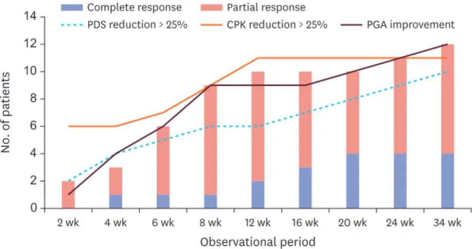
RTX administration and treatment response
Six (37.5%) patients received two doses of 1,000 mg RTX 2 weeks apart (total 2 g), 4 (25.0%) patients received two doses of 500 mg RTX 2 weeks apart (total 1 g), 3 (18.8%) patients received a single dose of 1,000 mg, one patient received 500 mg weekly for a total of four doses (total 2 g), 1 patient received 600 mg RTX 2 weeks apart (total 1.2 g), and one patient received a single dose of 200 mg RTX (Table 1).
The response rate to RTX treatment is shown in Fig. 1. At 12 weeks after RTX treatment, 11 (68.8%) and 9 (56.3%) patients achieved a 25% reduction in CPK and daily corticosteroid,
Table 1. Clinical characteristics of 16 patients with idiopathic inflammatory myopathies treated with RTX No. Sex Age, yr Disease
duration, mon Diagnosis Previous treatment Concomitant
treatment RTX dose, mg RTX cycles Responsea
1 F 56 187 DM AZA, Cys, TAC, HCQ, IVIG MTX, MMF 500 × 2 1 Y
2 F 54 169 DM MTX, MMF, Cys, TAC, CYC, LEF, HCQ, IVIG AZA 1,000 × 2 2 N
3 F 42 37 DM MTX, AZA, TAC, IVIG MMF, HCQ 500 × 2 1 N
4 M 59 20 DM AZA, Cys, CYC, HCQ MMF 500 × 2 1 Y
5 F 53 57 DM HCQ, IVIG MMF 1,000 × 2 2 N
6 F 33 186 PM AZA, CYC, IVIG MMF, MTX 500 × 2 2 Y
7 F 38 99 PM AZA, IVIG MTX 1,000 × 2 1 Y
8 F 57 13 DM CYC, HCQ, IVIG AZA 200 × 1 1 Y
9 F 63 84 PM CYC, IVIG AZA 500 × 4 1 Y
10 M 66 14 PM MMF, IVIG AZA 600 × 2 1 Y
11 F 41 171 DM AZA, MMF, Cys, IVIG 1,000 × 2 1 Y
12 F 57 62 DM MTX, AZA TAC 1,000 × 2 1 Y
13 F 44 70 PM MTX, AZA, MMF, TAC, IVIG MTX 1,000 × 2 1 Y
14 F 59 96 DM HCQ HCQ 1,000 × 1 1 N
15 F 45 144 PM MTX, AZA, MMF, Cys, TAC, IVIG 1,000 × 1 1 N
16 M 62 6 DM HCQ MTX 1,000 × 1 1 Y
DM = dermatomyositis, AZA = azathioprine, Cys = cyclosporin, TAC = tacrolimus, HCQ = hydroxychloroquine, IVIG = intravenous immunoglobulin, MTX = methotrexate, MMF = mycophenolate mofetil, PM = polymyositis, RTX = rituximab.
aClinical response at 24 weeks after RTX infusion.
respectively, and 6 (37.5%) patients showed improved physician's global assessment (PGA).
CR and PR were achieved by 2 and 6 patients, respectively, and the overall response rate was 62.5% (10 patients).
At 24 weeks after RTX treatment, 11 (68.8%) patients achieved a 25% reduction in
corticosteroid dose and CPK levels and 9 (56.3%) showed improved PGA. CR was achieved by 4 (25.0%) patients, and the overall response rate was 68.8% (11 patients). The median serum CPK level decreased from 421.5 (67.8–973.0) to 42.0 (interquartile range [IQR], 15.5–222.0) and the median dose of corticosteroid decreased from 20 mg/day (IQR, 15–45.0) to 13.8 mg (IQR, 10.6–15.2). At the end of follow-up (median 24 weeks, range 24–68 weeks), the overall response was 12 (75.0%) patients: 4 (25.0%) and 8 (50.0%) patients achieved CR and PR, respectively.
Table 2. Myositis activity at the time of rituximab treatment
Variables Values
Combined clinical manifestations
Muscle weakness 14 (87.5)
Myalgia 11 (68.8)
Skin rash 8 (50.0)
Fatigue 6 (37.5)
ILD 5 (31.3)
Dysphagia 4 (25.0)
Arthritis 4 (25.0)
Fever 3 (18.8)
Weight loss 3 (18.8)
Dyspnea 3 (18.8)
Hair loss 3 (18.8)
Itching 2 (12.5)
Coughing 2 (12.5)
Laboratory markers
CPK 421.0 (67.8–937.0)
LDH 405.0 (286.0–594.0)
Aldolase 15.9 (6.7–20.6)
ESR, mm/hr 20.5 (7.8–61.0)
CRP, mg/L 3.5 (1.9–4.3)
Daily corticosteroid dosage, mg, PDS equivalent dose 20.0 (15.0–45.0) Values are given as median (interquartile range) or number (%).
ILD = interstitial lung disease, CPK = creatine phosphokinase, LDH = lactate dehydrogenase, ESR = erythrocyte sedimentation rate, CRP = C-reactive protein, PDS = prednisolone.
0 2 6
2 wk
Observational period 12
14
10 8
4
4 wk 6 wk 8 wk 12 wk 16 wk 20 wk 24 wk 34 wk
No. of patients
Complete response Partial response
PDS reduction > 25% CPK reduction > 25% PGA improvement
Fig. 1. Response rate of RTX treatment.
CPK = creatine phosphokinase, PGA = physician's global assessment, PDS = prednisolone, RTX = rituximab.
Three patients were re-administered RTX after completion of the first cycle of RTX administration (at intervals of 27, 30, and 52 weeks) when IIM relapsed or was clinically indicated. Two patients were assessed to have no response in both the first and second cycles.
Factors associated with response to RTX treatment
A comparison between responders and non-responders at 24 weeks after RTX treatment is shown in Table 3. Baseline median muscle enzyme levels were higher in responders than non-responders: serum CPK, 692.0 vs. 64.0, P = 0.013; LDH, 530.0 vs. 286.0, P = 0.013; and aldolase, 18.1 vs. 6.7, P = 0.013. All patients with baseline serum CPK over 400 responded to treatment, but 2 out of 3 patients with CPK between 100 and 400, and only 1 out of 5 patients with CPK below 100 responded to RTX treatment. However, all other demographic and clinical characteristics, including patient age, sex, disease subtype, disease duration, RTX dose, ESR, and serum CRP, were not significantly different between the two groups.
We investigated autoantibody patterns and treatment response. Of the 16 patients, 10 patients tested ANA at the time of RTX administration, and ANA was detected in 6 patients.
The speckled pattern was the most common ANA pattern (4 out of 6 patients), followed by mixed and cytoplasmic patterns (1 patient each). All the 6 ANA–positive patients responded to RTX treatment (including all the 4 cases of CR), while only 2 cases achieved PR and the other 2 cases did not respond to RTX treatment among the 4 ANA-negative patients. Anti Jo-1 antibody was tested for in 3 patients and detected in 2 patients. All the 3 patients who tested positive for anti Jo-1 antibody achieved CR.
Table 3. Comparison between responders and non-responders at 24 weeks after RTX treatment
Variables at baseline Responders (n = 11) Non-responders (n = 5) P value
Age, yr 52.4 ± 11.3 50.6 ± 7.0 0.583
Disease duration 82.9 ± 70.4 100.6 ± 56.0 0.743
BMI 22.8 ± 4.3 23.6 ± 4.9 1.000
RTX dose/BSA 813.6 ± 339.3 890.0 ± 309.2 0.859
Daily corticosteroid 20.0 (15.0–37.5) 30.0 (20.0–30.0) 0.228
CPK 692.0 (363.0–1,104.0) 64.0 (63.0–79.0) 0.013
LDH 530.0 (405.0–599.0) 286.0 (258.0–309.0) 0.013
Aldolase 18.1 (15.9–22.3) 6.7 (6.7–8.3) 0.016
ESR 22.9 ± 20.1 54.0 ± 43.7 0.298
CRP 0.3 (0.2–0.4) 0.4 (0.3–0.4) 0.544
Sex 0.509
Male (n = 3) 3 (100.0) 0 (0.0)
Female (n = 13) 8 (61.5) 5 (38.5)
Disease subtype 0.588
DM (n = 10) 6 (60.0) 4 (40.0)
PM (n = 6) 5 (83.3) 1 (16.7)
Disease duration, yr 1.000
< 5 (n = 6) 4 (66.7) 2 (33.3)
5–10 (n = 5) 4 (80.0) 1 (20.0)
> 10 (n = 5) 3 (60.0) 2 (40.0)
Dose of RTX 1.000
Less than 2 g (n = 11) 7 (70.0) 3 (30.0)
2 g (n = 5) 4 (66.7) 2 (33.3)
Values are given as mean (standard deviation), median (interquartile rage) or number (%).
Normality was tested using Shapiro-Wilk normality test.
RTX = rituximab, BMI = body mass index, BSA = body surface area, CPK = creatine phosphokinase, LDH = lactate dehydrogenase, ESR = erythrocyte sedimentation rate, CRP = C-reactive protein, DM = cermatomyositis, PM = polymyositis.
Safety of RTX
In the 39.3 person-years of the observation period, we observed one adverse event (AE) (rate
= 25.4/1,000 person-years). One patient developed upper respiratory tract infection, but hospitalization was not required. No severe adverse event (SAE) or infusion reaction occurred.
DISCUSSION
In this multicenter study, RTX was effective in 75% of patients (12/16) at a median of 24 weeks after treatment for patients with refractory IIM who had responded poorly to conventional immunosuppressant treatment. The serum CPK level and daily dose of corticosteroid decreased significantly compared with the baseline after RTX in most patients, suggesting the clinical efficacy and safety of RTX in patients with refractory IIM.
Since the first classification criteria and 5 subsets for IIM were provided by Bohan and Peter17 in 1975, extended understanding and more clinical subsets of IIM have been suggested.
However, unlike the simple terminology “myopathy,” neither the pathophysiology nor phenotype of patients with IIM are straightforward. It is important to note that the diagnosis of IIM may be challenging, as the diagnosis of IIM should be made circumspectly after the exclusion of a variety of diseases that mimic IIM.19,20 Patient assessment also is complicated.
The pattern and severity of organ involvement is highly heterogeneous in each patient, and an objective measurement of muscular and extra-muscular involvement is complex.
Given the rarity and heterogeneity of IIM,19-21 outcome measure and response criteria have been available only in recent years, and evidence level for IIM treatment strategy is low. Unfortunately, a meaningful portion of patients with IIM responds incompletely to conventional immunosuppressive agents; therefore, patients with refractory IIM may be subjected to therapeutic strategies that have not been sufficiently investigated.
RTX, a selective CD20+ B lymphocyte-depleting agent, is a potential therapeutic option for the IIM treatment with evidence of prominent B lymphocyte infiltration in the muscles of patients with IIM. Infiltration of cytotoxic CD8+ T lymphocytes at the endomysium and/
or that of B lymphocytes accompanied by CD4+ T lymphocytes at perivascular sites are the pathologic hallmark of IIM.22,23 Although earlier studies suggested that B lymphocytes were considered to be more important in dermatomyositis than in polymyositis,24,25 subsequent studies identified the presence of plasma cells and immunoglobulin-transcribing genes in the endomysium of patients with polymyositis and inclusion body myositis.26
The strong association between MAA and clinical phenotype also supports the critical role of B lymphocytes in the pathophysiology of IIM. Antisynthetase syndrome (ASS) is a well-known subtype of IIM, characterized by a progressive disease course with severe ILD, mechanic's hand, a distinct histological pattern on muscle tissue, and ASS-specific autoantibodies, such as anti-Jo-1 antibody.27 Immune-mediated necrotizing myopathy (IMNM) is another particular subtype of IIM, associated with necrosis of microfibers without inflammatory cell infiltrates and IMNM-specific antibodies, including anti-3-hydroxy-3- methylglutaryl CoA reductase antibody and anti-signal recognition protein antibody.28-30 Anti-melanoma differentiation–associated gene 5 (MDA5) antibody is a novel and specific antibody associated with amyopathic dermatomyositis with rapidly progressive ILD.31,32 Anti- transcription intermediary factor 1γ antibody and the NM fragment of anti-nuclear matrix protein antibody have been described as related to cancer-associated myositis.33-35
Thus, evidence has started to accumulate regarding the efficacy of RTX in the management of IIM. A number of case series and small open-label trials have been published, and the majority of the studies have described patients with refractory and/or life-threatening IIM.
There also is a completed randomized, double-blinded, controlled clinical trial in patients with IIM (the RTX in myositis [RIM] trial).36 In total, 195 definite or probable patients with dermatomyositis or polymyositis or juvenile patients with dermatomyositis with poor response to glucocorticoids and at least one immunosuppressive agent were included in the RIM trial. Patients were randomly assigned to receive RTX at baseline (RTX early group) or 8 weeks later (RTX late group), and the time to achieving the International Myositis Assessment and Clinical Studies (IMACS) definition of improvement was compared between the two groups.37 At the end of the study (44 weeks after RTX treatment), 82.6% (161/195) of the patients met the definition of improvement although early and late treatment of RTX showed no significant differences for the primary and secondary end points.
It also is important to emphasize that previous reports show a considerable association between the clinical response and presence of MAA. The efficacy of RTX in patients with refractory ASS has been reported.38 Levine et al.39 reported that patients with the anti-Jo-1 antibody had a greater improvement in lung function after RTX treatment, and one recent study reported that the serum titer of MAA decreased after B cell depletion and was correlated with changes in disease activity.40 Aggarwal et al.41 evaluated predictors for the response to RTX from the RIM trial, and revealed that presence of MAA and less IIM-associated damage were associated with good prognosis and rapid clinical improvement. In this present study, we also revealed that ANA-positivity was associated with better clinical response to RTX treatment: all the 4 cases of CR were ANA–positive, while among the 4 ANA-negative patients, 2 cases achieved PR and the other 2 cases did not respond to RTX treatment.
When comparing responders and non-responders, ESR and serum CRP levels tended to be higher in non-responders (although not statistically significantly), despite the lower CPK levels in non-responders. This could be associated with the fact that the improvement in extra-muscular involvement was not fully reflected in the current response criteria. There were 2 patients who received RTX due to acute aggravation of organizing pneumonia, with normal serum CPK level (64 unit/dL and 79 unit/dL, respectively) and high ESR (116 mm/hr and 64 mm/hr, respectively). Unlike previous studies42,43 which reported especially favorable responses to pulmonary involvement, the 2 patients mentioned above with organizing pneumonia did not fully recover after RTX treatment, and they did not achieve CPK reduction nor by their physician's opinion. Contrarily, one patient with generalized and refractory skin lesions with normal serum CPK (32 unit/dL), CRP (0.15 mg/dL) and ESR levels (18 mm/hr) had almost fully recovered after RTX treatment and was classified as a responder. With the brief response criteria in this study, the data regarding extra-muscular lesions were simply handled by ‘physician's global assessment’, and further studies should apply a standardized assessment protocol for extra-muscular involvement such as the 2016 ACR-EULAR response criteria.44 Most of the patients did not receive additional immunosuppressive agents during the
observational period except one patient who started MTX 6 weeks after RTX administration. Our data reveals that the long-term clinical efficacy of RTX may persist, which is similar to previous studies,45 whereas reconstitution of peripheral blood B cell lineage occurs within a few months.46 Although this study describes the clinical efficacy of RTX in patients with refractory IIM, our study differs from the RIM trial as RTX is not approved for use in Korean patients with
IIM and our study is based on real-world data. Participants in our study were older (median, 51.8 years of age) with a longer disease duration (median, 7.4 years) than those in the RIM trial (mean age early forties, mean disease duration 5.3 years). However, the ratio of female participants (81%), the number of previous immunosuppressive agents administered prior to RTX treatment (median, 3.6), and the daily corticosteroid dose at RTX administration (median, 20.0 mg) were similar to that of the RIM trial (71% female patients, mean 3.1 immunosuppressive agents, approximately 20 mg of daily corticosteroid). The dosing regimen for RTX in IIM was also different. In the RIM trial, the dose of RTX used (total 2 g, as 2 × 1 g infusions) was the same as the FDA-approved dose for RA, GPA, and MPA.
The safety of RTX should also be discussed. We observed only one infection, and no SAE or infusion reaction occurred in this study; the rate of AEs in this study is lower than that of other studies. In a study that analyzed the efficacy and safety of RTX in Korean patients with active lupus nephritis, four out of 39 patients experienced an infusion reaction and three patients had an infection.12 In the RIM trial, there were 67 SAEs in 64 patients, and the RTX group showed significantly more infusion reactions than the placebo group. This underestimation of AE/SAEs may be related to the RTX dose, which was lower than that of other studies (less than half the patients received a total of 2 g RTX) and the collection of AE/
SAEs by retrospective chart review.
The relatively small number of study populations, lack of long term outcome and repeated treatment, incompletion of the autoantibody data and the lack of MAA and immunological data including B lymphocyte count are the limitations of our study.
Despite these limitations, it can be concluded that RTX is an effective and safe therapeutic option in patients with refractory IIM who have responded poorly to conventional treatment.
We believe that this study adds to the current knowledge of RTX in patients with IIM. Further prospective studies in Korean patients with IIM are required to provide a rationale for the application of RTX in patients with refractory IIM.
REFERENCES
1. Oddis CV, Aggarwal R. Treatment in myositis. Nat Rev Rheumatol 2018;14(5):279-89.
PUBMED | CROSSREF
2. Schiopu E, Phillips K, MacDonald PM, Crofford LJ, Somers EC. Predictors of survival in a cohort of patients with polymyositis and dermatomyositis: effect of corticosteroids, methotrexate and azathioprine.
Arthritis Res Ther 2012;14(1):R22.
PUBMED | CROSSREF
3. Taborda AL, Azevedo P, Isenberg DA. Retrospective analysis of the outcome of patients with idiopathic inflammatory myopathy: a long-term follow-up study. Clin Exp Rheumatol 2014;32(2):188-93.
PUBMED
4. Khoo T, Limaye V. Biologic therapy in the idiopathic inflammatory myopathies. Rheumatol Int 2019;40(2):191-205.
PUBMED | CROSSREF
5. Pierpont TM, Limper CB, Richards KL. Past, present, and future of rituximab-the world's first oncology monoclonal antibody therapy. Front Oncol 2018;8:163.
PUBMED | CROSSREF
6. Kinch MS. An overview of FDA-approved biologics medicines. Drug Discov Today 2015;20(4):393-8.
PUBMED | CROSSREF
7. Edwards JC, Szczepanski L, Szechinski J, Filipowicz-Sosnowska A, Emery P, Close DR, et al. Efficacy of B-cell- targeted therapy with rituximab in patients with rheumatoid arthritis. N Engl J Med 2004;350(25):2572-81.
PUBMED | CROSSREF
8. Joly P, Maho-Vaillant M, Prost-Squarcioni C, Hebert V, Houivet E, Calbo S, et al. First-line rituximab combined with short-term prednisone versus prednisone alone for the treatment of pemphigus (Ritux 3):
a prospective, multicentre, parallel-group, open-label randomised trial. Lancet 2017;389(10083):2031-40.
PUBMED | CROSSREF
9. Stone JH, Merkel PA, Spiera R, Seo P, Langford CA, Hoffman GS, et al. Rituximab versus cyclophosphamide for ANCA-associated vasculitis. N Engl J Med 2010;363(3):221-32.
PUBMED | CROSSREF
10. Arnold DM, Dentali F, Crowther MA, Meyer RM, Cook RJ, Sigouin C, et al. Systematic review: efficacy and safety of rituximab for adults with idiopathic thrombocytopenic purpura. Ann Intern Med 2007;146(1):25-33.
PUBMED | CROSSREF
11. Rovin BH, Furie R, Latinis K, Looney RJ, Fervenza FC, Sanchez-Guerrero J, et al. Efficacy and safety of rituximab in patients with active proliferative lupus nephritis: the Lupus Nephritis Assessment with Rituximab study. Arthritis Rheum 2012;64(4):1215-26.
PUBMED | CROSSREF
12. Bang SY, Lee CK, Kang YM, Kim HA, Suh CH, Chung WT, et al. Multicenter retrospective analysis of the effectiveness and safety of rituximab in korean patients with refractory systemic lupus erythematosus.
Autoimmune Dis 2012;2012:565039.
PUBMED | CROSSREF
13. Hu Y, Nie H, Yu HH, Qin C, Wu LJ, Tang ZP, et al. Efficacy and safety of rituximab for relapsing-remitting multiple sclerosis: A systematic review and meta-analysis. Autoimmun Rev 2019;18(5):542-8.
PUBMED | CROSSREF
14. Fasano S, Gordon P, Hajji R, Loyo E, Isenberg DA. Rituximab in the treatment of inflammatory myopathies: a review. Rheumatology (Oxford) 2017;56(1):26-36.
PUBMED | CROSSREF
15. Yang JA, Lee SJ, Park JW, Kwon HM, Moon JY, Ko DJ, et al. Rituximab treatment for the patients with refractory inflammatory myopathy. J Rheum Dis 2013;20(5):303-9.
CROSSREF
16. Bohan A, Peter JB, Bowman RL, Pearson CM. Computer-assisted analysis of 153 patients with polymyositis and dermatomyositis. Medicine (Baltimore) 1977;56(4):255-86.
PUBMED | CROSSREF
17. Bohan A, Peter JB. Polymyositis and dermatomyositis (first of two parts). N Engl J Med 1975;292(7):344-7.
PUBMED | CROSSREF
18. Couderc M, Gottenberg JE, Mariette X, Hachulla E, Sibilia J, Fain O, et al. Efficacy and safety of rituximab in the treatment of refractory inflammatory myopathies in adults: results from the AIR registry.
Rheumatology (Oxford) 2011;50(12):2283-9.
PUBMED | CROSSREF
19. Dalakas MC. Inflammatory muscle diseases. N Engl J Med 2015;372(18):1734-47.
PUBMED | CROSSREF
20. Lundberg IE, de Visser M, Werth VP. Classification of myositis. Nat Rev Rheumatol 2018;14(5):269-78.
PUBMED | CROSSREF
21. Bernatsky S, Joseph L, Pineau CA, Bélisle P, Boivin JF, Banerjee D, et al. Estimating the prevalence of polymyositis and dermatomyositis from administrative data: age, sex and regional differences. Ann Rheum Dis 2009;68(7):1192-6.
PUBMED | CROSSREF
22. Engel A. Myology: Basic and Clinical. New York, NY: McGraw-Hill Companies; 1994.
23. Vattemi G, Mirabella M, Guglielmi V, Lucchini M, Tomelleri G, Ghirardello A, et al. Muscle biopsy features of idiopathic inflammatory myopathies and differential diagnosis. Auto Immun Highlights 2014;5(3):77-85.
PUBMED | CROSSREF
24. Arahata K, Engel AG. Monoclonal antibody analysis of mononuclear cells in myopathies. I: quantitation of subsets according to diagnosis and sites of accumulation and demonstration and counts of muscle fibers invaded by T cells. Ann Neurol 1984;16(2):193-208.
PUBMED | CROSSREF
25. Grundtman C, Malmström V, Lundberg IE. Immune mechanisms in the pathogenesis of idiopathic inflammatory myopathies. Arthritis Res Ther 2007;9(2):208.
PUBMED | CROSSREF
26. Greenberg SA, Bradshaw EM, Pinkus JL, Pinkus GS, Burleson T, Due B, et al. Plasma cells in muscle in inclusion body myositis and polymyositis. Neurology 2005;65(11):1782-7.
PUBMED | CROSSREF
27. Greco M, García de Yébenes MJ, Alarcón I, Brandy-García AM, Rúa-Figueroa Í, Loza E, et al. Idiopathic inflammatory myopathies and antisynthetase syndrome: contribution of antisynthetase antibodies to improve current classification criteria. Ann Rheum Dis 2019;78(9):1291-2.
PUBMED | CROSSREF
28. Basharat P, Christopher-Stine L. Immune-mediated necrotizing myopathy: update on diagnosis and management. Curr Rheumatol Rep 2015;17(12):72.
PUBMED | CROSSREF
29. Musset L, Allenbach Y, Benveniste O, Boyer O, Bossuyt X, Bentow C, et al. Anti-HMGCR antibodies as a biomarker for immune-mediated necrotizing myopathies: a history of statins and experience from a large international multi-center study. Autoimmun Rev 2016;15(10):983-93.
PUBMED | CROSSREF
30. Ladislau L, Arouche-Delaperche L, Allenbach Y, Benveniste O. Potential pathogenic role of anti-signal recognition protein and anti-3-hydroxy-3-methylglutaryl-CoA reductase antibodies in immune-mediated necrotizing myopathies. Curr Rheumatol Rep 2018;20(9):56.
PUBMED | CROSSREF
31. Moghadam-Kia S, Oddis CV, Aggarwal R. Anti-MDA5 antibody spectrum in western world. Curr Rheumatol Rep 2018;20(12):78.
PUBMED | CROSSREF
32. Abe Y, Matsushita M, Tada K, Yamaji K, Takasaki Y, Tamura N. Clinical characteristics and change in the antibody titres of patients with anti-MDA5 antibody-positive inflammatory myositis. Rheumatology (Oxford) 2017;56(9):1492-7.
PUBMED | CROSSREF
33. Fiorentino DF, Chung LS, Christopher-Stine L, Zaba L, Li S, Mammen AL, et al. Most patients with cancer-associated dermatomyositis have antibodies to nuclear matrix protein NXP-2 or transcription intermediary factor 1γ. Arthritis Rheum 2013;65(11):2954-62.
PUBMED | CROSSREF
34. Yang H, Peng Q, Yin L, Li S, Shi J, Zhang Y, et al. Identification of multiple cancer-associated myositis- specific autoantibodies in idiopathic inflammatory myopathies: a large longitudinal cohort study. Arthritis Res Ther 2017;19(1):259.
PUBMED | CROSSREF
35. Hengstman GJ, Vree Egberts WT, Seelig HP, Lundberg IE, Moutsopoulos HM, Doria A, et al. Clinical characteristics of patients with myositis and autoantibodies to different fragments of the Mi-2 beta antigen. Ann Rheum Dis 2006;65(2):242-5.
PUBMED | CROSSREF
36. Oddis CV, Reed AM, Aggarwal R, Rider LG, Ascherman DP, Levesque MC, et al. Rituximab in the treatment of refractory adult and juvenile dermatomyositis and adult polymyositis: a randomized, placebo-phase trial. Arthritis Rheum 2013;65(2):314-24.
PUBMED | CROSSREF
37. Rider LG, Giannini EH, Brunner HI, Ruperto N, James-Newton L, Reed AM, et al. International consensus on preliminary definitions of improvement in adult and juvenile myositis. Arthritis Rheum 2004;50(7):2281-90.
PUBMED | CROSSREF
38. Marie I, Dominique S, Janvresse A, Levesque H, Menard JF. Rituximab therapy for refractory interstitial lung disease related to antisynthetase syndrome. Respir Med 2012;106(4):581-7.
PUBMED | CROSSREF
39. Levine TD. Rituximab in the treatment of dermatomyositis: an open-label pilot study. Arthritis Rheum 2005;52(2):601-7.
PUBMED | CROSSREF
40. Aggarwal R, Oddis CV, Goudeau D, Koontz D, Qi Z, Reed AM, et al. Autoantibody levels in myositis patients correlate with clinical response during B cell depletion with rituximab. Rheumatology (Oxford) 2016;55(6):991-9.
PUBMED | CROSSREF
41. Aggarwal R, Bandos A, Reed AM, Ascherman DP, Barohn RJ, Feldman BM, et al. Predictors of clinical improvement in rituximab-treated refractory adult and juvenile dermatomyositis and adult polymyositis.
Arthritis Rheumatol 2014;66(3):740-9.
PUBMED | CROSSREF
42. Doyle TJ, Dhillon N, Madan R, Cabral F, Fletcher EA, Koontz DC, et al. Rituximab in the Treatment of Interstitial Lung Disease Associated with Antisynthetase Syndrome: A Multicenter Retrospective Case Review. J Rheumatol 2018;45(6):841-50.
PUBMED | CROSSREF
43. Barsotti S, Cioffi E, Tripoli A, Tavoni A, D'Ascanio A, Mosca M, et al. The use of rituximab in idiopathic inflammatory myopathies: description of a monocentric cohort and review of the literature. Reumatismo 2018;70(2):78-84.
PUBMED | CROSSREF
44. Rider LG, Aggarwal R, Pistorio A, Bayat N, Erman B, Feldman BM, et al. 2016 American College of Rheumatology/European League against rheumatism criteria for minimal, moderate, and major clinical response in juvenile dermatomyositis: an International Myositis Assessment and Clinical Studies Group/
Paediatric Rheumatology International Trials Organisation Collaborative Initiative. Ann Rheum Dis 2017;76(5):782-91.
PUBMED | CROSSREF
45. de Souza FH, Miossi R, de Moraes JC, Bonfá E, Shinjo SK. Favorable rituximab response in patients with refractory idiopathic inflammatory myopathies. Adv Rheumatol 2018;58(1):31.
PUBMED | CROSSREF
46. Leandro MJ, Cambridge G, Ehrenstein MR, Edwards JC. Reconstitution of peripheral blood B cells after depletion with rituximab in patients with rheumatoid arthritis. Arthritis Rheum 2006;54(2):613-20.
PUBMED | CROSSREF