INTRODUCTION
Lower extremity deep venous thrombosis (DVT) is a seri- ous medical condition that can result in death or major dis- ability due to pulmonary embolism and post-thrombotic syn- drome (PTS). Although anticoagulation (heparin followed by oral anticoagulation) is currently considered the standard of care for the prevention of pulmonary embolism and recurrent DVT, this form of therapy does not protect the patient from the manifestations of PTS, which can appear months to years
after the acute episode of DVT. Early thrombus removal is a logical approach to improve the long-term outcome of DVT.
Two goals may be achieved by early thrombus removal: relief of venous outflow obstruction and preservation of valve func- tion, both of which are established determinants of PTS.
Treatment strategies of DVT have included systemic throm- bolytic therapy, surgical thrombectomy, and catheter-directed thrombolysis. Among them, catheter-directed thrombolysis is an attractive method, because it can potentially help achieve restoration of the lumen and removal of the thrombus lining
J Korean Soc Radiol 2011;65(4):337-344
Received April 19, 2011; Accepted July 19, 2011 Corresponding author: Jae Kyu Kim, MD
Department of Radiology, Chonnam National University Hospital, 42 Jebong-ro, Dong-gu, Gwangju 501-757, Korea.
Tel. 82-62-220-5746 Fax. 82-62-226-4380 E-mail: [email protected]
Copyrights © 2011 The Korean Society of Radiology
Purpose: To evaluate the venous patency in patients treated by catheter-directed thrombolysis with low-dose urokinase (UK) for symptomatic lower extremity deep venous thrombosis (DVT).
Materials and Methods: Eighty-nine consecutive patients (46 women and 43 men; mean age, 58.1 years), treated by catheter-directed thrombolysis with low- dose UK were included in this study. Immediate venous patency was evaluated in terms of technical success (successful restoration of antegrade in-line flow in the treated vein with residual stenosis rate of less than 30%) and clinical success (sig- nificant reduction of clinical symptoms before hospital discharge). Late venous pa- tency was evaluated in terms of primary patency rate and clinical success.
Results: Immediate technical success was achieved in all patients and immediate clinical success in 80 (90%) patients. There was no major systemic bleeding compli- cation. The primary patency rate at 6 months and 12 months was 84% and 79%, respectively. Fifty-six (63%) patients were asymptomatic after a median clinical fol- low-up of 18 months, eleven (12%) patients improved moderately, seven (8%) pa- tients remained unchanged, and fifteen (17%) patients had no clinical follow-up.
Conclusion: Short-term catheter-directed thrombolysis with low-dose UK can be an effective, safe method to manage DVT of the lower extremities.
Index terms
Catheter-Directed Thrombolysis Aspiration Thrombectomy
Lower Extremity Deep Venous Thrombosis
Short-Term Catheter-Directed Thrombolysis with Low-Dose
Urokinase Followed by Aspiration Thrombectomy for Treatment of Symptomatic Lower Extremity Deep Venous Thrombosis
1저용량 유로키나제를 이용한 단기간 혈전용해술과 혈전흡입술을 시행한 하지 심부정맥혈전증 환자의 치료 효과1
Se Hee Jung, MD
1, Jae Kyu Kim, MD
1,2, Nam Kyu Chang, MD
2,3, Jae Hoon Lim, MD
1,2,
Nam-Yeul Lim, MD
1,2, Jang-Hyeon Song, MD
1, Soo Jin Na Choi, MD
4, Sang Young Chung, MD
41Department of Radiology, Chonnam National University Hospital, Gwangju, Korea
2Department of Radiology, Chonnam National University Medical School, Gwangju, Korea
3Department of Radiology, Chonnam National University Hwasun Hospital, Hwasun, Korea
4Department of Surgery, Chonnam National University Hospital, Chonnam National University Medical School, Gwangju, Korea
vein, and the popliteal vein. Over time, the ipsilateral poplite- al venous approach became the access site of choice. In a typi- cal case, with the patient prone on the angiographic table, the popliteal vein was accessed under ultrasonographic guidance with a small gauge needle. Most commonly, a 5-F sheath was inserted and all subsequent catheter and wire exchanges were performed through it. Infusion was performed by using the end hole catheter and/or a multiple-side-hole infusion cathe- ter (Multi-sideport catheter infusion set, Cook, Bloomington, IN, USA). Before thrombolytic therapy, venography was once again performed to confirm the position and evaluate the infe- rior vena cava (IVC) thrombus. Prophylactic inferior vena cava filter placement was not routinely performed; the catheter was placed two thirds of the distance into the thrombosed venous segment and urokinase (Urokinase, Green Cross, Seoul, Korea) was administered either as a single bolus (100,000-150,000 IU) or as a continuous infusion (100,000-150,000 IU/h in split dos- es). Patients were systemically anticoagulated with a 5,000 IU bolus of intravenous heparin.
After catheter-directed thrombolysis, aspiration thrombec- tomy in 67 (75%) patients and/or mechanical thrombectomy in 30 (34%) patients were performed in an attempt to clear large volumes of softening thrombi. Aspiration thrombecto- my was performed in a to-and-fro fashion using an 8-10 F guiding catheter (Envoy catheter, Cordis, Miami, FL, USA), while maintaining negative pressure with a 50 mL syringe. To prevent body volume loss during aspiration thrombectomy, the same amount of heparinized saline was infused manually via the sheath side arm. A mechanical thrombectomy was performed using a Trerotola Percutaneous Thrombectomy Device (Arrow International, Reading, PA, USA) to fragment the organized thrombus.
The status of lysis was monitored at the venography at 30 minutes intervals. If only partial lysis was achieved, the infu- sion catheter was repositioned within the residual thrombus and the infusion was continued. The start and end times of thrombolysis, as well as the concentration and total amount of UK administered, were recorded. If UK was applied as a single bolus injection, the infusion time was less than 10 sec- onds. In patients with persistent occlusion of the iliac seg- ment but a patent femoro-popliteal segment, the iliac vein was recanalized by means of angioplasty only or followed by the venous valves (1-3). However, the risk of major bleeding
complications during venous thrombolysis has varied signifi- cantly in published studies and represents a significant impedi- ment to widespread acceptance of catheter-directed thrombol- ysis for the treatment of the DVT (1-3). Several studies have demonstrated that mechanical thrombectomy (MT) devices can remove venous thrombus quickly and effectively without the bleeding risks associated with pharmacologic thromboly- sis (4, 5).
The purpose of this study was to evaluate the safety and ef- ficacy of short-term catheter-directed thrombolysis using low-dose urokinase (UK) using an adjunctive method.
MATERIALS AND METHODS
Patients
This study was approved by the institutional review board.
The radiology information systems database was searched for patients who underwent catheter-directed thrombolysis for DVT between January 2006 and April 2010. Patients were in- cluded in this retrospective analysis if they had documented sonographic, computed tomographic, and/or venographic confirmation of deep vein thrombosis. Patients were excluded from this retrospective analysis if UK was administered con- tinuously overnight (> 8 hours). Inclusion criteria for this study were applied to low-dose UK as either a single intrave- nous bolus or as a continuous infusion for less than 8 hours.
Consequently, eighty-nine patients (46 women and 43 men;
age range, 16-86 years; mean 58.1 years) were evaluated. All patients did not have a contraindication for the use of antico- agulation, contrast media, or thrombolytic agents. Contrain- dications for thrombolytic agents included active internal bleeding, recent cerebrovascular accident, allergy to thrombo- lytic agents, recent major surgery, recent serious gastrointesti- nal bleeding, recent serious trauma, and coagulopathy. Initial symptoms, duration of symptoms, predisposing factors, and thrombus location were recorded for each treated extremity.
Endovascular Procedure
The physician who performed the procedure selected the catheterization technique for thrombolysis. The venous access sites included the internal jugular vein, the common femoral
patency (6). For patients who were lost to a more recent fol- low-up, the follow-up interval was defined as the time be- tween the venous intervention and the last time point at which information about the clinical limb status was clearly documented in the medical record. Clinical success was grad- ed as completely improved (asymptomatic), partially im- proved (moderately improved), unchanged, or worse.
Major bleeding was defined as intracranial bleeding, bleeding resulting in death, transfusion, surgery, or cessation of thrombo- lytic therapy, according to Society of Interventional Radiology reporting standards (10).
Data Analysis and Statistics
The technical success rates, clinical success rates, primary pa- tency rates, and incidences of major bleeding were expressed as percentages. The mean thrombolytic infusion times, total thrombolytic agent doses, and their standard deviations were calculated and the primary patency rate calculated at 6 and 12 months, respectively.
RESULTS
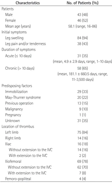
All patients were symptomatic with one or more of the fol- lowing: leg swelling, leg pain and/or tenderness (Table 1). In 31 (35%) patients, acute DVT (defined as symptom duration of 10 days or less) was treated. In 58 (65%) patients, chronic DVT (defined as symptom duration of greater than 10 days) was treated. At the time of treatment, nine (10%) patients had a known malignancy and one (1%) patient was pregnant. The location of the treated thrombosed venous segments was the left limb in 75 (84%) patients and the right limb in 14 (16%) patients. As determined at venography, iliofemoral DVT was present in 69 (78%) patients, in which 7 (8%) where the throm- bus extended into the IVC. Isolated femoro-popliteal DVT was found in 4 (4%) patients, whereas isolated iliac DVT was found in 16 (18%) patients.
Thirty-six (40%) patients were given a single bolus injection of UK (range, 4-14 × 104 IU, mean dose, 4.89 ± 2.51 × 10⁴ IU) and 53 (60%) patients had a continuous infusion of UK (range, 12-80, mean dose, 33.73 ± 16.42 × 10⁴ IU) for a mean of 168 minutes (range, 30-420 minutes) (Table 2). After cathe- ter-directed thrombolysis, aspiration thrombectomy with or stent deployment. Initial dilatation for reconstruction of the
iliac vein was achieved with a 8-12-mm-diameter balloon catheter (Cook, Bloomington, IN, USA). Stents were placed over a guide wire into the iliac vein and were typically be- tween 10 to 14 mm in diameter and 6 to 8 cm long. Zilver stents (Cook, Bloomington, IN, USA; n = 25) and Smart stents (Cordis, Miami, FL, USA; n = 37) were commonly used to re- construct the iliac vein. The deployed stents were fully dilated with the appropriate diameter angioplasty balloon catheter.
After endovascular therapy, 6 months of anticoagulation therapy with warfarin sodium was initiated and adjusted to maintain the international normalized ratio in the range of 2.0-3.0. Patients were subsequently fitted for a knee- or thigh- high venous compression hose.
Outcome Variables
The patients’ medical records, radiology reports, procedural data, and venograms were reviewed. Immediate venous pa- tency was evaluated in terms of technical success and clinical success. Assessment of immediate technical success was ret- rospectively performed by one radiologist on examination of the initial venographic images and the images obtained after completion of the interventional procedure. Technical success was defined as either the complete thrombolysis of clot, tech- nical restoration of normal venous blood flow, and less than 30% remaining luminal narrowing; or partial thrombolysis of clot that allows adjunctive methods (such as angioplasty, stent placement, or thrombectomy) to be used for the successful restoration of patency and flow (6-9). Immediate clinical suc- cess was defined as the presence of technical success in con- junction with considerable improvement in lower extremity swelling and/or pain before hospital discharge.
Late venous patency was evaluated in terms of primary pa- tency rate and/or clinical success. Because objective imaging follow-up to assess for recurrent DVT and valvular dysfunc- tion was not routinely obtained, late primary patency rate was evaluated in patients who underwent follow-up computed to- mography angiography (CTA) or Doppler sonography after 6 months and 12 months at the end of the catheter-directed thrombolysis. Primary patency was defined as the time from the intervention, to the first occurrence of either thrombosis of the treated segment or to an intervention for maintaining
without mechanical thrombectomy was performed in 67 (75%) patients. Subsequent angioplasty and stent placement was performed in 83 (93%) patients and 65 (73%) patients, re- spectively. The rate of aspiration thrombectomy with or without mechanical thrombectomy was higher in acute DVT patients (97%) than in chronic DVT patients (64%). Balloon angioplas- ty was performed in 30 (97%) acute DVT patients and stents were placed in 25 (81%) acute DVT patients. Balloon angio- plasty was performed in 53 (91%) chronic DVT patients and stents were placed in 40 (69%) chronic DVT patients.
Continuous UK infusion was performed in 15 (48%) acute DVT patients and 38 (66%) chronic DVT patients (Table 2).
In acute DVT patients, the mean amount of UK delivered in a single dose and continuous infusion was 4.8 ± 2.5 × 10⁴ IU and 29.3 ± 12.6 × 10⁴ IU, respectively, in chronic DVT pa- tients, the mean amount of UK delivered in a single dose and continuous infusion was 4.7 ± 2.2 × 10⁴ IU and 36.4 ± 18.2 × 10⁴ IU, respectively. The mean amount of continous UK infu- sion was higher in chronic DVT patients than in acute DVT patients. Also, both mean UK infusion time and total proce- dure time were higher in chronic DVT patients than in acute DVT patients.
Immediate technical success was achieved in all patients by completion venography. Clinical success, defined as a de- crease in swelling, pain and/or swelling of the affected ex- tremity within hospital discharge, was displayed in 80 (90%) patients. There was no major complication during or after the Table 1. Patient Characteristics with Deep Venous Thrombosis (n = 89)
Characteristics No. of Patients (%) Patients
Male 43 (48)
Female 46 (52)
Mean age (years) 58.1 (range, 16-86) Initial symptoms
Leg swelling 84 (94)
Leg pain and/or tenderness 38 (43) Duration of symptoms
Acute (≤ 10 days) 31 (35)
(mean, 4.9 ± 2.9 days, range, 1-10 days)
Chronic (> 10 days) 58 (65)
(mean, 181.1 ± 660.5 days, range, 11-3,500 days) Predisposing factors
Immobilization 29 (33)
May-Thurner syndrome 20 (22)
Previous operation 13 (15)
Malignancy 9 (10)
Pregnancy 1 (1)
Unknown 31 (35)
Location of thrombus
Left limb 75 (84)
Right limb 14 (16)
Iliac 16 (18)
Without extension to the IVC 14 (16) With extension to the IVC 2 (2)
Iliofemoral 69 (78)
Without extension to the IVC 62 (70) With extension to the IVC 7 (8)
Femoro-popliteal 4 (4)
Note.-IVC = inferior vena cava
Table 2. Procedure in 89 Patients with Deep Venous Thrombosis (n = 89)
Acute (≤ 10 days) Chronic (> 10 days) Procedures
UK thrombolysis (No, %) 89 (100) 31 58
Single-dose 36 (40) 16 (52) 20 (34)
Continuous infusion 53 (60) 15 (48) 38 (66)
Aspiration thrombectomy (No, %) 67 (75) 30 (97) 37 (64)
Mechanical thrombectomy (No, %) 30 (34) 12 (39) 18 (31)
Balloon angioplasty (No, %) 83 (93) 30 (97) 53 (91)
Stent (No, %) 65 (73) 25 (81) 40 (69)
UK dosage
Mean amount (× 104 IU, SD)
Single-dose 4.89 ± 2.51 (range, 4-14) 4.8 ± 2.5 4.7 ± 2.2
Continuous infusion 33.73 ± 16.42 (range, 12-80) 29.3 ± 12.6 36.4 ± 18.2
Mean time (minute, SD)
Continuous infusion 168 ± 94 (range, 30-420) 148 ± 74.5 181 ± 102.9
Note.-UK = urokinase
vice reported that in 2008, 5.31 per 100,000 of the population were treated for new or recurring DVT, and the incidence of this disease is increasing (11). Lower extremity DVT is a seri- ous medical condition that can result in death or major dis- ability due to pulmonary embolism and post-thrombotic syn- drome. Therefore, the goals of therapy in patients with DVT include prevention of pulmonary embolism, restoration of obstructed venous return, prevention of recurrent DVT, and preservation of venous valve function.
Systemic heparin followed by oral anticoagulation was considered the standard of care for the prevention of pulmo- nary embolism and recurrent DVT. Although anticoagulation is successful in preventing further propagation of thrombus, this form of therapy does not effectively treat the extensive thrombus. Consequently, there is slow relief of venous out- flow obstruction and damage of the existing valves, both of which play a role in post-thrombotic syndrome (12-14). Thera- pies for the existing thrombus include a surgical thrombecto- my, systemic thrombolytic therapy, and catheter-directed thrombolysis. Catheter-directed thrombolysis is a particularly attractive method because it has been proven to be effective in restoring venous patency and preserving of valve function. In a multicenter venous registry, complete lysis was seen in 31%
of patients with a 1-year patency rate of 79% (15). For 20 years, the plasminogen activator urokinase was the dominant thrombolytic agent in the catheter-directed treatment of ve- nous occlusions. However, several limitations have precluded its widespread use: (i) major bleeding complications in 11%
of patients in the only published multicenter study; (ii) long infusion time causing significant patient discomfort and procedure.
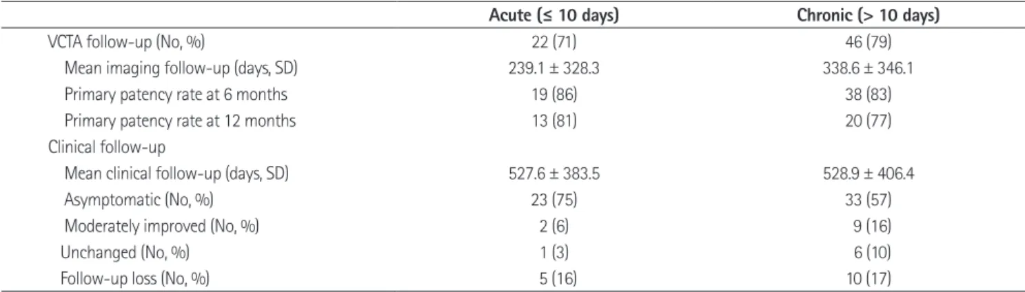
Late anatomic success was evaluated in patients who un- derwent follow-up CTA or Doppler sonography (Table 3).
The interval of the mean imaging follow-up was 10 months.
The primary patency rate at 6 months and 12 months was 84% and 77%, respectively. The primary patency rate at 6 months was 86% and 83% in acute DVT patients and chronic DVT patients, respectively. And primary patency rate at 12 months was 81% and 77%, respectively. Fifty-six of 89 (63%) patients were asymptomatic after a median clinical follow-up of 18 months, eleven (12%) patients were moderately im- proved, seven (8%) patients were unchanged, and fifteen (17%) patients had no clinical follow-up.
Rethrombosis of treated segments were observed in 3 pa- tients with acute DVT and 11 patients with chronic DVT during the follow-up period. All of them were symptomatic and had computer tomographic confirmation of rethrombosis.
All patients received a reintervention. Immediate technical suc- cess was achieved in all patients by completion venography of the reintervention. In all recurring patients, catheter-directed thrombolysis was performed ether as a single intravenous bo- lus or as a continuous infusion. Reintervention of acute DVT patients included ballon angioplasty (n = 2) and stent deploy- ment (n = 2). In chronic DVT patients, aspiration thrombec- tomy in 7 patients, balloon angioplasty in 6 patients, and/or stent deployment in 5 patients, were performed.
DISCUSSION
The Korean Health Insurance Review & Assessment Ser-
Table 3. Late Venous Patency: Anatomic and Clinical Results
Acute (≤ 10 days) Chronic (> 10 days)
VCTA follow-up (No, %) 22 (71) 46 (79)
Mean imaging follow-up (days, SD) 239.1 ± 328.3 338.6 ± 346.1
Primary patency rate at 6 months 19 (86) 38 (83)
Primary patency rate at 12 months 13 (81) 20 (77)
Clinical follow-up
Mean clinical follow-up (days, SD) 527.6 ± 383.5 528.9 ± 406.4
Asymptomatic (No, %) 23 (75) 33 (57)
Moderately improved (No, %) 2 (6) 9 (16)
Unchanged (No, %) 1 (3) 6 (10)
Follow-up loss (No, %) 5 (16) 10 (17)
Note.-VCTA = venous computed tomography angiography
ferent patients who are evaluated using different criteria and varying thrombolysis protocols in which variable combina- tions of initial lacing dose of thrombolytic agents, balloon maceration, use of mechanical thrombectomy devices, and timing of stent placement have been used. Nevertheless, the high overall immediate clinical success rate (90%), low com- plication rate (0%), and low severity of late venous disability observed in patients in this study certainly provides support for the hypothesis that short-term catheter-directed throm- bolysis with low-dose UK and adjunctive method followed by early stent placement, is a safe and effective endovascular method for symptomatic DVT with reduction of the number of thrombolytic agents, infusion times, and complications.
REFERENCES
1. Comerota AJ, Throm RC, Mathias SD, Haughton S, Mewis- sen M. Catheter-directed thrombolysis for iliofemoral deep venous thrombosis improves health-related quality of life. J Vasc Surg 2000;32:130-137
2. Patel NH, Stookey KR, Ketcham DB, Cragg AH. Endovascu- lar management of acute extensive iliofemoral deep ve- nous thrombosis caused by May-Thurner syndrome. J Vasc Interv Radiol 2000;11:1297-1302
3. Ouriel K, Katzen B, Mewissen M, Flick P, Clair DG, Benenati J, et al. Reteplase in the treatment of peripheral arterial and venous occlusions: a pilot study. J Vasc Interv Radiol 2000;11:849-854
4. Delomez M, Beregi JP, Willoteaux S, Bauchart JJ, Janne bleeding complication during treatment, and (iii) significant
hospital costs because of the need to closely monitor patients receiving thrombolytic infusions (15).
Aspiration thrombectomy and mechanical thrombectomy are therefore an attractive approach for the treatment of a DVT because these methods can provide a rapid and effective means of DVT treatment without the risk of bleeding, followed by high dosage or long time thrombolytic agent administra- tion. In our study, after short-term catheter-directed throm- bolysis, aspiration thrombectomy using a pullback technique and mechanical thrombectomy, were performed in 67 (75%) patients and in 30 (34%) patients, respectively. The pullback technique is defined as a dynamic to-and-fro movement of the aspiration catheter that occurs while maintaining negative pressure using a syringe. For example, an aspiration catheter is introduced into a thrombus-filled vein through the sheath and is withdrawn with to-and-fro movement at the throm- bus-filled vein while negative pressure is applied and main- tained using a syringe. Using this technique, a large thrombus can be fragmented into smaller sizes and can be easily aspirat- ed via an aspiration catheter.
In this study, the mean UK infusion time of continuous in- fusion was 168 ± 94 minutes, which is shorter than other published studies (Table 4). The mean total UK dose of a sin- gle dose and continuous infusion was 4.89 ± 2.51 × 10⁴ IU and 33.73 ± 16.42 × 10⁴ IU, respectively, which is lower than other published studies. In addition, there was no major systemic bleeding complication. However, a comparison between this study and other published DVT studies is confounded by dif- Table 4. Literature Review with Mean Total Dose and Thrombolysis Time
No. of Patients Mean Total Dose (× 104 IU)
Mean Thrombolysis Time (Hours)
Primary Patency Rate at 1 Year (%) UK alone
Bjarnason (16) 87 1,000 75.0 63
Mewissen (15) 312 680 48.8 60
O’ Sullivan (10) 31 610 40.0 79
Patel (2) 10 590 52.0 90
UK with MT
Kasirajan (17) 9 Not available 20.2 52
Suh (18) 24 180 23.3 85
Jung (present study) 77
Single-dose 36 4.9 Less than 10 seconds
Continous infusion 53 33.7 2.8
Note.-MT = mechanical thrombectomy, UK = urokinase
12. Akesson H, Brudin L, Dahlström JA, Eklöf B, Ohlin P, Plate G. Venous function assessed during a 5 year period after acute ilio-femoral venous thrombosis treated with antico- agulation. Eur J Vasc Surg 1990;4:43-48
13. Johnson BF, Manzo RA, Bergelin RO, Strandness DE Jr. Re- lationship between changes in the deep venous system and the development of the postthrombotic syndrome af- ter an acute episode of lower limb deep vein thrombosis:
a one- to six-year follow-up. J Vasc Surg 1995;21:307- 312; discussion 313
14. Elsharawy M, Elzayat E. Early results of thrombolysis vs anticoagulation in iliofemoral venous thrombosis. A ran- domised clinical trial. Eur J Vasc Endovasc Surg 2002;24:
209-214
15. Mewissen MW, Seabrook GR, Meissner MH, Cynamon J, Labropoulos N, Haughton SH. Catheter-directed throm- bolysis for lower extremity deep venous thrombosis: re- port of a national multicenter registry. Radiology 1999;
211:39-49
16. Bjarnason H, Kruse JR, Asinger DA, Nazarian GK, Dietz CA Jr, Caldwell MD, et al. Iliofemoral deep venous thrombosis:
safety and efficacy outcome during 5 years of catheter- directed thrombolytic therapy. J Vasc Interv Radiol 1997;8:
405-418
17. Kasirajan K, Gray B, Ouriel K. Percutaneous AngioJet thrombectomy in the management of extensive deep ve- nous thrombosis. J Vasc Interv Radiol 2001;12:179-185 18. Suh SH, Lee DY, Won JY. Endovascular treatment of left il-
iofemoral deep vein thrombosis using urokinase throm- bolysis and adjunctive aspiration thrombectomy. J Korean Soc Radiol 2010;63:19-28
d’Othée B, Asseman P, et al. Mechanical thrombectomy in patients with deep venous thrombosis. Cardiovasc Inter- vent Radiol 2001;24:42-48
5. Gandini R, Maspes F, Sodani G, Masala S, Assegnati G, Simonetti G. Percutaneous ilio-caval thrombectomy with the Amplatz device: preliminary results. Eur Radiol 1999;
9:951-958
6. Vedantham S, Grassi CJ, Ferral H, Patel NH, Thorpe PE, An- tonacci VP, et al. Reporting standards for endovascular treatment of lower extremity deep vein thrombosis. J Vasc Interv Radiol 2009;20:S391-S408
7. Sacks D, Marinelli DL, Martin LG, Spies JB; Society of In- terventional Radiology Technology Assessment Commit- tee. Reporting standards for clinical evaluation of new peripheral arterial revascularization devices. J Vasc Interv Radiol 2003;14:S395-S404
8. Vedantham S, Vesely TM, Sicard GA, Brown D, Rubin B, Sanchez LA, et al. Pharmacomechanical thrombolysis and early stent placement for iliofemoral deep vein thrombo- sis. J Vasc Interv Radiol 2004;15:565-574
9. Grossman C, McPherson S. Safety and efficacy of cathe- ter-directed thrombolysis for iliofemoral venous thrombo- sis. AJR Am J Roentgenol 1999;172:667-672
10. O’Sullivan GJ, Semba CP, Bittner CA, Kee ST, Razavi MK, Sze DY, et al. Endovascular management of iliac vein com- pression (May-Thurner) syndrome. J Vasc Interv Radiol 2000;11:823-836
11. Jang MJ, Bang SM, Oh D. Incidence of venous thrombo- embolism in Korea: from the Health Insurance Review and Assessment Service database. J Thromb Haemost 2011;9:
85-91
저용량 유로키나제를 이용한 단기간 혈전용해술과 혈전흡입술을 시행한 하지 심부정맥혈전증 환자의 치료 효과1
정세희
1· 김재규
1,2· 장남규
2,3· 임재훈
1,2· 임남열
1,2· 송장현
1· 최수진나
4· 정상영
4목적: 저용량 유로키나제를 이용하여 단기간 혈전용해술을 시행한 유증상 하지 심부정맥혈전증 환자의 정맥 개통성에 대해 알아본다.
대상과 방법: 2006년 1월부터 2010년 4월까지 저용량 유로키나제를 이용하여 혈전용해술을 시행한 89명을 대상으로 하였다. 즉각 정맥 개통성은 기술적인 측면과 임상적인 측면에서 평가하였고 후기 정맥 개통성은 일차 혈관 개통률 측면 과 임상적인 측면에서 평가하였다. 일차 혈관 개통률은 시술 6개월과 12개월 이후에 정맥혈관조영술을 시행한 환자를 대 상으로 하였다.
결과: 즉각적인 기술적 및 임상적 성공률은 각각 100%, 90%이며 시술 중 또는 시술 직후에 발생한 주요 합병증은 없었 다. 추적기간 동안에 6개월과 12개월의 일차 혈관 개통률은 각각 84%와 79%였다. 18개월의 중앙추적기간 동안 무증 상은 63%, 중등도의 증상 개선은 12%, 변화 없는 경우는 8%였으며 17%의 환자가 임상적으로 추적 관찰되지 않았다.
결론: 저용량 유로키나제를 이용한 단기간 혈전용해술은 하지 심부정맥혈전증의 치료에 효과적이고 안전한 치료방법이다.
1전남대학교병원 영상의학과, 2전남대학교 의과대학 영상의학과학교실,
3화순전남대학교병원 영상의학과, 4전남대학교 의과대학 전남대학교병원 외과학교실