Introduction
Progress in materials technology and manufacturing procedures has extended the clinical use of the ceramic restorations. The increased popularity of ceramic restorations as an alternative to met- al- ceramic restorations is attributed to their excellent esthetics, chem- ical stability, and biocompatibility.1Recently, the development of advanced dental ceramics, which can be produced from a computer- assisted design/computer-aided manufacture (CAD/CAM) sys- tem, has led to the application of partially stabilized zirconia in restora- tive dentistry.2
The internal and marginal adaptation of ceramic restorations is par- ticularly important for their clinical success and longevity.3,4Clinical situations have proven the importance of the fit of restorations for clinical success. Inadequate fit could damage the abutment teeth and the periodontal tissues also, causing deterioration of the luting agent in the gap and permitting the percolation of bacteria.
Additional complications such as dental caries and periodontal disease also can occur.5,6
Many authors have reported several factors that influence the mar- ginal discrepancy of ceramic restorations.7-15While some investigations have assessed the clinical indicators such as location of the margin,
서로 다른 두 가지 스캔법을 이용하여 제작된 지르코니아 보철물의 적합도에 대한 비교
최현석∙조진현*
경북대학교 치과대학 치과보철학교실
Assessment of the fit of zirconia-based prostheses fabricated with two different scan methods
Hyun-Suk Choi, Jin-Hyun Cho*
Department of Prosthodontics, School of Dentistry, Kyoungpook National University, Daegu, Republic of Korea
Purpose: This research was conducted to compare the marginal and internal fit of zirconia prostheses fabricated with the model scan method and the intraoral scan method.
Materials and methods: In this study, 20 extracted human mandibular first molar was used in the preparation of abutment tooth for the fabrication of zirconia prostheses. In the first group, the model scan method was applied on 10 prepared teeth. In the other group, the intraoral scan method was used on other 10 prepared teeth. Datum of both groups were transmitted to the software system. Afterwards, 20 zirconia prostheses were fabricated using the Ceramill system. Weight technique was used to evaluate the internal gap of the zirconia prostheses. In the Replica technique, marginal gap of the zirconia prostheses were analyzed by optical microscopy. Statistical analysis was based on one-way ANOVA. Results: Model scan group showed lower average weight than intraoral scan group when weight technique was applied, which has significance (P < .05). Also, mod- el scan group showed significantly lower figures in all 5 measurements of replica technique than intraoral scan group (P < .05). Conclusion: Zirconia prostheses of both groups demonstrated clinically acceptable margin and internal fit. However, model scanned zirconia prostheses showed higher marginal and internal fit than intraoral scanned crowns.
(J Korean Acad Prosthodont 2017;55:135-43)
Keywords: Zirconia-based prostheses; Model scan method; Intraoral scan method; Weight technique; Replica technique
c cc
2017 The Korean Academy of Prosthodontics
This is an Open Access article distributed under the terms of the Creative Commons Attribution Non-Commercial License (http://creativecommons.org/licens- es/by-nc/3.0) which permits unrestricted non-commercial use, distribution, and reproduction in any medium, provided the original work is properly cited.
*Corresponding Author: Jin-Hyun Cho
Department of Prosthodontics, School of Dentistry, Kyoungpook National University 2177, Dalgubeol-daero, Jung-gu, Daegu 41940, Republic of Korea
+82 (0)53 600 7651: e-mail, [email protected]
Article history: Received October 5, 2016 / Last Revision November 16, 2016 / Accepted November 23, 2016
geometry of the tooth preparation or type of cement, others have eval- uated the dental laboratory fabrication techniques used.8In this regard, many authors have compared the marginal or internal fit of CAD/CAM-fabricated restorations with those of conventionally made restorations.9,16-22However, the results of such studies vary widely because of differences in sample size, measurements per specimen, measurement method, cement space, and the CAD/CAM systems used. Several studies reported a larger marginal or internal gap for CAD/CAM-fabricated restorations than for conventionally made restorations.9,16In contrast, other studies showed that CAD/CAM fab- ricated restorations also had a clinically acceptable marginal fit com- pared with conventionally made restorations.17
There are several scan methods to obtain digital data from the pre- pared tooth for fabrication of ceramic restorations, especially zir- conia prostheses. First, a chairside intraoral scanner can be used to scan the prepared teeth.3,4This method offers the advantage of obtaining digital dental models directly from the patient without the need for dental impressions.3The other is a model scan method, in which rubber impression material is used to make an impression of the prepared tooth. Digital data are then obtained by scanning the master die optically with a guided sensor.23
There are few studies comparing the adaptation of zirconia prostheses fabricated with the two different scan methods mentioned above; furthermore, there is no clear evidence that one method of fabrication provides a consistently superior fit. Therefore, the pur- pose of this study was to evaluate the fit of zirconia prostheses fab- ricated with the model scan method and the intraoral scan method.
The weight technique (WT) and replica technique (RT) were used to evaluate the internal and marginal fit. The null hypothesis for this experiment was that there would be no difference in the internal and marginal fit of zirconia prostheses made with two different scan meth- ods.
Materials and Methods Fabrication of experiment models
Extracted human mandibular first molars were used in this study.
For preparation of the abutment tooth, a preparation design with a 1 mm-wide, pronounced chamfer, free of any irregularities, with a 90-degree cavosurface angle around the circumference of the tooth, a total occlusal convergence of 6 degrees, and an occlusal reduction of 2.0 mm at the center of the occlusal surface was executed. An axi- al surface height of 7 mm was maintained on all surfaces (Fig. 1).
For the model scan group, 10 individual impressions were taken using light-body silicone (Aquasil Ultra XLV, Dentsply, Milford, DE, USA) and heavy-body silicone (Monophage, Dentsply) impression
material. Then, one gypsum die per impression was made with a class IV die stone (Fuji Rock, GC, Leuven, Belgium) to fabricate the mas- ter die (n = 10) (Fig. 2).
For the intraoral scan group, the prepared abutment teeth (n = 10) were scanned with a chairside oral scanner (TRIOS Standard, 3shape, Copenhagen, Denmark). All digital scanning procedures were performed by the same operator and according to the manufactur- er's guidelines.
Fabrication of zirconia prostheses
For the model scan group, 10 master dies were sprayed with scan spray (Arti-Scan CAD/CAM Spray, Bausch Gmbh & Co. KG, Köln, Germany) and then scanned using a Ceramill map400 (Amann Girrbach AG, Koblach, Austria). The data for the model scan group and the intraoral scan group were then transferred to a design software program (Ceramill Mind, Amann Girrbach AG) (Fig.
3). In the intraoral scan group, a 40-㎛ space was set on the software program for the cement space. The manufacturing process of the zir- conia prostheses for both group was carried out as follows. First, a partially sintered zirconia block (Ceramill Zolid FX, Amann Girrbach AG) was mounted to a 5-axis milling machine (Ceramill
Fig. 1. Prepared abutment tooth.
Fig. 2. Fabrication of 10 master dies.
Motion 2, Amann Girrbach AG) for dry milling. The design of the zirconia prostheses for each group was kept uniform by one dental laboratory technician using the Ceramill Mind design software pro- gram (Fig. 4). Diamond rotary cutting burs with diameters of 2.5, 1.0 and 0.6 mm were used in the milling of the partially sintered zir- conia block. Zirconia prostheses were then removed from the block and finally sintered with a sintering furnace (Ceramill Therm, Amann Girrbach AG) at a temperature of 1450。C for two hours with a heating rate of 8。C/min.
Measurement of the marginal and internal gap
Zirconia prostheses were replaced on the prepared abutment teeth to measure the marginal and internal gaps without the need for adjustment by the dental technician (Fig. 5). Measurements of the marginal and internal gap were performed on prepared abutment tooth which were used to accommodate all manufactured zirconia prostheses using the RT and WT.
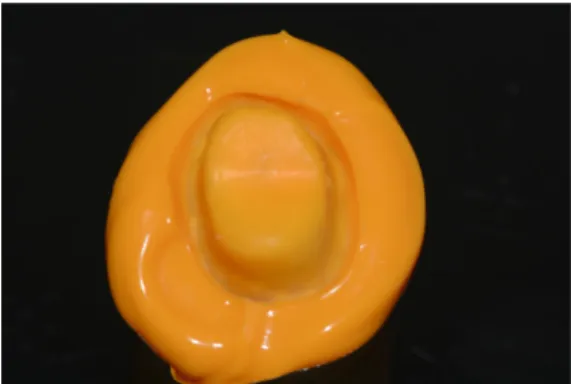
For the WT, each crown was applied to the prepared abutment tooth using a light-body silicone impression material (Aquasil Ultra XLV) instead of using luting cement, simulating the clinical application of
a crown. The prepared abutment tooth was lubricated with spraying separating fluid (Microfilm, Kerr Italia Srl, Salerno, Italy) before this procedure in order to prevent the impression material from sticking to the tooth. After the removal of excess impression material at the mar- gin, finger pressure was applied for 4 minutes of setting time. After polymerization of the impression material, crowns were removed from the prepared abutment teeth and the light-body silicone impression material was carefully removed and weighed on an analytical balance (Mettler AJ 100, Mettler-Toledo, Oakland, CA, USA). The weight was measured three times for each specimen from both groups and all mea- surements were performed by the same operator.
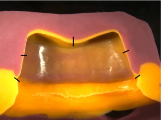
The RT was used to measure the marginal and internal fitting accu- racy. Light-body silicone impression material of very low viscos- ity (Aquasil Ultra XLV) was applied to the crown interior, after which the crown was set onto the prepared abutment tooth with finger pres- sure in the occlusal direction. After polymerization of the impres- sion material, the crowns were removed from the prepared abutment teeth. The thin silicone film remaining on the abutment tooth rep- resented the space between the abutment tooth and the zirconia pros- thesis (Fig. 6). After wax-relief of the prepared abutment tooth, an individual tray (Ostron 100, GC Corporation, Tokyo, Japan) was fab- Fig. 3. Scan images from each group. (A) Intraoral scan, (B) model scan.
Fig. 4. Design of the zirconia prostheses for each group. (A) Intraoral scan, (B) model scan.
Fig. 5. Zirconia prosthesis placed on the prepared abutment tooth.
Fig. 6. Silicone film representing the space between the abutment tooth and the zirconia prosthesis.
ricated. After applying light-body silicone impression material (Aquasil Ultra XLV) to the abutment tooth, using a plastic syringe, an individual tray filled with heavy-body silicone impression material (Aquasil Ultra Monophase, Dentsply) was placed to sta- bilize the silicone film. After completion of polymerization of the silicone impressions, the resulting replicas were placed in a set- ting jig and bisected in the middle of the mesial axial wall with a No.
10 surgical blade in the bucco-palatal direction (Fig. 7). The bisected replicas were then measured and photographed using a stereo- scopic microscope with surface illumination (MZ-16FA, Leica Microsystems, Wetzlar, Germany) by using a Leica microscope soft- ware at 5 pre-determined points on the buccal and lingual margins, buccal and lingual axial walls and occlusal area (Fig. 8). The hor- izontal marginal discrepancy (x), the vertical marginal discrepan- cy (y), the absolute marginal discrepancy (a), and the internal gap (b) were evaluated (Fig. 9). Measurements were performed by the same operator, as recommended by Holmes et al.24The horizontal marginal discrepancy is defined as the horizontal misfit between the outermost portions of the crown margin and the preparation edge of the abutment teeth, measured perpendicularly to the path of draw
of the restoration. The internal gap is defined as the perpendicular measurement from the internal surface of the crown to the axial wall of the prepared abutment tooth.
Statistical analysis
Statistical analysis was performed using SPSS Statistics for Windows (Version 20.0, IBM Corp., Armonk, NY, USA). The mean values and the standard deviations for each group were cal- culated. The Shapiro-Wilk test was used to confirm that the marginal gaps were normally distributed. Levene's test was performed to eval- uate the equality of variances. One-way analysis of variance (ANOVA) was used to assess the influence of the impression methods on the marginal and internal gaps. The level of significance was established as 0.05.
Results
The means and standard deviations of weight measurement for the two groups (n = 10, for each) are presented in Table 1 and Fig.
10. The mean silicone weights of the intraoral scan group and model scan groups were 198.59 ± 11.93 mg and 134.57 ± 5.86 mg, respectively. The mean weight was significantly higher for the intraoral scan group than for the model scan group (P < .05).
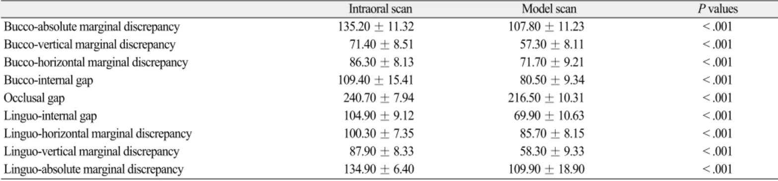
The mean values and standard deviations for the marginal gap and internal gap width measurements are presented in Table 2 and Fig. 11. In the intraoral scan group, the internal gap width measurements of the bucco-axial surface, occlusal surface, and linguo-axial sur- face were 109.40 ± 15.41 ㎛, 240.70 ± 7.94 ㎛ and 104.90 ± 9.12
㎛, respectively. In the model scan group, the internal gap width mea- surements were 80.50 ± 9.34 ㎛, 216.50 ± 10.31 ㎛, and 69.90
± 10.63 ㎛, respectively.
Fig. 8. Schematic representation of the five predetermined measuring points in the cross-sectional cut of the replica.
Fig. 9. Microscopic cross-sectional photograph of a replica. Definition of measuring distances for marginal accuracy: x, horizontal marginal discrepancy; y, vertical mar- ginal discrepancy; a, absolute marginal discrepancy; b, internal gap.
Fig. 7. Stabilized replica after segmentation in the bucco-palatal direction.
x a y
b
Marginal discrepancies were measured in three aspects: horizontal, vertical and absolute marginal discrepancy. In the intraoral scan group, the horizontal, vertical and absolute marginal discrepancies at the buccal margin were 86.30 ± 8.13 ㎛, 71.40 ± 8.51 ㎛, and 135.20 ± 11.32 ㎛, respectively. At the lingual margin, they were 71.70 ± 9.21 ㎛, 57.30 ± 8.11 ㎛ and 107.80 ± 11.23 ㎛, respectively. In the model scan group, the horizontal, vertical and absolute marginal discrepancies at the buccal margin were 71.70 ± 9.21 ㎛, 57.30 ± 8.11 ㎛, and 107.80 ± 11.23 ㎛, respectively. At the lingual margin, they were 85.70 ± 8.15 ㎛, 58.30 ± 9.33 ㎛, and 109.90 ± 18.90 ㎛, respectively.
Table 1. Means ± SD value of silicone weight measurements for the intraoral scan group and model scan group (n = 10, for each), with independent t test results.
Silicone weight (mg)
Mean SD Min Max P value
Intraoral scan 198.59 11.93 185.30 224.00
< .001
Model scan 134.57 5.86 125.70 143.00
Fig. 10. Means of silicone weight measurements for the intraoral scan group and model scan group (n = 10, for each). Error bars represent ± SD.
Fig. 11. Means of marginal and internal gaps for the intraoral scan group and model scan groups (n = 10, for each). Error bars represent ± SD.
Table 2. Means ± SD value of the marginal and internal gaps for the intraoral scan group and model scan group (n = 10, for each), in ㎛, with independent t test results
Intraoral scan Model scan P values
Bucco-absolute marginal discrepancy 135.20 ± 11.32 107.80 ± 11.23 < .001
Bucco-vertical marginal discrepancy 71.40 ± 8.51 57.30 ± 8.11 < .001
Bucco-horizontal marginal discrepancy 86.30 ± 8.13 71.70 ± 9.21 < .001
Bucco-internal gap 109.40 ± 15.41 80.50 ± 9.34 < .001
Occlusal gap 240.70 ± 7.94 216.50 ± 10.31 < .001
Linguo-internal gap 104.90 ± 9.12 69.90 ± 10.63 < .001
Linguo-horizontal marginal discrepancy 100.30 ± 7.35 85.70 ± 8.15 < .001
Linguo-vertical marginal discrepancy 87.90 ± 8.33 58.30 ± 9.33 < .001
Linguo-absolute marginal discrepancy 134.90 ± 6.40 109.90 ± 18.90 < .001
An independent t test indicated that the marginal and internal gaps were significantly higher at all measured points in the intraoral scan group than in the model scan group (P < .05).
Discussion
The aim of this study was to compare the marginal and internal fit of zirconia prostheses fabricated using different scan methods:
intraoral scan and model scan. Internal fit was evaluated by weigh- ing light-body silicone impression material and the cement thick- ness of the crowns on prepared abutment teeth was measured to eval- uate the marginal and internal fits.
Extracted human mandibular first molar was prepared in this study, compared to some of previous studies that used typodont resin teeth.
In addition, the marginal and internal gaps of zirconia prostheses from the two groups were measured on the prepared abutment tooth instead of on master die, to better reflect the clinical situation.
There was a significant difference in the marginal and internal gap between the two fabrication methods; therefore, the null hypothe- sis was rejected. The fit of the intraoral scan group was better than that of the model scan group in all measured areas (Table 1 and Table 2). Flügge et al.25compared the precision of intraoral digital scanning to model scanning, finding intraoral scanning to be less accu- rate; this corresponds closely to the results of the present study. The Flügge et al. study also suggested that intraoral conditions (saliva, limited space) could contribute to the inaccuracy of a scan; however, these conditions were not relevant to our study. Instead, other factors, such as the accuracy of the intraoral scanner and data transmitting system, could have influenced the results.
In previous studies, there is no consensus on a clinically accept- able marginal gap for fixed restorations. The values reported in the literature range from 50 to 200 ㎛.2,26McLean and von Fraunhofer reported a clinically acceptable marginal gap of within 120 ㎛.27 Fransson et al.28and McLean and von Fraunhofer27reported that the clinically acceptable marginal gap should be less than 150 μm and 120 ㎛, respectively. Moldovan et al.29suggested that 100 ㎛ of marginal misfit is considered as "good" and 200 - 300 ㎛ as "clin- ically acceptable". In the present study, the marginal discrepancies of the crowns manufactured using both methods were under 140 ㎛ which is in the clinically acceptable range according to the previ- ous studies. In addition, the occlusal internal gap was significant- ly higher than the axial internal gap. This finding is also consistent with previous studies.30
Sorensen reported that standardized methods for determination of crown margin include the direct view technique, the cross-sec- tioning technique, the impression replica technique, and other methods (e.g., the WT).31
The RT was first introduced by Molin and Karlsson,32who used this technique to compare the fitness between gold inlays and ceramic inlays. This is a popular method for evaluating marginal dis- crepancies between the crown and the abutment tooth; therefore, defor- mation of the margin, which can occur during cutting, is prevent- ed. Furthermore, the number of measuring sites can easily be increased, and measurements can be repeated. However, this tech- nique should be carefully applied in the evaluation of prostheses with good fitness because there is danger of damaging the impression mate- rial when separating the crown from the master die, which would cause inaccurate results.21,33
The WT is another non-destructive method used to evaluate the total internal gap of the restorations. For the WT, the weight of impression material is measured, instead of measuring certain points as is done for the RT. The value obtained represents the total thickness of the internal gap between the restoration and the prepared abutment tooth.34This technique requires low cost, easy and less time- consuming, compared with RT. However, with this technique, it is impossible to evaluate the marginal fit or the area where the largest gaps are found. Therefore, both techniques were used in this study, thereby effectively measuring the adaptation of restorations.26
This study proved that zirconia prostheses fabricated with two dif- ferent scan methods have clinically acceptable marginal and inter- nal gap thicknesses. In addition, zirconia prostheses fabricated with the model scan method had significantly lower marginal and internal gaps than did those fabricated with the intraoral scan method. However, this study has some limitations. For measurement of the marginal and internal gap, light-body silicone impression mate- rial was used instead of dental luting cement, which is actually used in clinical situations; therefore, the results of this study do not entirely reflect the final marginal fit of the restoration. In addition, crowns were seated on the prepared abutment tooth with finger pres- sure. Even though this method simulates the clinical cementation of crowns, it should be emphasized that finger pressure may be vari- able in each trial.
Conclusion
Within the limitations of this study, the following conclusions may be drawn: the marginal and internal fits of zirconia prostheses fabricated with both the model scan and intraoral scan methods were clinically acceptable. However, in the fabrication of zirconia pros- theses, the model scan method results in better marginal and inter- nal gap at all measured areas than does the intraoral scan method.
Therefore, the intraoral scan method requires improvement of the scanning procedure, data transfer, and crown fabrication in order to be a competitive alternative to the conventional method.
ORCID
Jin-Hyun Cho http://orcid.org/0000-0002-2453-9372
References
1. Conrad HJ, Seong WJ, Pesun IJ. Current ceramic materials and systems with clinical recommendations: a systematic review.
J Prosthet Dent 2007;98:389-404.
2. Martl′nez-Rus F, Sua′rez MJ, Rivera B, Pradl′es G. Evaluation of the absolute marginal discrepancy of zirconia-based ceramic cop- ings. J Prosthet Dent 2011;105:108-14.
3. Patzelt SB, Lamprinos C, Stampf S, Att W. The time efficiency of intraoral scanners: an in vitro comparative study. J Am Dent Assoc 2014;145:542-51.
4. Cho YB, Chung CH, Kim HJ. Marginal and internal fit of copings made by CAD/CAM using different scanning methods.
J Dent Rehab App Sci 2013;29:366-76.
5. Jacobs MS, Windeler AS. An investigation of dental luting cement solubility as a function of the marginal gap. J Prosthet Dent 1991;65:436-42.
6. Felton DA, Kanoy BE, Bayne SC, Wirthman GP. Effect of in vi- vo crown margin discrepancies on periodontal health. J Prosthet Dent 1991;65:357-64.
7. Holden JE, Goldstein GR, Hittelman EL, Clark EA. Comparison of the marginal fit of pressable ceramic to metal ceramic restora- tions. J Prosthodont 2009;18:645-8.
8. Quintas AF, Oliveira F, Bottino MA. Vertical marginal dis- crepancy of ceramic copings with different ceramic materials, fin- ish lines, and luting agents: an in vitro evaluation. J Prosthet Dent 2004;92:250-7.
9. Tan PL, Gratton DG, Diaz-Arnold AM, Holmes DC. An in vit- ro comparison of vertical marginal gaps of CAD/CAM titanium and conventional cast restorations. J Prosthodont 2008;17:378- 83.
10. Pera P, Gilodi S, Bassi F, Carossa S. In vitro marginal adaptation of alumina porcelain ceramic crowns. J Prosthet Dent 1994;72:585- 90.
11. Kohorst P, Brinkmann H, Dittmer MP, Borchers L, Stiesch M.
Influence of the veneering process on the marginal fit of zirco- nia fixed dental prostheses. J Oral Rehabil 2010;37:283-91.
12. Stappert CF, Dai M, Chitmongkolsuk S, Gerds T, Strub JR. Marginal adaptation of three-unit fixed partial dentures constructed from pressed ceramic systems. Br Dent J 2004;196:766-70.
13. Wolfart S, Wegner SM, Al-Halabi A, Kern M. Clinical evalua- tion of marginal fit of a new experimental all-ceramic system be- fore and after cementation. Int J Prosthodont 2003;16:587-92.
14. Balkaya MC, Cinar A, Pamuk S. Influence of firing cycles on the margin distortion of 3 all-ceramic crown systems. J Prosthet Dent 2005;93:346-55.
15. Fonseca JC, Henriques GE, Sobrinho LC, de Go′es MF. Stress- relieving and porcelain firing cycle influence on marginal fit of commercially pure titanium and titanium-aluminum-vanadi- um copings. Dent Mater 2003;19:686-91.
16. Wettstein F, Sailer I, Roos M, Hämmerle CH. Clinical study of
the internal gaps of zirconia and metal frameworks for fixed par- tial dentures. Eur J Oral Sci 2008;116:272-9.
17. Gonzalo E, Sua′rez MJ, Serrano B, Lozano JF. A comparison of the marginal vertical discrepancies of zirconium and metal ce- ramic posterior fixed dental prostheses before and after ce- mentation. J Prosthet Dent 2009;102:378-84.
18. Valderrama S, Van Roekel N, Andersson M, Goodacre CJ, Munoz CA. A comparison of the marginal and internal adaptation of titanium and gold-platinum-palladium metal ceramic crowns.
Int J Prosthodont 1995;8:29-37.
19. Baig MR, Tan KB, Nicholls JI. Evaluation of the marginal fit of a zirconia ceramic computer-aided machined (CAM) crown system. J Prosthet Dent 2010;104:216-27.
20. Han HS, Yang HS, Lim HP, Park YJ. Marginal accuracy and in- ternal fit of machine-milled and cast titanium crowns. J Prosthet Dent 2011;106:191-7.
21. Reich S, Wichmann M, Nkenke E, Proeschel P. Clinical fit of all- ceramic three-unit fixed partial dentures, generated with three dif- ferent CAD/CAM systems. Eur J Oral Sci 2005;113:174-9.
22. Jesu′s Sua′rez M, Lozano JF, Paz Salido M, Martl′nez F. Marginal fit of titanium metal-ceramic crowns. Int J Prosthodont 2005;18:390-1.
23. Castillo de Oyagüe R, Sa′nchez-Jorge MI, Sa′nchez Turrio′n A, Monticelli F, Toledano M, Osorio R. Influence of CAM vs.
CAD/CAM scanning methods and finish line of tooth prepara- tion in the vertical misfit of zirconia bridge structures. Am J Dent 2009;22:79-83.
24. Holmes JR, Bayne SC, Holland GA, Sulik WD. Considerations in measurement of marginal fit. J Prosthet Dent 1989;62:405-8.
25. Flügge TV, Schlager S, Nelson K, Nahles S, Metzger MC.
Precision of intraoral digital dental impressions with iTero and extraoral digitization with the iTero and a model scanner. Am J Orthod Dentofacial Orthop 2013;144:471-8.
26. Vojdani M, Torabi K, Farjood E, Khaledi A. Comparison the mar- ginal and internal fit of metal copings cast from wax patterns fab- ricated by CAD/CAM and conventional wax up techniques. J Dent (Shiraz) 2013;14:118-29.
27. McLean JW, von Fraunhofer JA. The estimation of cement film thickness by an in vivo technique. Br Dent J 1971;131:107- 11.
28. Fransson B, Oilo G, Gjeitanger R. The fit of metal-ceramic crowns, a clinical study. Dent Mater 1985;1:197-9.
29. Moldovan O, Rudolph H, Quaas S, Bornemann G, Luthardt RG.
Internal and external fit of CAM-made zirconia bridge frameworks- a pilot study. Dtsch Zahnärztl Z 2006;61:38-42.
30. Sakrana AA. In vitro evaluation of the marginal and internal dis- crepancies of different esthetic restorations. J Appl Oral Sci 2013;21:575-80.
31. Sorensen JA. A standardized method for determination of crown margin fidelity. J Prosthet Dent 1990;64:18-24.
32. Molin M, Karlsson S. The fit of gold inlays and three ceramic in- lay systems. A clinical and in vitro study. Acta Odontol Scand 1993;51:201-6.
33. Kokubo Y, Nagayama Y, Tsumita M, Ohkubo C, Fukushima S, Vult von Steyern P. Clinical marginal and internal gaps of In-Ceram crowns fabricated using the GN-I system. J Oral Rehabil 2005;
32:753-8.
34. Colpani JT, Borba M, Della Bona A. Evaluation of marginal and internal fit of ceramic crown copings. Dent Mater 2013;29:174- 80.
서로 다른 두 가지 스캔법을 이용하여 제작된 지르코니아 보철물의 적합도에 대한 비교
최현석∙조진현*
경북대학교 치과대학 치과보철학교실
목적: 연구의 목적은 동일한 지대치에서 각각 모델스캔 방법과 구강 내 스캔 방법으로 제작된 지르코니아 보철물의 변연 및 내면적합도를 비교하 는 것이다.
재료 및 방법: 발거된 사람의 하악 제1대구치 20개에 대해 지르코니아 보철물을 위한 지대치 형성이 이루어졌다. 첫 번째 그룹에서는 모델 스캔법으 로 10개의 지대치를 스캔하였고, 두 번째 그룹에서는 나머지 10개에 대해 구강 내 스캔법을 사용하였다. 각각의 데이터들은 소프트웨어 시스템으로 전송되었고 Ceramill CAD/CAM system을 이용하여 20개의 지르코니아 보철물을 제작하였다. 먼저 weight technique을 사용하여 내면 적합도를 평가하 였다. 다음으로 replica technique에서는 광학현미경을 통해 변연 적합도를 평가하였다. 통계처리는 일원배치분산분석을 이용하였다.
결과: 먼저 weight technique에서 모델 스캔법으로 제작한 그룹에서 구강 내 스캔법으로 제작한 그룹에 비해 평균적인 무게 값이 낮게 나타났고, 이는 유의성이 있었다(P < .05). 또한, replica technique에서도 측정된 5개의 영역 모두에서 구강 내 스캔법으로 제작한 그룹에 비해 모델 스캔법으로 제작한 그룹에서 변연 및 내면의 gap에 대한 거리 값이 유의하게 낮게 나타났다(P < .05).
결론: 두 그룹의 방법으로 제작한 지르코니아 보철물은 모두 임상적으로 받아들여질 만한 변연 및 내면 적합성을 보였다. 하지만 모델 스캔법으로 제작한 그룹에서 구강 내 스캔법으로 제작한 그룹에 비해 높은 변연 및 내면 적합성을 보였다. (대한치과보철학회지 2017;55:135-43)
주요단어: 지르코니아 보철물; 모델 스캔법; 구강 내 스캔법; Weight technique, Replica technique
*교신저자: 조진현
41940 대구 중구 달구벌대로 2177 경북대학교 치과대학 치과보철학교실 053 600 7651: e-mail, [email protected]
원고접수일: 2016년 10월 5일 / 원고최종수정일: 2016년 11월 16일 / 원고채택일:
2016년 11월 23일
2017 대한치과보철학회
이 글은 크리에이티브 커먼즈 코리아 저작자표시-비영리 3.0 대한민국 라이선스에 따라 이용하실 수 있습니다.
c cc