Congenital middle ear cholesteatoma (CMEC) is a rare disease entity in otolaryn- gology. However, we try to assess the characteristic features and recurrences of CMEC in pediatric patients according to stages, and to determine the value of pre- operative computed tomography (CT) scan. Retrospective review of 35 cases of CMEC under the age of 15 yr that had been treated at the tertiary referral center from 1995 through 2006. The main outcome measures were CT findings, surgical findings, recurrence rate and hearing assessment. Preoperative CT scan accurate- ly predicted the extent of the cholesteatoma seen during surgery in 30/35 (85.7%).
The recurrence rate of CMEC was 5.7% (2/35) and all of recurred cases were stage IV. In recurred cases, cholesteatomas were extended to sinus tympani and facial recess at revisional operation as well as initial operation. So we concluded that pre- operative CT scan is essential in defining the extent of existing pathology. The intra- operative CMEC extension and location influence the outcome of surgery. In the higher stages, careful eradication of disease, particularly in the region of sinus tym- pani and facial recess is recommended.
Key Words : Congenital; Cholesteatoma; Tomography, Radiography Computed
INTRODUCTION
Congenital middle ear cholesteatoma (CMEC) manifests as a whitish mass in the middle ear with intact tympanic membrane in young ages, and it is a relatively rare disease consisting of approximately 2% of the entire cholesteatoma cases (1). Recently, with the increase of interests on this dis- ease, its detection rate is increased, and thus the ratio of con- genital cholesteatoma to the entire cholesteatomas has been reported to be from 3.7% to 24% (2).
The symptoms are diverse depending on the location and the extent of lesions from asymptomatic to the conductive hearing loss, labyrinthitis, facial palsy, sensorineural hearing loss, intracranial complications, etc. The representative hy- potheses of the etiology are the defect of the tympanic ring, epidermoid formation, epithelial metaplasia and implanta- tion theory (2-4). Nevertheless, none of the hypotheses ex- plain accurate mechanisms. Recently, together with studies on the progression of congenital cholesteatoma, their classi- fication has been reported. Koltai et al. (5), Nelson et al. (6), and Potsic et al. (7) have suggested the disease stage based on the presence or absence of the invasion to the ossicle and the invasion to the mastoid.
In the treatment of CMEC, it is very important to classify the pattern of cholesteatoma and to determine the lesion ac- cording to it, and to evaluate accurately the damage of im-
portant anatomical structures and complications prior to surgery. It is thought that the prognosis may be different depending on the location of CMEC, its disease stage, and surgical methods. Therefore, in pediatric CMEC underwent surgical treatment, we analyzed the association of the com- puted tomography (CT) stage, the disease stage at the time of surgery (surgical stage), surgical methods, the result of audiologic test and recurrence rate.
MATERIALS AND METHODS
On 35 pediatric CMEC who were treated surgically in the department of otolaryngology head and neck surgery at the Catholic university hospital from 1995 to 2006. Follow up period were ranged from 9 to 128 months, average 47 months.
And we reviewed clinical symptoms, physical findings, audi- ologic test, radiologic test and surgical findings retrospec- tively.
The patients were children younger than the age of 15 yr, and the diagnosis of CMEC was made based on the diagnos- tic criteria suggested by Levenson et al. (8) that 1) a whitish mass present in the middle ear cavity with normal tympan- ic membrane, 2) the pars flaccida and pars tensa of tympan- ic membrane showed normal findings, 3) without the past history of otorrhea and perforation, 4) without the history of
126
Kyoung-Ho Park, Shi-Nae Park, Ki-Hong Chang, Min-Kyo Jung, and Sang-Won Yeo
Department of Otolaryngology Head & Neck Surgery, College of Medicine, the Catholic University of Korea, Seoul, Korea
Address for correspondence Sang-Won Yeo, M.D.
Department of Otolaryngology Head & Neck Surgery, Kangnam St. Mary’s Hospital, the Catholic University of Korea, 505 Banpo-dong, Seocho-gu, Seoul 137-701, Korea
Tel : +82.2-2258-6210, Fax : +82.2-595-1354 E-mail : [email protected]
DOI: 10.3346/jkms.2009.24.1.126
Congenital Middle Ear Cholesteatoma in Children; Retrospective Review of 35 Cases
Received : 29 November 2007 Accepted : 12 April 2008
otological surgery, 5) the occulusion of the external canal, intramembranous cholesteatoma and giant cholesteatoma were excluded, and 6) the past history of otitis media was not precluded.
Regarding the classification of CMEC according to their location, based on the handle of malleus, it was divided into the lesion in the anterior and posterior area.
As the disease stage of cholesteatoma, based on the classi- fication suggested by Potsic et al. (7), the CT stages and sur- gical stages were compared (Tabel 1). Depending on the dis- ease stage, tympanoplasty, epitympanoplasty, canal wall up mastoidectomy, canal wall down mastoidectomy, and if need- ed, ossiculoplasty was performed.
As audiologic test, according to the guideline of the Amer- ican Academy of Otolaryngology Head and Neck Surgery (1995), the mean hearing level at 500 Hz, 1,000 Hz, 2,000 Hz, and 3,000 Hz was used. The analysis of hearing test after surgery was performed based on the most recent audiologic test, 2-3 months after the surgery. The hearing change after surgery was defined as the difference of the air bone conduc- tion threshold gap prior to surgery and the air bone conduc- tion threshold gap after surgery.
The statistical significance of the hearing change after surgery was assessed by paired t-test and Wilcoxon signed rank test, and a significant level was p<0.05.
RESULTS The study population
The age of patients was from 2 to 13 yr old and their mean age was 6.2. The boy was 27 patients, the girl was 8, and the ratio of the male and the female was 3.4:1. CT stage according to Potsic et al. (7), the stage 1 was 2 patients, the stage 2 was 4, the stage 3 was 10, and the stage 4 was 17.
The mean age at the time of surgery in the stage 1 was 4.5 yr old, the stage 2 was 6.3, the stage 3 was 5.1, and the stage 4 was 7.3.
Clinical symptoms and physical findings
All patients had a unilateral lesion, developed in the left
side was in 15 cases (43.0%), and in the right side in 20 (57.0%). Asymptomatic cases discovered were 16 cases (45.0
%). And in symptomatic cases, hearing impairment was 10 (29.0%), which was most prevalent, otalgia was 7 (20.0%), tinnitus was 4 (11.4%), and ear fullness was 3 (8.5%), and in others 1 case (2.8%), a symptom similar to postauricular swelling. In the past history, the cases who had acute otitis media or otitis media with effusion were 11 patients (31.4
%). Concerning the otoscopic findings, normal findings were 7 patients (20.0%), a whitish mass in the normal eardrum was detected in 21 (60.0%), and bulging of posterior part of tympanic membrane was detected in 7 (20.0%), and none of patients had a perforation in the tympanic membrane.
Audiologic test
Among 35 patients, preoperative audiologic test was per- formed on 23 patients. The air bone conduction threshold gap prior to surgery in the CT stage 1 & 2 was 10.0 dB, in the stage 3 was 27.0 dB, in the stage 4 was 37.5 dB, and a trend of the increase with advanced disease stage was detect- ed. Patients underwent audiologic test prior to surgery and 2-3 months after surgery in the stage 3 were 4 cases and the stage 4 was 10. The improvement of the air bone conduc- tion threshold gap in the stage 3 was 2.5 dB and the stage 4 was 6.7 dB, but statistically significant improvement was not detected (Table 2).
Radiological test
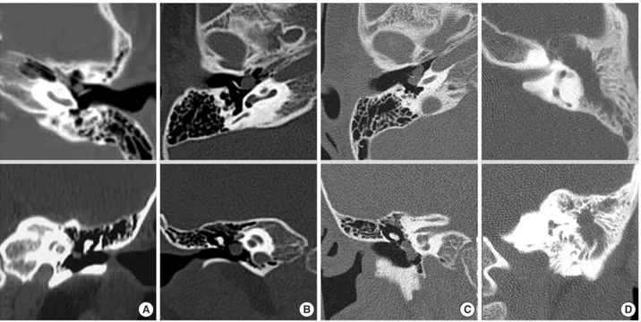
Based on the disease stage by Potsic et al. (7), 35 patients were classified according to their disease stage based on pre- operative CT findings (Fig. 1) and surgical findings, and com- pared. It was found that the cases whose CT stage correspond- ing to surgical stages were 30 cases (85.7%), the cases whose CT stage was underestimated in comparison with surgical stages were 2 (5.7%), and the overestimated cases were 3 (8.6%) (Table 3).
Surgical findings & surgical methods
Regarding the disease stage at the time of surgery, the sur- gical stage 1 was 4 cases (11.4%), the stage 2 was 4 (11.4%), the stage 3 was 10 (28.6%) and the stage 4 was 17 (48.6%).
Stage 1 Single quadrant: no ossicular involvement or mastoid extension
Stage 2 Multiple quadrant: no ossicular involvement or mastoid extension
Stage 3 Ossicular involvement: includes erosion of ossicles and surgical removal for eradication of disease; no mastoid involvement
Stage 4 Mastoid extension (regardless of findings elsewhere) Table 1. Staging system for congenital cholesteatoma (Potsic et al., 2002)
PR, preoperative ; AB gap, Air-bone gap; PO, postoperative.
Surgical stage
PR AB gap (dB)
p value PO AB gap change (dB) PO AB
gap (dB)
Stage I (n=4) 10.1 (n=2) 10.1 (n=1) 0 (n=1) 0.501 Stage II (n=4) 10.1 (n=1) 10.0 (n=1) 0 (n=1) 0.302 Stage III (n=10) 27.0 (n=5) 22.5 (n=4) 2.5 (n=4) 0.211 Stage IV (n=17) 36.8 (n=14) 35.5 (n=10) 6.7 (n=9) 0.413 Table 2. Air-bone gap change after operation
4 cases of the surgical stage 1 were restricted to the antero- supeior area, and 2 cases of the stage 2 were limited to the anterior area and the others of the stage 2 were limited to pos- terior area. It was found that in 26 cases of the stage 3 and 4, none of cases was limited to the anterior area, the posteri- or area was 3 cases, and the both anterior and postrior was 24 (Table 4).
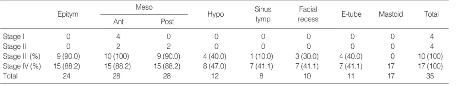
In the stage 3 and 4 invaded to more than the middle ear cavity, the cases invaded up to the epitympanum was 9 cases (90.0%) in the stage 3 and 15 cases (88.2%) in the stage 4, and the cases expanded to the hypotympanum in the stage 3 was 4 cases (40.0%) and 8 cases (47.0%) in the stage 4.
The invasion to the sinus tympani in the stage 3 was 1 case (10.0%) and 7 cases (41.1%) in the stage 4. Invasion to the facial recess in the stage 3 was 3 cases (30.0%) and 7 cases (41.1%) in the stage 4. And invasion to the Eustachian tube in the stage 3 was 4 cases (40.0%) and the stage 4 was 7 cases (41.1%). As the stage was advanced, the invasion to the sinus tympani, the facial recess and the Eustachian tube was in- creased (Table 5). In the cases of invasion into the facial recess and sinus tympasni, we tried to remove cholesteatoma com-
pletely using many microinstruments, especially with micro- mirror.
Regarding the invasion into the ossicles, the malleus was 9 cases, the incus was 26 and the stapes was 20. The dam- age of the facial canal in the stage 3 was 1 case and the stage 4 was 6, the dura exposure in the stage 3 was 1 and the stage 4 was 2, and none of patients had the exposure in the lateral semicircular canal. As the disease stage is advanced, the expo- sure of facial canal or dural exposure becomes more frequent (Table 6).
Regarding the surgical procedures, tympanoplasty was done for the patients with stage 1 & 2. And there was no recurrence. As for the stage 3, tympanoplasty was done for 1 case, tympanoplasty & epitympanoplasty for 2 cases, canal wall up mastoidectomy with tympanoplasty for 6 cases and canal wall down mastoidectomy with tympanoplasty for 1 case. And ossiculoplasty was done at the time surgery for 5 cases (5/10) with partial ossicular replacement prothesis (PORP). As for stage 4, canal wall up mastoidectomy with
SF, surgical findings.
CT\SF Stage I Stage II Stage III Stage IV
Stage I 3 0 0 0
Stage II 1 4 0 0
Stage III 0 0 8 2
Stage IV 0 0 2 15
Table 3. Stage according to CT findings and surgical findings
A&P, anterior and posterior.
Anterior A&P Posterior Total
Stage I 4 0 0 4
Stage II 2 0 0 4
Stage III 0 9 1 10
Stage IV 0 15 2 17
Total 6 24 5 35
Table 4. Location of cholesteatoma in the middle ear according to the surgical stage
Fig. 1. Stage according to CT findings. (A) Stage I. Cholesteatoma occupies a single quadrant without ossicular extension. (B) Stage II.
Cholesteatoma occupies at least two quadrants without mastoid extension. (C) Stage III. Cholesteatoma involve the ossicle. (D) Stage IV.
Cholesteatoma extends into the mastoid.
A B C D
tympanoplasty was done for 10 cases and canal wall down mastoidectomy with tympanoplasty for 7 cases. Ossiculo- plasty was done at the time of the first surgery for 4 cases (4/17) using PORP.
Recurrence
It recurred in 2 cases among 35 patients (5.7%). They were all the surgical stage 4 and underwent canal wall up mastoidectomy. As revisional surgery, canal wall down mas- toidectomy was performed in 1 case, and revision canal wall up mastoidectomy was performed in the other. Ossiculo- plasty was performed in all cases. At the time of revisional surgery, in all 2 cases, the invasion of cholesteatoma in the sinus tympani and the facial recess were observed, and they were the patients who had the invasion to the sinus tympa- ni and the facial recess also at the first surgery. Until the last follow up observation, findings suspicious of recurrence were not detected in 33 patients, and the second look operation was not performed.
DISCUSSION
House (9) reported the case of CMEC for the first time in 1953, and in 1965, Derlacki and Clemis (10) established the diagnostic criteria for congenital cholesteatoma. Leven- son et al. (8) confirmed the diasease history of acute otitis media in 70% of infants younger than the age of 2 yr in the U.S.A. and thus did not preclude the past history of acute
otitis media from the diagnostic criteria. Similarly, we did not preclude the past history of otitis media according to the diagnostic criteria of Levenson et al. (8) and patients who had acute otitis media or otitis media with effusion were 11 cases (31.4%), which was not greatly different from the pre- vious report 9-55% (11).
Clinical symptoms of CMEC manifest diversely depend- ing on the advanced stage. Generally, asymptomatic cases are most prevalent and the symptoms such as conductive hearing loss, ear fullness, otalgia, tinnitus, dizziness, headache, paral- ysis of facial nerves, etc. can be detected. House and Sheehy (12) have reported that in the cases with the unilateral con- ductive hearing loss which is difficult to explain by unilater- al otitis media with effusion, regardless of the age of patient, the possibility of CMEC should be considered. In our study, asymptomatic cases discovered incidentally were 45%, and the cases who did not show any specific findings with oto- scope were 20%. And at the time of surgery, the advanced lesion equivalent to the disease stage 3 or 4 was detected in most cases. In addition, it was found that as the disease stage become advanced, the frequency of the invasion to the epi- tympanum, the hypotympanum, the sinus tympani, the facial recess, the facial canal and the dura was increased. Consider- ing that CMEC is discovered frequently in its advanced stage, it is thought that for its early diagnosis, a screening program including otoscopic examination, hearing test and CT for chil- dren may be required.
As the etiology of CMEC, defect in the tympanic ring, epi- dermoid formation, epithelial metaplasia and implantation theory have been proposed. Nonetheless, none of hypotheses
M, malleus; I, incus; S, stapes; LSCC, lateral semicircular canal.
Ossicle erosion
M I S
Eroded facial canal
Eroded LSCC Exposed
dura
Stage I 0 0 0 0 0 0
Stage II 0 0 0 0 0 0
Stage III 3 9 8 1 1 0
Stage IV 6 17 12 6 2 0
Total 9 26 20 7 3 0
Table 6. Invasion of ossicles, facial canal, dura and lateral semi- circular canal according to the surgical stage
Tymp, tympanoplasty; Epitymp, epitympanoplasty; CWU M&T, canal wall up mastoidectomy and tympanoplasty; CWD M&T, canal wall down mastoidectomy and tympanoplasty.
Tymp Tymp &
epitymp CWU M&T
CWD M&T
Ossiculo- plasty
Stage I 4 0 0 0 0
Stage II 4 0 0 0 0
Stage III 1 2 6 1 5
Stage IV 0 0 10 7 4
Total 9 2 16 8 9
Table 7. Surgical procedure according to the surgical stage Epitym, epitympanum; Meso, mesotympanum; Ant, anterior; Post, posterior; Hypo, hypotympanum; Sinus Tymp, sinus tympani.
Meso
Ant Post
Epitym Hypo Sinus
tymp
Facial
recess E-tube Mastoid Total
Stage I 0 4 0 0 0 0 0 0 4
Stage II 0 2 2 0 0 0 0 0 4
Stage III (%) 9 (90.0) 10 (100) 9 (90.0) 4 (40.0) 1 (10.0) 3 (30.0) 4 (40.0) 0 10 (100)
Stage IV (%) 15 (88.2) 15 (88.2) 15 (88.2) 8 (47.0) 7 (41.1) 7 (41.1) 7 (41.1) 17 17 (100)
Total 24 28 28 12 8 10 11 17 35
Table 5. Cholestatoma extent according to the surgical stage
could explain the etiology completely (2-4).
Concerning the location of CMEC, Levenson et al. (8) have reported that it is located primarily in the anterior area of the tympanic cavity and Karmarkar et al. (13) have reported that the posterior area of the tympanic cavity is the site where it is developed preferentially. But recently, it has been report- ed to be developed in the posterior area of the tympanic cavi- ty more frequently (11). In our study, among 35 cases, 6 cases (17.1%) were limited to the anterior area, 5 cases (14.2%) were limited to the posterior area, and in 24 cases (68.5%), the anterior and posterior area were involved simultaneously.
Nelson et al. (6) classified CMEC according to the pres- ence or absence of the invasion to the ossicles and the mas- toid. The lesion limited to the middle ear is the type 1, with the invasion to the ossicles is the type 2, invasion to the mas- toid is the type 3. And toward to the type 3, its recurrence was increased. Potsic et al. (7) and Kazahaya and Potsic (14) have reported that in the classification according to Nelson et al. (6), the cases without invasion to the ossicle were divid- ed again into two groups and classified to total 4 groups.
The risk of residual cholesteatoma after surgery in the dis- ease stage 1 was 13% and the stage 4 was 67%. Hence, the risk of residual cholesteatoma was increased as the disease stage advanced, and proposed the disease stage system that could predict the prevalence rate of residual cholesteatoma.
Surgical methods vary depending on the progression of the lesion. According to Nelson et al. (6), the type 1 could be treated sufficiently only by tympanoplasty, the type 2 may require extended tympanoplasty, canal wall up mastoidecto- my, ossiculoplasty, and second look operation is required occa- sionally. And the type 3 are similar to type 2 cases, nonethe- less, it may require canal wall down mastoidectomy. How- ever, canal wall down mastoidectomy has a shortcoming that continuous ear treatment is required throughout the life and the possibility of developing cavity problem is high. And thus it should be performed only in the cases with the defect in the posterior wall of the external auditory canal, invasion of cholesteatoma into the inner ear or the petrous apex, and the cases whose follow ups are difficult (15). Ueda et al. (16) also recommended to perform canal wall up mastoidectomy if possible, and to choose the one-stage surgery for the cases that on presurgical CT findings, the epitympanum and the mastoid were aerated, and without the damage in the malleus and the incus, to perform sequential tympanoplasty. Because such principle increases the possibility of the aeration of the epitympanum and the mastoid, if residual cholesteatoma could be detected early by CT, the formation of an attic retrac- tion pocket could be prevented. Gocmen et al. (17) also rec- ommended the canal wall up mastoidectomy for limited lesions and the canal wall down mastoidectomy for choles- teatoma in a wider area.
The results of recurrence rate, improvement of hearing and hearing deterioration were not different between the cases performed the wall down mastoidectomy or the wall up mas-
toidectomy. In our cases of advanced stage, we did one stage operation with ossiculoplasty. We thought that we removed the cholesteatoma properly and tried to reduce hearing dif- ficulty of patients and concerns of second operation.
The rate of residual cholesteatoma at the time of second surgery is very diverse, 8-81% (18). In our study, recurrence was observed 2 cases (5.7%) and a relatively low recurrence rate was shown, which is thought to be the reason that the number of patients was 35 cases which was not large, and the follow-up period in some patients was not long. Two recurred cases were the disease stage 4 at the time of first surgery, with the invasion to the sinus tympanic and the facial recess, and the canal wall up mastoidectomy was per- formed. And at the time of recurrence, the recurrence in the sinus tympani and facial recess could be observed. It is con- sidered that such result was obtained due to the failure of the complete removal of cholesteatoma by the canal wall up mastoidectomy. And it is thought that for the cases with invasion in these areas, to prevent recurrence, the canal wall down mastoidectomy is required.
To perform CT prior to surgery considered as a standard preoperative examination nowadays. Levenson et al. (8) accept- ed the usefulness of CT as presurgical test, nevertheless, they preferred micro-otoscopy. Friedberg (2) have reported that imaging tests were a great help to understand the extent of lesion and to decide surgical approach. And El-Bitar et al.
(19) claimed that CT is of help to understand the extent of cholesteatoma and to evaluate invasion to the ossicles, and mediates an effect on surgery outcomes. By using CT, Kim et al. (20) detected the invasion to the middle ear cavity in 80%, invasion to the mastoid in 76.5%, and invasion to the sinus tympani in 50%. And as the prediction of the ossicle destruction, the malleus, the incus and the stapes was shown to be 75%, 65%, and 85%, respectively. In our study, the result showed that the CT stage corresponded to surgical stage in 85.7%. It is thought that by predicting the location of lesion and the spread route by CT imagings prior to sur- gery, it is of help to plan the treatment methods and reduc- tion of complications for cholesteatoma patients.
In pediatric congenital middle ear cholesteatoma, it is thought that screening tests including CT for its early detec- tion are required. Considering the preoperational evaluation, CT showed a high corresponding rate to the surgical stage determined by Potsic. CT is thought to be a useful tool for the presurgical evaluation of cholesteatoma, its treatment method and follow ups. In the cases that the cholesteatoma was large and invaded to the sinus tympani or the facial re- cess, the canal wall down mastoidectomy may be required to reduce recurrence.
REFERENCES
1. McDonald TJ, Cody DT, Ryan RE Jr. Congenital cholesteatoma of
the ear. Ann Otol Rhinol Laryngol 1984; 93: 637-40.
2. Friedberg J. Congenital cholesteatoma. Laryngoscope 1994; 104:
1-24.
3. Aimi K. Role of the tympanic ring in the pathogenesis of congenital cholesteatoma. Laryngoscope 1983; 93: 1140-6.
4. Michaels L. An epidermoid formation in the developing middle ear:
possible source of cholesteatoma. J Otolaryngol 1986; 15: 169-74.
5. Koltai PJ, Nelson M, Castellon RJ, Garabedian EN, Triglia JM, Roman S, Roger G. The natural history of congenital cholesteatoma.
Arch Otolaryngol Head Neck Surg 2002; 128: 804-9.
6. Nelson M, Roger G, Koltai PJ, Garabedian EN, Triglia JM, Roman S, Castellon RJ, Hammel JP. Congenital cholesteatoma: classifica- tion, management, outcome. Arch Otolaryngol Head Neck Surg 2002;
128: 810-4.
7. Potsic WP, Samadi DS, Marsh RR, Wetmore RF. A staging system for congenital cholesteatoma. Arch Otolaryngol Head Neck Surg 2002; 128: 1009-12.
8. Levenson MJ, Michaels L, Parisier SC. Congenital cholesteatomas of the middle ear in children: origin and management. Otolaryngol Clin north Am 1989; 22: 941-54.
9. House HP. An apparent primary cholesteatoma, case report. Laryn- goscope 1953; 63: 712-13.
10. Derlacki EL, Clemis JD. Congenital cholesteatoma of the middle ear and mastoid. Ann Otol Rhinol Laryngol 1965; 74: 706-27.
11. Zappia JJ, Wiet RJ. Congenital cholesteatoma. Arch Otolaryngol
Head Neck Surg 1995; 121: 19-22.
12. House JW, Sheehy JL. Cholesteatoma with intact tympanic mem- brane: A report of 41 cases. Laryngoscope 1980; 90: 70-6.
13. Karmarkar S, Bhatia S, Khashaba A, Saleh E, Russo A, Sanna M.
Congenital cholesteatomas of the middle ear: a different experience.
Am J Otol 1996; 17: 288-92.
14. Kazahaya K, Potsic WP. Congenital cholesteatoma. Curr Opin Oto- laryngol Head Neck Surg 2004; 12: 398-403.
15. Rizer FM, Luxford WM. The management of congenital choleste- atoma: surgical results of 42 cases. Laryngoscope 1988; 98: 254-6.
16. Ueda H, Nakashima T, Nakata S. Surgical strategy for cholesteatoma in children. Auris Nasus Larynx 2001; 28: 125-9.
17. Gocmen H, Kilic R, Ozdek A, Kizilkaya Z, Safak MA, Samim E.
Surgical treatment of cholesteatoma in children. Int J Pediatr Otorhi- nolaryngol 2003; 67: 867-72.
18. Darrouzet V, Duclos JY, Portmann D, Bebear JP. Congenital mid- dle ear cholesteatomas in children: our experience in 34 cases.
Otolaryngol Head Neck Surg 2002; 126: 34-40.
19. El-Bitar MA, Choi SS, Emamian SA, Vezina LG. Congenital mid- dle ear cholesteatoma: need for early recognition-role of computed tomography scan. Int J Pediatr Otorhinolaryngol 2003; 67: 231-5.
20. Kim CN, Chung SM, Kim SM, Kim YJ, Park MH, Ju MS. Study of the correlation with the temporal bone CT and operative findings in chronic otitis media with cholesteatoma. Korean J Otolaryngol 1993; 36: 313-20.