INTRODUCTION
Cellular injury can trigger a variety of responses, such as adaptation, repair, proliferation and cell death. Apoptosis is a programmed cell death that has been implicated in the regulation of tissue homeostasis, and the pathophysiologic process of neurologic disorders, such as Alzheimer’s disease, Parkinson’s disease, Huntington’ disease, amyotrophic later- al sclerosis, and ischemic insults (1, 2).
It is generally believed that the mechanism of apoptotic cell death is different from necrosis. By electron microscopic analysis, apoptotic cell death demonstrates specific morpho- logical changes including cell shrinkage, chromatin conden- sation, DNA fragmentation, membrane blebbing, apoptotic body formation, while necrosis is consistent with cell swelling and destruction of cell morphology with the release of intra- cellular organelles. During the early phase of apoptosis, phos- phatidylserine (PS), which is normally almost totally confined to the inner layer of phospholipid plasma membrane is moved to the outer leaflet of the plasma membrane by the decreased ATP-dependent translocase (flippase) activity and the increased calcium-dependent scramblase activity while maintaining the plasma membrane integrity (2-4). Fluorescein isothiocyanate (FITC)-conjugated annexin-V, which is a Ca2+dependent phos-
pholipid binding protein, is used as a reliable marker for PS exposure (3, 5, 6). During the late phase of apoptosis, a shift from tightly to loosely packing of the plasma membrane phos- pholipids with PS externalization allows propidium iodide (PI) penetration into the cell and binding with DNA. Dou- ble staining of cells with FITC-annexin-V and PI in associa- tion with flow cytometry has been proven to be a valuable method to discriminate between early and late phase of apop- tosis. Changes in morphology of the apoptotic cell can also be monitored by flow cytometry as a change in light-scatter- ing properties, forward scattering (FSC) and side scattering (SSC) (5, 7).
Central nervous system (CNS) neurons are extremely sensi- tive to metabolic changes and physio-chemical injuries. Resto- ration of neuronal viability after apoptotic or necrotic cell death has been suggested to be impossible, therefore, it is hoped that more focused therapies to prevent apoptotic cell death could be developed. Many clinicians are reluctant to use ketamine in head injury patients in the clinical setting due to an increase in the cerebral metabolic rate, cerebral blood flow, and intra- cranial pressure. However, the protective effect of ketamine in the amelioration of ischemic insults of CNS has been report- ed in several animal and cellular experiments. Ketamine was reported to suppress excessive excitatory amino acid release,
Soo Joo Choi, Myung Hee Kim, Seung Woon Lim*, Mi Sook Gwak
Department of Anesthesiology and Pain Medicine, Samsung Medical Center, Sungkyunkwan University School of Medicine, Seoul; Department of Anesthesiology and Pain Medicine*, Chungbuk National University, Cheongju, Korea
Address for correspondence Myung Hee Kim, M.D.
Department of Anesthesiology and Pain Medicine, Samsung Medical Center, Sungkyunkwan University School of Medicine, 50 Irwon-dong, Kangnam-gu, Seoul 135-710, Korea
Tel : +82.2-3410-2461, Fax : +82.2-3410-0361 E-mail : mhkim @smc.samsung.co.kr
113
Effect of Ketamine on Apoptosis by Energy Deprivation in Astroglioma Cells using Flow Cytometry System
Apoptosis is a programmed, physiologic mode of cell death that plays an important role in tissue homeostasis. As for the central nervous system, ischemic insults can induce pathophysiologic cascade of apoptosis in neurophils. Impairment of astroc- tye functions during brain ischemia can critically influence neuron survival by neuron- glia interactions. We aimed to elucidate the protective effect of ketamine on apop- tosis by energy deprivation in astrocytes. Ischemic insults was induced with iodoac- etate/carbonylcyanide m-chlorophenylhydrazone (IAA/CCCP) 1.5 mM/20 M or 150 M/2 M for 1 hr in the HTB-15 and CRL-1690 astrocytoma cells. Then these cells were reperfused with normal media or ketamine (0.1 mM) containing media for 1 hr or 24 hr. FITC-annexin-V staining and propidium iodide binding were determined by using flow cytometry. Cell size and granularity were measured by forward and side light scattering properties of flow cytometry system, respectively. An addition of keta- mine during reperfusion increased the proportion of viable cells. Ketamine alleviated cell shrinkage and increased granularity during the early period, and ameliorated cell swelling during the late reperfusion period. Ketamine may have a valuable effect on amelioration of early and late apoptosis in the astrocytoma cells, even though the exact mechanism remains to be verified.
Key Words : Apoptosis; Astrocytes; Flow Cytometry; Ketamine
Received : 28 June 2004 Accepted : 20 August 2004
attenuate the development of focal brain edema, and amelio- rate neurological dysfunction after neuronal injury in animal experiments (8-12).
Recent studies have revealed that glial cells are regarded to play many important roles, such as regulating extracellu- lar concentrations of ions, metabolites, and neurotransmitters, and participated in synaptic functions with neurons in CNS.
Therefore, damage of the most abundant glial cells, astrocytes, can bring about crucial influence on the neuronal survival (13-15). It is now well known that signals between astroglial cells and neurons go back and forth to modulate synaptic activity (16-18).
Here, under in vitro conditions, we investigated the effect of ketamine and reperfusion on the apoptosis of the astrocy- toma cells following energy depletion with glycolysis inhibitor iodoacetate (IAA) and blocker of oxidative phosphorylation carbonylcyanide m-chlorophenylhydrazone (CCCP), by flow cytometric analysis.
MATERIALS AND METHODS Experimental cells
Human astrocytoma cell line HTB-15 and CRL-1690 cells were cultivated in T-tube flask. Dulbecco’s modified minimal essential medium (DMEM) for HTB-15 cells and DMEM with 1.5 g/L sodium bicarbonate, 1.0 mM sodium pyruvate was used for CRL-1690 cells. The medium was supplement- ed with 10% fetal calf serum and 100 IU penicillin G and 50 g/mL streptomycin. The cells were incubated in a 5%
CO2chamber at a humidified atmosphere. Once the cells were grown confluent to the flask, the cells were harvested for the experiment with 0.1% trypsin-EDTA in a phosphate buffer solution (PBS) and washed twice thereafter. The addition of DMEM blocked the trypsin activity, and then suspension cells were transferred to the other T-tube. For the flow cytometric analysis, 2-3 mL of cell suspension were again transferred to the cuvette. In order to control the metabolic rate of experi- mental cells carefully, harvested cells only in the 3-5 passages were used.
Induction of cell damage and effect of ketamine
To mimic the ischemic damage in vitro, cells were treated with IAA/CCCP 1.5 mM/20 M or 150 M/2 M for 1 hr.
And then, to observe reperfusion-induced cellular damage or the effect of ketamine during the reperfusion, experimental protocols called for cells to be exposed to normal media or ketamine (0.1 mM or 0.5 mM)-containing media for 1 hr or 24 hr after wash out of agonists. Also, to investigate the effect of ketamine itself on the PI binding, ketamine 0.1 mM or 0.5 mM was added to the perfusion media without ischemic insult.
Measurement of cellular viability by Annexin-V and PI binding
For the measurement of surface exposure of PS and plasma membrane leakage of experimental cells with flow cytometry system, 105cells were incubated with FITC-annexin-V (1 g/
mL) for 15 min and PI (2 g/mL) for 2 min in a dark area at room temperature, and then washed with calcium-containing phosphate buffered solution to remove excess fluorescent dyes.
We used commercially available FITC-annexin-V and PI (Tre- vigen Inc, Gaithersburg, MD, U.S.A.), and those were dilut- ed to 1:50, 1:10 with calcium buffer solution, respectively, in this experiment.
Flow cytometry
All experiments were performed using a FACStar Plus Flow Cytometer (Becton Dickinson, Sunnyvale, CA, U.S.A.) equipp- ed with a single argon ion laser exciting at 488 nm. FITC- annexin-V fluorescence was collected through a 530 nm band pass filter while PI fluorescence was measured through 639 nm band pass filter (19). It was designed specifically to detect cellular changes of less than 1%. Prior to every experiment, the flow cytometry was calibrated electrically and mechani- cally by latex beads with already known diameter. Quanti- tative analysis of cell viability of 104cells was performed in 20 sec. The mean value of cellular changes was obtained by the computer analysis of cytogram. The X, Y-axis indicated the fluorescence of PI and annexin-V, respectively. By the gate of the 4 areas, it was possible to detect and quantitatively com- pare normal cells (annexin-V-/PI-), early apoptosis (annexin- V+/PI-), late apoptosis or necrosis (annexin-V+/PI+) in the whole cell population.
Alterations of cellular characteristics were also analyzed by the degree and direction of light scattering through laser beam of flow cytometry. FSC determines cell volume, SSC for cel- lular granularity, density and folding. Decrease in FSC and increase in SSC indicative of cell shrinkage and increased cell granularity, respectively, indicate early phase of apoptosis. In- crease in FSC and decrease in SSC are associated with late phase of apoptosis. Therefore, flow cytometry was also utilized to assess viable cells, early or late phase of apoptosis according to cell volume and degree of cellular granularity. In order to analyze better morphological changes with treatment, we expressed cell volume and granularity as % of those of the control cells without any treatment.
Statistical analysis
Experimental data were expressed as mean±SD. To com- pare between the two groups the unpaired t-test was perform- ed. p value less than 0.05 was considered statistically signifi- cant. Jandel Sigma Stat (version 2.0, Jandel Corporation, Chi- cago, IL, U.S.A.) was used for statistical analysis.
RESULTS
The effect of ketamine on the annexin-V binding and PI uptake during reperfusion for 1 hr
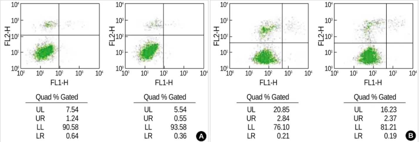
Annexin-V binding and PI uptake according to the con- centrations of IAA/CCCP were observed in CRL-1690 cells.
The portion of annexin-V positive cells and PI positive cells increased from 4.9% and 0.8% in control cells to 8.8% and 1.9% in cells treated with IAA/CCCP 150 M/2 M for 1 hr, respectively (Fig. 1A). In a different condition, annexin-V pos- itivity and PI positivity changed from 12.3% and 3.8% to 23.7% and 3.1%, respectively after treatment with IAA/
CCCP 1.5 mM/20 M (Fig. 1B). These results demonstrat- ed the relationship between intracellular energy depletion and PS redistribution.
The administration of ketamine 0.1 mM during the reper- fusion induced an increase in the portion of vital cells (annex-
in-V and PI negative cells) from 90.6% to 93.6% (Fig. 2A), and from 76.1% to 81.2%, respectively, after the insult with IAA/CCCP 150 M/2 M, and 1.5 mM/20 M (Fig. 2B).
These showed that ketamine plays a potential role in the inhi- bition of PS redistribution during intracellular energy deple- tion.
The effect of ketamine on the annexin-V binding and PI uptake during reperfusion for 24 hr
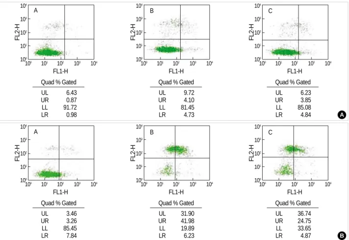
Only cellular debris was found following treatment with a higher concentration of IAA/CCCP for 1 hr and reperfusion for 24 hr. The experimental results after treatment with a low concentration of IAA/CCCP, 150 M/2 M, for 1 hr and reper- fusion for 24 hr exhibited similar patterns of responses as with other experiments. The viable cells decreased from 91.7%
to 81.5% after reperfusion, but the administration of keta- mine 0.1 mM during the reperfusion caused an increase in
Fig. 1.Bivariate PI/annexin-V analysis of the CRL-1690 cells during the reperfusion for 1 hr after IAA/CCCP treatment. Quadrant % gated in this assay identify the different cell populations, i.e. region UL: PI-positive/ annexin-V-negative, UR: PI-positive/ annexin-V-positive, LL:
PI-negative/annexin-V-negative, LR: PI-negative/ annexin-V-positive. (A) and (B) indicate the effect of different concentrations of IAA/CCCP (150 M/2 M vs. 1.5 mM/20 M) on the cell apoptosis.
FL2-H
104 103 102 101 100
100 101 102 103 104 FL1-H
Quad % Gated
UL 4.43
UR 0.44
LL 94.74
LR 0.39
FL2-H
104 103 102 101 100
100 101 102 103 104 FL1-H
FL2-H
104 103 102 101 100
100 101 102 103 104 FL1-H
FL2-H
104 103 102 101 100
100 101 102 103 104 FL1-H
Quad % Gated
UL 7.54
UR 1.24
LL 90.58
LR 0.64
Quad % Gated
UL 9.61
UR 2.70
LL 86.64
LR 1.05
Quad % Gated UL 20.85
UR 2.84
LL 76.10
LR 0.21 B
A
Fig. 2.The effect of ketamine on the apoptosis of the CRL-1690 cells during the reperfusion for 1 hr Ketamine 0.1 mM addition during the reperfusion increased cell viability after both low (A) and high concentration (B) IAA/CCCP treatment.
FL2-H
104 103 102 101 100
100 101 102 103 104 FL1-H
Quad % Gated
UL 7.54
UR 1.24
LL 90.58
LR 0.64
FL2-H
104 103 102 101 100
100 101 102 103 104 FL1-H
FL2-H
104 103 102 101 100
100 101 102 103 104 FL1-H
FL2-H
104 103 102 101 100
100 101 102 103 104 FL1-H
Quad % Gated
UL 5.54
UR 0.55
LL 93.58
LR 0.36
Quad % Gated UL 20.85
UR 2.84
LL 76.10
LR 0.21
Quad % Gated UL 16.23
UR 2.37
LL 81.21
LR 0.19 B
A
the viable cells to 85.1% (Fig. 3A). In the other experiments with a viability of 85.5%, reperfusion left only 19.9% of cells viable. The addition of ketamine in this condition also saved cell viability of up to 33.7% (Fig. 3B). Based on these results,
we found that the addition of ketamine during the reperfu- sion protected cells from the progression of apoptosis in any condition.
Fig. 3.Bivariate PI/annexin V analysis of the CRL-1690 cells during the reperfusion for 24 hr. A, B, C indicate cells in normal media for 24 hr, in reperfusion for 24 hr after IAA/CCCP (150 M/2 M) treatment for 1 hr and in reperfusion with ketamine 0.1 mM containing media after IAA/CCCP treatment, respectively. (A) and (B) are examples of many experiments.
FL2-H
104 103 102 101 100
100 101 102 103 104 FL1-H
Quad % Gated A
UL 6.43
UR 0.87
LL 91.72
LR 0.98
FL2-H
104 103 102 101 100
100 101 102 103 104 FL1-H
FL2-H
104 103 102 101 100
100 101 102 103 104 FL1-H
Quad % Gated
UL 9.72
UR 4.10
LL 81.45
LR 4.73
Quad % Gated
UL 6.23
UR 3.85
LL 85.08
LR 4.84 A
FL2-H
104 103 102 101 100
100 101 102 103 104 FL1-H
Quad % Gated
UL 3.46
UR 3.26
LL 85.45
LR 7.84
FL2-H
104 103 102 101 100
100 101 102 103 104 FL1-H
FL2-H
104 103 102 101 100
100 101 102 103 104 FL1-H
Quad % Gated UL 31.90 UR 41.98 LL 19.89
LR 6.23
Quad % Gated UL 36.74 UR 24.75 LL 33.65
LR 4.87 B
Fig. 4.The effect of ketamine on the changes of size and granularity of the HTB-15 cells. Forward light scattering properties measured the size of cells in the normal media for 1 hr (1), in reperfusion for 1 hr after IAA/CCCP (150 M/2 M) treatment for 1 hr (2), and in reperfusion with ketamine 0.1 mM containing media for 1 hr after IAA/CCCP treatment (3), respectively (A). Side light scattering properties measured the granularity of cells (B) in the normal media for 1 hr (1), in reperfusion for 1 hr after IAA/CCCP (150 M/2 M) treatment for 1 hr (2), and in reperfusion with ketamine 0.1 mM containing media for 1 hr after IAA/CCCP treatment (3).
A
Counts
50 40 30 20 10
0
0 200 400 600 800 1,000
FSC-H
key Name Parameter Gate
B
Counts
100 80 60 40 20
0
0 200 400 600 800 1,000
FSC-H
3 FSC-H G2
2 FSC-H G2
1 FSC-H G2
key Name Parameter Gate
3 SSC-H G1
2 SSC-H G1
1 SSC-H G1
B C
A B C
The effect of ketamine on cell volume and granularity
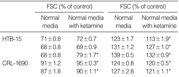
Fig. 4 clearly showed the effect of ketamine 0.1 mM on the change of cellular volume and granularity following IAA/
CCCP (150 M/2 M) treatment in HTB-15 cells. The addi- tion of ketamine during the reperfusion ameliorated the de- crease in. Compared to the control condition, cell volume de- creased to 71, 68, 68%, while cell granularity increased to 123, 131, 139%, respectively, during the reperfusion for 1 hr after energy depletion in HTB-15 cells. These cellular changes were indicative of apoptosis. An addition of ketamine 0.1 mM dur- ing the reperfusion reduced cell shrinkage to 72, 69, 79%
of control cells and increased granularity to 113, 127, 132%
of control cells, respectively. CRL-1690 cells also showed sim- ilar effect of ketamine on the cellular changes after energy depletion (Table 1).
During reperfusion for 24 hr after being exposed to IAA/
CCCP 150 M/2 M for 1 hr, however, both HTB-15 and CRL-1690 cells showed cell swelling, rather than cell shrink- age. This indicated morphological changes from the early phase of apoptosis to late stage of apoptosis. The addition of ketamine during the reperfusion protected cells from swelling.
However, the changes of cellular granularity was not consis- tent, thereby, the effect of ketamine on the cellular granular- ity after reperfusion for 24 hr was not observed (Table 2).
The effect of ketamine on the membrane integrity
This experiment was performed to investigate the effect of ketamine on the PI binding during the reperfusion for 24 hr following IAA/CCCP treatment in CRL-1690 cells. PI pos- itive cells increased 7.4% in control cells to 80.8% in cells treated with IAA/CCCP 150 M/2 M. The addition of ke- tamine at both concentrations of 0.1 mM and 0.5 mM during the reperfusion significantly decreased PI binding to 73.1%
and 65.2%, respectively. PI positive cells with ketamine 0.1 mM or 0.5 mM were 6.6% and 8.4%, respectively, which showed no difference with the control data. Therefore, we found that ketamine protected cells from increased plasma membrane permeability due to energy depletion, rather than affect normal cell membrane integrity (Table 3).
DISCUSSION
Ischemia-induced pathologic processes in the CNS are one of the leading causes of primary mortality and morbidity. ATP is required for a number of vital neuronal processes and energy failure evokes numerous pathologic events including apoptosis.
Many previous studies reported that astrocyte function could critically influence neuronal survival during ischemia. There- fore, we mimicked ischemic condition in vitro by using IAA/
CCCP for depleting intracellular energy. In this study, we de- monstrated the evidence of ketamine effect on the reduction of apoptosis in astrocytoma cells by using flow cytometry system.
In this experiment, we used Annexin-V and PI as indica- tors for apoptosis. Recently Yu et al. (20) demonstrated that the mechanism of astroglial cell death due to ischemic insults is apoptosis by using annexin-V staining. Annexin-V cannot penetrate the double layer of cell membrane and interact with PS. PS mostly faces the cytosol by outside-inside PS translo- case, using ATP, in the normal cells. Having damages to the cells including hypoxia or ischemia, internally located PS moves over the outer leaflet of the plasma membrane; hence allow- ing the binding of annexin-V (21).
The cells were reperfused with normal media or ketamine 0.1 mM con- taining media for 1 hr after IAA/CCCP (150 M/2 M) treatment for 1 hr.
Values are mean±SD. *p <0.05 compared with reperfusion with normal media.
FSC (% of control) Normal media with ketamine Normal
media
FSC (% of control) Normal media with ketamine Normal
media
HTB-15 71±0.8 72±0.7 123±1.7 113±1.9*
68±0.8 69±0.9 131±1.2 127±1.0*
68±0.8 79±1.7* 139±0.5 132±0.9*
CRL-1690 91±1.2 95±0.3* 124±0.8 120±0.5*
87±1.8 90±1.1* 127±2.8 121±1.1*
Table 1.The esffect of ketamine on the changes of FSC and SSC during reperfusion for 1 hr after IAA/CCCP treatment
The cells were reperfused with normal media or ketamine 0.1 mM con- taining media for 24 hr after IAA/CCCP (150 M/2 M) treatment for 1 hr.
Values are mean±SD. *p <0.05 compared with reperfusion with normal media.
FSC (% of control) Normal media
with ketamine Normal
media
FSC (% of control) Normal media with ketamine Normal
media
HTB-15 107±1.4 103±0.3* 122±2.7 129±0.6*
105±1.4 99±2.0* 106±1.1 103±1.0*
CRL-1690 124±2.7 108±2.5* 105±2.0 107±2.2 115±3.8 104±1.0* 82±2.2 79±2.3*
Table 2.The effect of ketamine on the changes of FSC and SSC during reperfusion for 24 hr after IAA/CCCP treatment
Values are mean±SD. *p <0.05 compared with control; �p <0.05 com- pared with reperfusion after IAA/CCCP.
Treatment PI (%)
Control 7.4±0.5
Reperfusion after IAA/CCCP 80.8±2.5*
Reperfusion with ketamine 0.1 mM after IAA/CCCP 73.1±1.6� Reperfusion with ketamine 0.5 mM after IAA/CCCP 65.2±3.7�
Ketamine 0.1 mM 6.6±0.6
Ketamine 0.5 mM 8.4±0.4
Table 3.The effect of ketamine on PI binding in the CRL-1690 cells during reperfusion for 24 hr after IAA/CCCP (150 M/2 M) treatment for 1 hr
Annexin-V assay with flow cytometry for assessing apopto- sis is simple and useful. The observation with electron micro- scopic analysis to discriminate between apoptosis and necro- sis is elaborate, time consuming, and cumbersome for quan- titative analysis (5, 6). Although terminal deoxynucleotidyl transferase (TdT)-mediated dUTP-biotin nick-end labeling (TUNEL) analysis shows a high level of specificity, experimen- tal method is complicated and requires 3-4 hr to complete (22). Flow cytometry using annexin-V fluorescence allows rapid quantitative measurement in 5-10 min, and is relatively easy because it does not require enzyme or fixation (23). In addition, DNA fragmentation is not an early event in apop- tosis, and not always associated with apoptosis. Apoptosis is a programmed, physiologic mode of cell death, which is char- acterized by structural nuclear changes, but also relatively by preserved plasma membrane and organelle components. Re- cently the externalization of PS has been demonstrated in the absence of nuclear damage (24). This indicates that a cytoplas- mic apoptotic regulator may be responsible for apoptosis in the early phase. Castedo et al. (25) suggested that the redistri- bution of PS was not affected by increased intracellular cal- cium ion or reactive oxygen species (ROS), but disruption of mitochondrial membrane potential. When lymphocytes are exposed to CCCP, breakdown of mitochondrial potential was demonstrated in a few minutes, followed by PS exposure on the outer membrane layer in 60 min.
The addition of pharmacological agents which stabilize mitochondrial membrane potential or inhibit disruption of mitochondrial potential, protected PS exposure (26). There- fore, aberrant PS movement may be a secondary effect sub- sequent to the disruption of mitochondrial membrane poten- tial. Moreover, the functional impact of mitochondrial respi- ratory chain may be significant during the reperfusion period than during the ischemic insult, by the rapid ionic movement through the damaged membrane (27). Changes of mitochon- drial membrane potential are irreversible and appear in sev- eral cells in the early event of apoptosis, without exception.
Less than 5% of annexin-V positive cells contain normal nucle- ar morphology. Loss of plasma membrane asymmetry by the cell surface exposure of PS precedes loss of membrane integri- ty, increased membrane permeability and nuclear condensa- tion (3, 5, 24, 28).
Annexin-V is commonly used to analyze apoptotic cell death in suspending cells. But, approximately 1-5% of cells in cul- ture medium show apoptosis (5). Trypsinization to detach adherent cells from the culture plate may cause membrane damage by handling and following annexin-V staining. In the human lung carcinoma cell line MR-65, approximately 25% of the suspension cells showed annexin-V binding and a lack of PI uptake after trypsin treatment (5). About 10%
of control cells were positive for PI in PC-12 cell line (4). In this experiment, the portion of the HTB-15 and CRL-1690 cells showing both annexin-V and PI negativity after trypsi- nization ranged from 85 to 95%. Therefore these cells were
appropriate for performing experiment on the apoptotic process.
Apoptosis has been described to occur under several stim- ulations. We found that in this experiment, the degree of re- distribution of PS depended on the concentration of IAA/
CCCP: higher concentration of IAA/CCCP is associated with increased annexin-V and PI uptake during the reperfusion (Fig. 1). The addition of ketamine 0.1 mM during the reper- fusion increased viable cells with both annexin-V and PI neg- ative staining, compared to reperfusion without ketamine (Fig. 2, 3). This finding strongly suggests that ketamine was able to restore intracellular energy, maintain membrane integri- ty by the inhibition of phospholipid membrane peroxidation, consequently protect cells from PS redistribution and PI entry intracellulary. Zhou et al. (29) suggested that the addition of ketamine 0.1-10 mM inhibited chemical- or ischemia-induced lipid peroxidation and ischemic glutathione depletion.
For mimicking energy depletion we used CCCP in this experiment. As an uncoupler of mitochondrial respiratory chain, CCCP decreases mitochondrial membrane potential, causes alterations of energy metabolism, produces membrane peroxidation and disrupt membrane integrity due to structural and functional alterations (25, 26). Table 1, 2 represents the results concerning the changes of cell volume and granularity due to intracellular energy depletion, by using forward and side light scattering on flow cytometry. Reperfusion for 1 hr causes cell shrinkage and increased granularity, but in the late apoptotic phase a progressive water influx causes cell swelling and increased the release of intracellular organelles, which can be related to decreased granularity. It is evident that the progression of early apoptosis to secondary necrotic cell death is dependent on the intensity of the stimulus and the energy status of the cell (30, 31). CsA-induced apoptosis in the renal proximal tubular cell line, LLC-PK1showed increased FITC- annexin-V affinity and cell granularity, and decreased cell volume with a low dose, while decreased membrane integrity and cell granularity, and increased cell volume in higher con- centration (19). In the apoptosis of HL-60 cells induced by UV exposure for 30 min, morphological change showing cel- lular swelling, the phenomenon of secondary cellular necro- sis, was observed after 6 hr under the electronic microscopy (1). As time passes, the entry of PI was increased in fresh cells without stimuli, although cell morphology was well preserved under electronic microscopy (32).
We found that ketamine addition during the reperfusion for 1 hr ameliorated morphological changes of apoptosis, such as cellular shrinkage and increased cellular granularity (Table 1), and ketamine addition during the reperfusion for 24 hr also ameliorated cellular necrosis accompanying cellular swelling (Table 2). Chen and Simard (33) also demonstrated that hy- poxia-ischemia and ATP depletion are associated with glial swelling and blebbing. Ketamine 0.1 mM itself was found not to induce apoptosis by the observation that annexin-V affinity or PI uptake was not increased. Thus, ketamine was effective in two types of cell death, both early stage of apop-
tosis and necrosis.
As a non-competitive NMDA receptor blocker (34), keta- mine produced considerable neuroprotection after mechani- cal trauma or ischemic injury. Until very recently, NMDA receptor was demonstrated only in the neuronal cell mem- brane, however, Krebs et al. revealed the expression of func- tional NMDA receptor in astrocytes under the ischemic in- sults by immunohistochemistry (35). Ketamine may become a valuable agent in restoring vulnerable astrocytes (36). The potential neuroprotective effect of ketamine was proved at the cellular level (9, 32). Many studies revealed that ketamine stabilized neuronal and glial electrophysiological functions by regulating Ca2+, Na+, K+conductance, thus maintaining intracellular ion homeostasis (32, 37-39). Administration of ketamine before anoxic-hypoxic insults preserved neuronal action potential in the hippocampus and protected cellular energy status, maintaining two-thirds of ATP production (9, 10). Related to experiments mimicking ischemic insult or head trauma in rats, ketamine improved neurological out- comes by inhibiting pathophysiologic NO production as a potential blocker for NMDA receptor (11, 32), decreasing catecholamine level (12) and ameliorating brain swelling (8).
Using harvested cells, which have similar characteristics of primary cells, is relatively easy in vitro experiment. We used astroglioma cells instead of primary astrocytes in the current study. Recently, the capacity of the CNS has been revealed in many studies. As the most abundant cell type in the cen- tral nervous system, astrocytes play very important and diverse roles, interacting with neurons. Astrocytes provide structural, trophic, and metabolic support to neurons and coordinate with neurons in neurotransmission. A large body of studies has demonstrated on the relationship between neurons and astro- cytes survival. The addition of astrocyte-conditioned medium in the serum free medium allows neurons to be attached to the bottom of the coverslip and neuritis outgrowth, whereas in the absence of astrocyte-conditioned medium impairment of neuronal survival was shown (17). In the condition of co- culture of neurons and astrocytes, removal of astrocytes caused neuronal apoptosis (16). To prevent hypoxia-ischemia induced neuronal degeneration, astrocytes survival is crucial (37).
In conclusion, we have demonstrated the occurrence of early and late phase of apoptosis during the reperfusion after intra- cellular energy depletion in astrocytoma cells by using flow cytometry system. Ketamine addition during the reperfusion protected astrocytoma cells from annexin-V binding and PI uptake, and changes of cell volume and granularity. Consid- ering the intimate functional and structural interaction of astrocyte with neuron in the CNS, the presence of astrocyte is very important for neuronal survival.
REFERENCES
1. Reno F, Burattini S, Rossi S, Luchetti F, Columbaro M, Santi S, Papa
S, Falcieri E. Phospholipid rearrangement of apoptotic membrane does not depend on nuclear activity. Histochem Cell Biol 1998; 110:
467-76.
2. Walton M, Sirimanne E, Reutelingsperger C, Williams C, Gluckman P, Dragunow M. Annexin V labels apoptotic neurons following hypox- ia-ischemia. Neuroreport 1997; 8: 3871-5.
3. Martin SJ, Reutelingsperger CP, McGahon AJ, Rader JA, van Schie RC, LaFace DM, Green DR. Early redistribution of plasma membrane phosphatidylserine is a general feature of apoptosis regardless of the initiating stimulus: inhibition by overexpression of Bcl-2 and Abl. J Exp Med 1995; 182: 1545-56.
4. Rimon G, Bazenet CE, Philpott KL, Rubin LL. Increased surface phosphatidylserine is an early marker of neuronal apoptosis. J Neu- rosci Res 1997; 48: 563-70.
5. van Engeland M, Ramaekers FC, Schutte B, Reutelingsperger CP. A novel assay to measure loss of plasma membrane asymmetry during apoptosis of adherent cells in culture. Cytometry 1996; 24: 131-9.
6. Bose S, Tuunainen I, Parry M, Medina OP, Mancini G, Kinnunen PK. Binding of cationic liposomes to apoptotic cells. Anal Biochem 2004; 331: 385-94.
7. Telford WG, King LE, Fraker PJ. Rapid quantitation of apoptosis in pure and heterogeneous cell populations using flow cytometry. J Immunol Methods 1994; 172: 1-16.
8. Shapira Y, Lam AM, Eng CC, Laohapraist V, Michel M. Therapeu- tic time window and response of the beneficial effects of ketamine in experimental head injury. Stroke 1994; 25: 1637-43.
9. Rothman SM, Thurston JH, Hauhart RE, Clark GD, Solomon JS.
Ketamine protects hippocampal neurons from anoxia in vitro. Neu- roscience 1987; 21: 673-8.
10. Pophprecki R, Becker GL, Reilly PJ, Landers DF. Ketamine, MK-801 or calmidazolium protects rat hippocampal energy status during in vitro ischemia. Ann NY Acad Sci 1991; 625: 818-20.
11. Lin SZ, Chiou AL, Wang Y. Ketamine antagonizes nitric oxide release from cerebral cortex after middle cerebral artery ligation in rats.
Stroke 1996; 27: 747-52.
12. Hoffman WE, Peligrino D, Werner C, Kochs E, Albrecht RF, Schulte am Esch J. Ketamine decreases plasma catecholamines and improves outcome from incomplete cerebral ischemia in rats. Anesthesiology 1992; 76: 755-62.
13. Chen Y, Swanson RA. Astrocytes and brain injury. J Cereb Blood Flow Metab 2003; 23: 137-49.
14. Aschner M, Sonnewald U, Tan KH. Astrocyte modulation of neuro- toxic injury. Brain Pathol 2002; 12: 475-81.
15. Tamatani M, Ogawa S, Niitsu Y, Tohyama M. Involvement of Bcl-2 family and caspase-3 like protease in NO-mediated neuronal apop- tosis. J Neurochem 1998; 71: 1588-96.
16. Ohgoh M, Kimura M, Ogura H, Katayama K, Nishizawa Y. Apoptotic cell death of cultured cerebral cortical neurons induced by withdraw- al of astroglial trophic support. Exp Neurol 1998; 149: 51-63.
17. Wang XF, Cynader MS. Effects of astrocytes on neuronal attachment and survival shown in a serum-free co-culture system. Brain Res Pro- toc 1999; 4: 209-16.
18. Araque A, Carmignoto G, Haydon PG. Dynamic signaling between astrocytes and neurons. Annu Rev Physiol 2001; 63: 795-813.
19. Healy E, Dempsey M, Lally C, Ryan MP. Apoptosis and necrosis:
mechanisms of cell death induced by cyclosporine A in a renal prox- imal tubular cell line. Kidney Int 1998; 54: 1955-66.
20. Yu AC, Wong HK, Yung HW, Lau LT. Ischemia-induced apoptosis in primary cultures of astrocytes. Glia 2001; 35: 121-30.
21. Gummadi SN, Hrafnsdottir S, Walent J, Watkins WE, Menon AK.
Reconstitution and assay of biogenic membrane-derived phospholipid flippase activity in proteoliposomes. Methods Mol Biol 2003; 228:
271-9.
22. Gavrieli Y, Sherman Y, Ben-Sasson SA. Identification of programmed cell death in-situ via specific labeling of nuclear DNA fragmentation.
J Cell Biol 1992; 119: 493-501.
23. Zhang G, Gurtu V, Kain SR, Yan G. Early detection of apoptosis using a fluorescent conjugate of annexin-V. Biotechniques 1997; 23:
525-31.
24. Chan A, Reiter R, Wiese S, Fertig G, Gold R. Plasma membrane phospholipid asymmetry precedes DNA fragmentation in different apoptotic cell models. Histochem Cell Biol 1998; 110: 553-8.
25. Castedo M, Hirsch T, Susin SA, Zamzami N, Marchetti P, Macho A, Kroemer G. Sequential acquisition of mitochondrial and plasma mem- brane alterations during early lymphocyte apoptosis. J Immunol 1996;
157: 512-21.
26. van Engeland M, Nieland LJ, Ramaekers FC, Schutte B, Reutelings- perger CP. Annexin V-affinity assay: a review on an apoptosis detec- tion system based on phosphatidylserine exposure. Cytometry 1998;
31: 1-9.
27. Smis NR, Anderson MF. Mitochondrial contributions to tissue dam- age in stroke. Neurochem Int 2002; 40: 511-26.
28. Wolvetang EJ, Johnson KL, Krauer K, Ralph SJ, Linnane AW. Mito- chondrial respiratory chain inhibitors induce apoptosis. FEBS Lett 1994; 339: 40-4.
29. Zhou M, Ma T, Tseng MT. Effects of taurine and ketamine on bovine
retinal membrane lipid peroxidation. Neuroscience 1991; 45: 461-5.
30. Bonfoco E, Kraine D, Ankarerona M, Nicotera P, Lipon SA. Apop- tosis and necrosis: two distinct events induced, respectively, by mild and intense insults with N-methyl-D-aspartate or nitric oxide/super- oxide in cortical cell cultures. Proc Natl Acad Sci USA 1995; 92:
7162-6.
31. Leist M, Single B, Castoldi AF, Kuhnle S, Nicotera P. Intracellular adenosine triphosphate (ATP) concentration: A switch in the decision between apoptosis and necrosis. J Exp Med 1997; 185: 1481-6.
32. Pfenninger E, Himmelseher S. Neuroprotection by ketamine at the cellular level. Anaesthesist 1997; 46: S47-54.
33. Chen M, Simard JM. Cell swelling and a nonselective cation chan- nel regulated by internal Ca2+and ATP in native reactive astrocytes from adult rat brain. J Neurosci 2001; 21: 6512-21.
34. Kohrs R, Durieux ME. Ketamine: teaching an old drug new tricks.
Anesth Analg 1998; 87: 1186-93.
35. Krebs C, Fernandes HB, Sheldon C, Raymond LA, Baimbridge KG.
Functional NMDA receptor subtype 2B is expressed in astrocytes after ischemia in vivo and anoxia in vitro. J Neurosci 2003; 23: 3364-72.
36. Kim MH, Hahm TS, Ahn HJ. Regulation of astroglial volume by ketamine in glutamate induced cellular volume changes. Korean J Anesthesiol 1997; 33: 1005-11.
37. Chan PH, Chu L. Ketamine protects cultured astrocytes from gluta- mate-induced swelling. Brain Res 1989; 487: 380-3.
38. Brau ME, Sander F, Vogel W, Hempelmann G. Blocking mechanisms of ketamine and its enantiomers in enzymatically demyelinated periph- eral nerve as revealed by single-channel experiments. Anesthesiology 1997; 86: 394-404.
39. Silver IA, Erecinska M. Intracellular and extracellular changes of Ca2+in hypoxia and ischemia in rat brain in vivo. J Gen Physiol 1990;
95: 837-66.