INTRODUCTION
Extramedullary hematopoiesis (EMH) is occasionally report- ed in idiopathic myelofibrosis and is generally found in the liver, spleen, and lymph nodes (1). Involvement of other sites, such as the intrathoracic cavity, kidney, and thyroid, has been reported (1, 2). However, involvement of the dura mater of the spinal cord, resulting in spinal cord compression, has rarely been described between two and 20 yr after diagnosis of myelofibrosis (2-6), and myelofibrosis presenting as spinal cord compression, resulting from EMH tissue, is also very rare (7). We report the first case of EMH due to chronic idio- pathic myelofibrosis in the lumbar spinal canal in Korea and present the surgical outcome.
CASE REPORT
A 39-yr-old man presented with back pain, subjective weakness and numbness in both legs that had progressed over 2 weeks. His medical history was not significant except that he received anti-tuberculosis medication at the age of 12 yr for pulmonary tuberculosis. There was no history of
trauma or primary malignancy.
On physical examination, his blood pressure was 110/70 mm Hg, pulse 94 beats per minutes, and respiratory rate was 18 breaths per minute. There was no pallor or jaundice, nor clinically palpable hepatosplenomegaly. His neurological status was unremarkable except for paresthesia at bilateral L5 and S1 sensory dermatomes and decreased deep tendon reflexes on ankle. He had normal mental function and no abnormality of the cranial nerves. Laboratory workup showed a normal hemoglobin level of 16.9 g/dL (range, 13-17 g/dL), mean corpuscular volume of 83.5 fL (range, 95-105 fL), mean corpuscular hemoglobin of 28.6 pg (range, 25-35 pg), hyper- leukocytosis of 23,800/ L (range, 4,000-10,000 g/L), and platelet count 111,000/ L without any other abnormal find- ings. Liver and renal function tests were also normal.
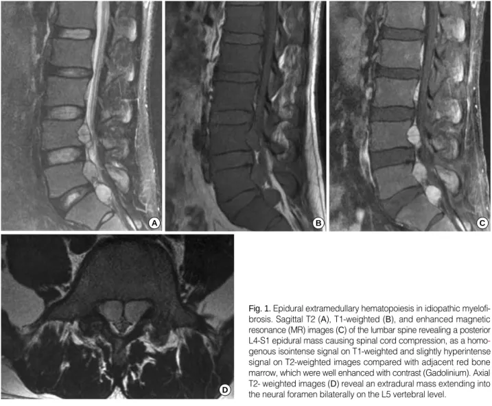
Chest and lumbar radiography was normal. Sagittal mag- netic resonance imaging (MRI) showed multiple anterior epidural mass extending from L4 to S1 with compression of the thecal sac and nerve root (Fig. 1A-C). Axial images reveal- ed an extradural mass extending into the neural foramen bilaterally (Fig. 1D), which gave slightly high signal on T2- weighted images; isointense on T1-weighted images and enhanced strongly with gadolinium. The mass seemed en-
1090
Duck-Ho Goh, Sun-Ho Lee, Dae-Chul Cho, Seong-Hyun Park, Jeong-Hyun Hwang, Joo-Kyung Sung
Department of Neurosurgery, School of Medicine, Kyungpook National University, Daegu, Korea
Address for correspondence Sun-Ho Lee, M.D.
Department of Neurosurgery, School of Medicine, Kyungpook National University, 50 Samduk-2 ga, Jung-gu, Daegu 700-721, Korea
Tel : +82.53-420-6524, Fax : +82.53-423-0504 E-mail : [email protected]
J Korean Med Sci 2007; 22: 1090-3 ISSN 1011-8934
DOI: 10.3346/jkms.2007.22.6.1090
Copyright � The Korean Academy of Medical Sciences
Chronic Idiopathic Myelofibrosis Presenting as Cauda Equina
Compression due to Extramedullary Hematopoiesis: A Case Report
Extramedullary hematopoiesis (EMH) is occasionally reported in idiopathic myelofi- brosis and is generally found in the liver, spleen, and lymph nodes several years after diagnosis. Myelofibrosis presenting as spinal cord compression, resulting from EMH tissue is very rare. A 39-yr-old man presented with back pain, subjective weak- ness and numbness in both legs. Sagittal magnetic resonance imaging showed multiple anterior epidural mass extending from L4 to S1 with compression of cauda equina and nerve root. The patient underwent gross total removal of the mass via L4, 5, and S1 laminectomy. Histological analysis showed islands of myelopoietic cells surrounded by fatty tissue, consistent with EMH, and bone marrow biopsy performed after surgery revealed hypercellular marrow and megakaryocytic hyper- plasia and focal fibrosis. The final diagnosis was chronic idiopathic myelofibrosis leading to EMH in the lumbar spinal canal. Since there were no abnormal hemato- logical findings except mild myelofibrosis, additional treatment such as radiothep- ary was not administered postoperatively for fear of radiotoxicity. On 6 month fol- low-up examination, the patient remained clinically stable without recurrence. This is the first case of chronic idiopathic myelofibrosis due to EMH tissue in the lumbar spinal canal in Korea.
Key Words : Hematopoiesis, Extramedullary; Myelofibrosis; Spinal Canal
Received : 23 August 2006 Accepted : 20 November 2006
Cauda Equina Compression due to EMH with Myelofibrosis 1091
capsulated and showed no invasion or communication with neuronal structures.
For neurological resuscitation and histological confirma- tion, the patient underwent decompressive surgery via L4, 5 and S1 total laminectomy and gross total removal of the epidu- ral masses which located in the ventral portion of thecal sac.
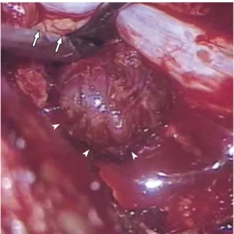
The intraoperative finding included several round and lobu- lated reddish tumors that was partially adherent to the ventral dura mater (Fig. 2). Four masses were removed in total. Two were removed from both sides of L5 vertebra and two from the right side of L4 and S1 vertebra. Each mass was inde- pendent from each other without connection or adhesion.
Histological analysis showed islands of myelopoietic cells surrounded by fatty tissue, consistent with EMH (Fig. 3).
Bone marrow aspiration and biopsy performed after surgery revealed hypercellular marrow and megakaryocytic hyper- plasia and focal fibrosis. The final diagnosis was chronic idio- pathic myelofibrosis (agnogenic myeloid metaplasia) lead- ing to EMH on lumbar spinal canal.
Postoperatively, there was marked improvement of neu-
rological symptom within a few days. Since there was no abnormal hematological finding except mild myelofibrosis, additional treatment such as radiothepary was not adminis- tered in consideration of radiotoxicity. On 6-month follow- up examination, the patient remained clinically stable and the MR imaging studies demonstrated no disease recurrence in the epidural space.
DISCUSSION
EMH is usually seen as a reactive process in various hema- tological diseases, with the liver and spleen as primary man- ifestation sites (1, 8-14). Rarely, EMH has been found local- ized in dura matter of spinal cord, and can result in spinal cord compression (2-14). EMH causing spinal cord compres- sion was first reported in 1956 by Close et al. in a patient with thalassemia minor (8). Although this most often occurs in thalassemia, it has also been described in myeloprolifera- tive diseases such as polycythemia vera, sideroblastic anemia,
Fig. 1.Epidural extramedullary hematopoiesis in idiopathic myelofi- brosis. Sagittal T2 (A), T1-weighted (B), and enhanced magnetic resonance (MR) images (C) of the lumbar spine revealing a posterior L4-S1 epidural mass causing spinal cord compression, as a homo- genous isointense signal on T1-weighted and slightly hyperintense signal on T2-weighted images compared with adjacent red bone marrow, which were well enhanced with contrast (Gadolinium). Axial T2- weighted images (D) reveal an extradural mass extending into the neural foramen bilaterally on the L5 vertebral level.
A B C
D
1092 D.-H. Goh, S.-H. Lee, D.-C. Cho, et al.
pyruvate kinase deficiency, and myelofibrosis disorders (8-14).
In our patient, unusually, there was no predisposing hema- tological disorder at presentation, and the diagnosis of EMH and idiopathic myelofibrosis was made retrospectively. Anoth- er atypical feature was the relatively minor degree of spleno- megaly at presentation. In case of myelofibrosis with EMH a far greater degree of splenomegaly would usually be expect- ed (2-6, 8).
The exact origin of EMH in the dura mater is unknown.
Some previous reports indicated the dura mater has a hema- topoietic capacity in the fetus and EMH may develop from primitive rests (2, 3). However, it is unlikely that EMH aris- es from extrusion of vertebral bone marrow in the absence of bony erosion or fractures. Symptomatic spinal cord compres- sion secondary to EMH shows a preference for localization in the middle and lower thoracic spine. Some authors related this to the narrow spinal diameter at this level (3, 5-14).
Radiologic features of intraspinal EMH are well defined especially on MR imaging. Some authors emphasized the use of MR imaging because of the notable isointense T1- and T2-weighted images secondary to the deposition of iron.
The principal MR imaging characteristics of EMH include the followings: an extramedullary mass (more often in the posterior location of the epidural space and in the thoracic region), with homogeneous signal intensity on T1-weight- ed images, slightly higher intensity of signal than the adja- cent red marrow of the vertebral bodies on T2-weighted images, and enhancement after Gadolinium injection (2, 5).
However, the diagnosis can be confirmed by fine-needle aspi- ration cytology or open biopsy in case of unusual imaging
findings or clinical history (1, 6, 11-14). The lesion is com- posed of various numbers of hematopoietic cells from all three hematologic cell lines.
Finally, management of patients with epidural EMH re- mains controversial, and there are no widely accepted treat- ment guidelines. Regarding the pathophysiology of the dis- ease, low doses of radiotherapy or blood transfusion has been recommended as the initial treatment during the past two decades (3, 5, 6, 9). However, the risks of radiotherapy in the treatment of cord compression in such patients include the lack of any tissue for histological diagnosis and the risks in- volved in radiation exposure, and the transfusion also has a risk such as infectious disease, iron overload, formation of antibodies (10). Recently, hydroxyurea has been proposed as a complementary approach to increase the efficiency of ery- thropoiesis and reduce the volume of transfusion required (7).
These non-surgical approaches can be undertaken in patients in whom the diagnosis of EMH is highly expected, based on clinical presentation, known chronic anemia, and MR imaging features. Although they can avoid surgery-related complications such as the high risk of bleeding due to the lesion’s vascularity or difficulty in transfusion due to anti- bodies and cardiopulmonary stress, disadvantages are they do not allow definitive diagnosis and have a high rate of recur- rence. In cases of symptomatic spinal cord compression, sur- gical decompression and subsequent low-dose radiotherapy have been recommended (2, 11-14). Decompessive surgery is immediately effective in most cases and radiotherapy is introduced as an adjunct to surgery because of prevention of recurrence, incomplete excision, or persisting neurological
Fig. 2.Intraoperative photograph showing the discrete, flesh-col- ored, elastic, and extradural mass (arrowheads) between lumbar nerve roots (arrows).
Fig. 3.Microscopic photograph of the extradural mass revealing cellular tissue of hematopoietic origin with small megakaryocytes (H&E, ×400).
Cauda Equina Compression due to EMH with Myelofibrosis 1093
deficit. As an alternative option, radiation can be saved as a salvage procedure for recurrence, if complete removal was accomplished (10). We decid-ed to treat our patient with surgical removal without adjuvant therapy to prevent com- plication of adjuvant therapy, and the postoperative result was favorable during the 6-month follow-up period.
In conclusion, authors successfully treated a rare case of EMH on spinal canal with surgical management. Although medical treatment should be first advised after discussion with the hematologist, surgical approach may be successfully used in case of severe neurologic deficit, failure of medical treat- ment, or uncertain diagnosis.
REFERENCES
1. Kwak HS, Lee JM. CT findings of extramedullary hematopoiesis in the thorax, liver and kidneys, in a patient with idiopathic myelofi- brosis. J Korean Med Sci 2000; 15: 460-2.
2. Cook G, Sharp RA. Spinal cord compression due to extramedullary haemopoiesis in myelofibrosis. J Clin Pathol 1994; 47: 464-5.
3. De Klippel N, Dehou MF, Bourgain C, Schots R, De Keyser J, Ebinger G. Progressive paraparesis due to thoracic extramedullary hematopoiesis in myelofibrosis. Case report. J Neurosurg 1993; 79:
125-7.
4. McDonald AC, Cook G, Sharp RA, Bissett D. Spinal cord compres- sion in myelofibrosis--a case report. Acta Oncol 1993; 32: 692-3.
5. Guermazi A, Miaux Y, Chiras J. Imaging of spinal cord compres- sion due to thoracic extramedullary haematopoiesis in myelofibro- sis. Neuroradiology 1997; 39: 733-6.
6. de Haas KP, van de Loosdrecht AA, Daenen SM. Intraspinal extramedullary haematopoiesis in a patient with myelofibrosis. Neth J Med 2002; 60: 256-9.
7. Horwood E, Dowson H, Gupta R, Kaczmarski R, Williamson M.
Myelofibrosis presenting as spinal cord compression. J Clin Pathol 2003; 56: 154-6.
8. Close AS, Taira Y, Cleveland DA. Spinal cord compression due to extramedullary hematopoiesis. Ann Intern Med 1958; 48: 421-7.
9. Saghafi M, Shirdel A, Lari SM. Extramedullary hematopoiesis with spinal cord compression in beta-thalassemia intermedia. Eur J Intern Med 2005; 16: 596-7.
10. Salehi SA, Koski T, Ondra SL. Spinal cord compression in beta- thalassemia: case report and review of the literature. Spinal Cord 2004; 42: 117-23.
11. Jalbert F, Chaynes P, Lagarrigue J. Asymptomatic spherocytosis presenting with spinal cord compression: case report. J Neurosurg Spine 2005; 2: 491-4.
12. Shin KH, Sharma S, Gregoritch SJ, Lifeso RM, Bettigole R, Yoon SS. Combined radiotherapeutic and surgical management of a spinal cord compression by extramedullary hematopoiesis in a patient with hemoglobin E beta-thalassemia. Acta Haematol 1994;
91: 154-7.
13. Ohta Y, Shichinohe H, Nagashima K. Spinal cord compression due to extramedullary hematopoiesis associated with polycythemia vera--case report. Neurol Med Chir (Tokyo) 2002; 42: 40-3.
14. Niggemann P, Krings T, Hans F, Thron A. Fifteen-year follow-up of a patient with beta thalassaemia and extramedullary haematopoi- etic tissue compressing the spinal cord. Neuroradiology 2005; 47:
263-6.