INTRODUCTION
Veno-occlusive disease (VOD) of the liver is a severe com- plication of intensive chemotherapy and hematopoietic stem cell transplantation (HSCT). The incidence rate of VOD ranges from 5% to 60%, and the mortality rate is as high as 50%
in some studies (1-4). In children who have undergone HSCT, the incidence of VOD ranges from 22% to 28%, and the mor- tality rate is reported to be up to 47% (5-8). VOD is charac- terized by right upper quadrant pain, hepatomegaly, fluid retention, ascites, weight gain and hyperbilirubinemia occur- ring by day 20 after HSCT (1).
Reported risk factors for developing VOD include pre- HSCT liver dysfunction, poor performance status, female gen- der, advanced disease status at the time of transplantation, prior hepatic irradiation, vancomycin or amphotericin B use in the early post transplantation period, HLA mismatch-relat- ed or -unrelated donor transplantation, and use of busulfan (Bu) or total body irradiation (TBI) as a conditioning regimen (2, 5-11). An important new risk factor for VOD is adminis- tration of gemtuzumab ozogamicin (Mylotarg), an anti-CD33 monoclonal antibody (12-17).
The prevention of fatal VOD is an obvious priority. The
prophylactic use of drugs such as heparin or prostaglandin E1 (PGE1) has a beneficial effect in preventing VOD development.
Some reports show that continuous infusion of low dose hep- arin is highly effective in preventing VOD after HSCT with- out an added risk of bleeding (18, 19). PGE1is a vasodilator with inhibitory effects on platelet aggregation and prothrom- bolytic activity (20). Gluckman et al. reported that the use of prophylactic PGE1via continuous intravenous infusion dec- reased the overall incidence of VOD, although they did not use lipo-PGE1(21). Cho et al. reported the possible beneficial effect of lipo-PGE1to prevent development of VOD, whereas they did not compare the efficacy with control group (22).
The purpose of the present single center study was to iden- tify VOD risk factors and the effect of prophylactic low-dose heparin or PGE1on pediatric patients undergoing allogeneic HSCT.
MATERIALS AND METHODS Patients
Eighty-nine pediatric patients undergoing allogeneic HSCT Joon Sup Song, Jong Jin Seo,
Hyung Nam Moon, Thad Ghim*, Ho Joon Im
Division of Hematology/Oncology/Bone Marrow Transplantation, Department of Pediatrics, Asan Medical Center, University of Ulsan College of Medicine, Seoul; Pediatric Hematology-Oncology Center*, Korean National Cancer Center, Goyang, Korea
Address for correspondence Ho Joon Im, M.D.
Department of Pediatrics, University of Ulsan College of Medicine, Asan Medical Center, 388-1 Pungnap-2dong, Songpa-gu, Seoul 138-736, Korea Tel : +82.2-3010-3371, Fax : +82.2-473-3725 E-mail : [email protected]
897
Prophylactic Low-dose Heparin or Prostaglandin E
1may Prevent Severe Veno-occlusive Disease of the Liver after Allogeneic Hematopoietic Stem Cell Transplantation in Korean Children
Studies investigating the effect of prophylactic drugs on hepatic veno-occlusive dis- ease (VOD) development are rare in children that have undergone allogeneic he- matopoietic stem cell transplantation (HSCT). This study examined risk factors for VOD, the effect of prophylactic low-dose heparin or lipo-prostaglandin E1(lipo-PGE1) and the survival rate at day +100 in children undergoing allogeneic HSCT. Eighty five children underwent HSCT between June 1997 and September 2004. Patients were diagnosed and classified as having mild, moderate or severe VOD according to Seattle clinical criteria. Among 85 patients, 25 (29%) developed VOD. VOD occurr- ed more frequently in patients receiving busulfan-based conditioning (24/65, 37%) than in those receiving TBI-based (1/10, 10%) or other (0/10, 0%) regimens (p<0.05).
The incidence of VOD was lower in patients with non-malignant disease compared to those with malignant disease (p<0.05). Survival at day +100 for non-VOD patients was better than that for VOD patients (92% vs. 76%, p<0.05). No patients receiving prophylactic heparin or lipo-PGE1were found to develop severe VOD, whereas 5 of 35 patients not receiving such prophylaxis developed severe VOD. Given severe VOD is associated with a high mortality rate, this study indicates that prophylactic heparin or lipo-PGE1may decrease mortality in children undergoing HSCT.
Key Words : Hepatic Veno-occlusive Disease; Heparin; Prostaglandins; prevention and control; Prophylaxis;
Hematopoietic Stem Cell Transplantation; Child
Received : 26 October 2005 Accepted : 16 February 2006
at the Asan Medical Center between June 1997 and Septem- ber 2004 were enrolled. Four patients who died of causes other than VOD before day +21 were subsequently excluded. The medical records of the remaining 85 patients were retrospec- tively reviewed.
Risk factors for development of VOD
The following parameters were evaluated to determine their status as risk factors for the development of VOD: 1) patient- related factors: age at time of HSCT, disease status at the time of transplantation, viral serology results in both donor and recipient, and liver function before HSCT; 2) transplantation- related factors: stem cell source, HLA matching, condition- ing regimen and prophylactic treatment for VOD.
Risk factors for the severity of VOD
The following factors were evaluated to determine their effect on the severity of VOD: maximum level of total serum bilirubin, maximum level of serum aspartate aminotransferase (AST), alanine aminotransferase (ALT), alkaline phosphatase (ALP), maximum level of serum blood urea nitrogen (BUN), serum creatinine (Cr) and post-transplantation weight gain.
Classification of disease status
Diseases were divided into three groups according to type and status at the time of transplantation: 1) non-malignant diseases, including severe aplastic anemia (SAA), pure red cell anemia (PRCA), chronic granulomatous disease (CGD) and hemophagocytic lymphoproliferate histiocytosis (HLH) in second remission, 2) early stage hematologic malignancies, including acute lymphoblastic leukemia (ALL) in the first or second complete remission, acute myeloid leukemia (AML) in the first complete remission, myelodysplastic syndrome (MDS), chronic myeloid leukemia (CML) in the chronic phase, acute mixed lineage leukemia (AMLL) in the first complete remission and juvenile myelomonocytic leukemia (JMML), 3) advanced stage hematologic malignancies, including ALL in greater than the second complete remission, CML in the accelerated phase, AMLL beyond the first complete remission and acute leukemia in partial remission.
VOD definition and severity
Definition: Seattle clinical criteria were used for VOD diag- nosis. A diagnosis of VOD required two of the following within 20 days of transplantation; serum bilirubin >2 mg/
dL (34 M/L), hepatomegaly and weight gain >2% over baseline.
Severity: The severity of VOD was classified as mild, mod- erate or severe. Mild VOD was defined as the absence of adverse effects of liver dysfunction with complete resolution of symp-
toms and signs. Moderate VOD was characterized as the ad- verse effects of liver dysfunction requiring therapy such as diuretics for fluid retention and analgesics for pain. Severe VOD was defined as the persistence of symptoms after day 100 or death before day 100 with ongoing VOD.
Prophylactic treatment for VOD
Prophylactic heparin (Heparin sodium, Hanlim, Seoul, Korea) was administered using the dose 100 U/kg/day in con- tinuous intravenous infusion, starting on day -7 until day +30. Heparin was discontinued if testing found the activated partial thromboplastin time (aPTT) had increased by 1.2-fold over normal, or if there were any major bleeding episodes.
Lipo-PGE1(Eglandin; alprostadil, Welfide, Osaka, Japan) was continuously intravenously infused at a dose of 1 g/kg/day from day +1 until day +30. Patients were assigned to receive or not receive prophylaxis according to the decision of each physician.
Statistical methods
Associations between VOD, potential risk factors and pro- phylaxis were tested by univariate analyses using the chi-square test or Fisher’s exact test for comparing categorical variables, and t tests for comparing continuous variables. Variables asso- ciated with a p<0.1 in univariate analysis were fitted to a logis- tic regression model for multivariate analysis. Analysis of sur- vival was calculated using the Kaplan-Meier method and log- rank test. All data were analyzed using SPSS version 11.5.
RESULTS Patient characteristics
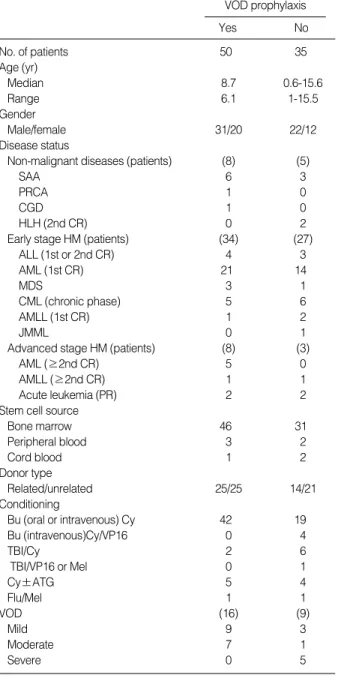
Patient characteristics at the time of transplantation are shown in Table 1. The median age at HSCT was 7.3 yr (range:
0.6 to 15.6 yr). Of a total of 85 patients, 11 received trans- plantation for non-malignant disease and 74 for malignant disease. Of the 74 patients with malignant disease, 11 were transplanted in the advanced stage of the disease. Condition- ing regimens were Bu-based for 65 patients (oral Bu for 33, intravenous Bu for 32), TBI-based for 10, and other regimens in 10 patients that included a case of non-myeloablative con- ditioning.
Incidence of VOD
VOD developed in 25 of 85 patients (29%) with the medi- an diagnosis day being 11.4 (range: day -4 to day +21). Of 25 patients, 12 had mild VOD (48%), 8 had moderate VOD (32%) and 5 had severe VOD (20%).
Risk factors for the development of VOD
Univariate analysis was performed to identify risk factors for VOD (Table 2). Age at transplantation, stem cell source and HLA matching were found not to be risk factors. Simi- larly, viral status including CMV, HSV, HBV and HCV for
either donor or recipient at transplantation had no significant influence on development of VOD. In contrast, disease sta- tus was identified as a risk factor, with none of 13 non-malig- nant disease patients developing VOD, while 20 of 61 patients with early stage hematologic malignancy (36%) and 5 of 11 patients with advanced stage hematologic malignancy (45%) developing VOD (p<0.03). The type of conditioning regimen was also found to affect VOD development (p=0.02), with
VOD prophylaxis
Yes No
No. of patients 50 35
Age (yr)
Median 8.7 0.6-15.6
Range 6.1 1-15.5
Gender
Male/female 31/20 22/12
Disease status
Non-malignant diseases (patients) (8) (5)
SAA 6 3
PRCA 1 0
CGD 1 0
HLH (2nd CR) 0 2
Early stage HM (patients) (34) (27)
ALL (1st or 2nd CR) 4 3
AML (1st CR) 21 14
MDS 3 1
CML (chronic phase) 5 6
AMLL (1st CR) 1 2
JMML 0 1
Advanced stage HM (patients) (8) (3)
AML (≥2nd CR) 5 0
AMLL (≥2nd CR) 1 1
Acute leukemia (PR) 2 2
Stem cell source
Bone marrow 46 31
Peripheral blood 3 2
Cord blood 1 2
Donor type
Related/unrelated 25/25 14/21
Conditioning
Bu (oral or intravenous) Cy 42 19
Bu (intravenous)Cy/VP16 0 4
TBI/Cy 2 6
TBI/VP16 or Mel 0 1
Cy±ATG 5 4
Flu/Mel 1 1
VOD (16) (9)
Mild 9 3
Moderate 7 1
Severe 0 5
Table 1.Patient characteristics at the time of transplantation
SAA, severe aplastic anemia; PRCA, pure red cell anemia; CGD, chronic granulomatous disease; ALL, acute lymphoblastic leukemia; AML, acute myeloid leukemia; MDS, myeloysplastic syndrome; CML, chronic myeloid leukemia; AMLL, acute mixed lineage leukemia; JMML, juvenile myelo- monocytic leukemia; HLH, hemophagocytic lymphoproliferate syndrome;
HM, hematologic malignancy; CR, complete remission; PR, partial remis- sion; Bu, busulfan; Cy, cyclophosphamide; VP16, Etoposide; Mel, mel- phalan; Flu, fludarabin; ATG, anti-thymocyte globulin; VOD, veno-occlu- sive disease.
Risk factor No. with VOD/
No. evaluated (%) p value
Age 0.63
<3 yr 5/17 (29%)
3-10 yr 10/40 (25%)
>10 yr 10/28 (34%)
Stem cell source 0.45
Bone marrow 24/77 (31%)
Peripheral blood 1/5 (20%)
Cord blood 0/3 (0%)
Donor type 0.98
Related 12/39 (31%)
Unrelated 13/46 (28%)
HLA matching 0.96
Match 20/70 (29%)
Mismatch 5/15 (33%)
CMV status, donor 0.70
Negative 4/10 (40%)
Positive 18/64 (28%)
CMV status, recipient 0.73
Negative 3/10 (30%)
Positive 22/74 (29%)
HSV serology, donor 0.66
Negative 0/1 (0%)
Positive 19/62 (31%)
HSV serology, recipient
Negative 25/85 (29%)
HBV serology, donor 0.62
Negative 24/84 (29%)
Positive 1/1 (100%)
HBV serology, recipient
Negative 25/85 (29%)
HCV serology, donor
Negative 25/85 (29%)
HCV serology, recipient 0.62
Negative 25/84 (30%)
Positive 0/1 (0%)
Disease status 0.03
Non-malignant disease 0/13 (0%) Early malignant disease 20/61 (36%) Advanced malignant disease 5/11 (45%)
Conditioning regimen 0.02
Busulfan-based 24/65 (37%)
TBI-based 1/10 (10%)
Other 0/10 (0%)
VOD prophylaxis 0.53
Lipo-PGE1 14/40 (35%)
Heparin 2/10 (20%)
None 9/35 (26%)
Table 2.Risk factors for VOD development
Bu-based regimens associated with an incidence of 37%, TBI- based regimens associated with an incidence of 10%, and other conditioning regimens associated with an incidence of 0%. There was no difference in VOD incidence according to the formulation of Bu (41% for oral Bu vs. 34% for intra- venous Bu, p=0.80). Prophylactic treatment with either low- dose heparin (2/10, 20%) or lipo-PGE1(14/40, 35%) showed no difference on development of VOD (p=0.53).
Pre-transplantation liver function
Pre-transplantation liver function and albumin levels were similar for both VOD and non-VOD patients (Table 3). The incidence of VOD in patients with normal liver function (AST
<40 IU/L, ALT <40 IU/L, total bilirubin <1.2 mg/dL and albumin >3 g/dL) was 27% (18/68) and that in patients with
abnormal liver function was 42% (7/17). However there was no significant difference in the incidence of VOD between two groups (p=0.37).
Severity of VOD
The median for development of mild/moderate VOD was day +12 (range: -4 to +21), while the median for severe VOD was day +7 (range: +2 to +15) (p>0.05). There were no sig- nificant differences in maximum levels of AST, ALT, total bilirubin and ALP after transplantation between patients with mild/moderate VOD and those with severe VOD. While the raw data suggested higher total bilirubin levels in severe VOD compared to mild/moderate VOD patients, statistical analysis did not find this difference significant. Ascites developed in 8 of 20 (40%) mild/moderate VOD patients and 4 of 5 (80%) severe VOD patients (p>0.05). There was no significant differ- ence between the two groups in terms of maximum serum BUN or Cr after transplantation. While renal dysfunction that elevated serum Cr more than twice the baseline level or re- quired dialysis was more frequent in patients with severe VOD, the difference from mild/moderate VOD patients was not statistically significant. However, renal dysfunction was less frequent in patients with mild VOD compared to patients with moderate or severe VOD (p=0.03). The occurrence of pulmonary dysfunction was more common in severe VOD compared to mild/moderate VOD patients (Table 4).
Pre-transplantation
liver function Value (mean±SD) p value
AST (IU/L) 0.16
Without VOD 29.3±13.5
With VOD 38.1±29.5
ALT (IU/L) 0.13
Without VOD 28.3±23.11
With VOD 46.2±55.3
Albumin (g/dL) 0.08
Without VOD 4.0±0.5
With VOD 3.8±0.4
Table 3.Pre-transplantation liver function in VOD and non-VOD patients
Mild/moderate Severe p value Day of diagnosis of VOD 12 (-4-21) 7 (2-15) 0.55
(post-transplant)*
Maximal AST (IU/L)* 163 (75-1678) 163 (65-1936) 0.38 Maximal ALT (IU/L)* 201 (37-2847) 124 (84-5065) 0.53 Maximal total bilirubin 3.4 (1.4-19) 26 (1.8-58) 0.11
(mg/dL)*
Maximal BUN (mg/dL)* 38 (17-132) 110 (23-198) 0.16 Maximal Cr (mg/dL)* 0.9 (0.3-3.8) 2.5 (0.3-3.3) 0.53 Maximal ALP (IU/L)* 188 (91-604) 182 (60-359) 0.70
Ascites 0.16
No 12/20 (60%) 1/5 (20%)
Yes 8/20 (40%) 4/5 (80%)
Pulmonary dysfunction 0.04
No 14/20 (70%) 1/5 (20%)
Effusion/edema/ 4/20 (20%) 1/5 (20%) O2requirement
Intubation 2/20 (10%) 3/5 (60%)
Renal dysfunction 0.37
No 11/20 (55%) 1/5 (20%)
Serum Cr >2-fold of 7/20 (35%) 3/5 (60%) baseline
Dialysis 2/20 (10%) 1/5 (20%)
Table 4.Patient characteristics as a function of VOD severity
Hematologic malignancy disease p=0.14
Early stage 4/60 (7%)
Advanced stage 1/11 (9%)
Prophylaxis p=0.02
Lipo-PGE1 0/40 (0%)
Heparin 0/10 (0%)
None 5/35 (56%)
Table 5.Development of severe VOD
Survival
1.0
0.8
0.6
0.4
0.2
0.0
0 10 20 30 40 50 60 70 80 90 100
Days post-transplantation
Severe VOD Mild/moderate VOD No VOD
p<0.01
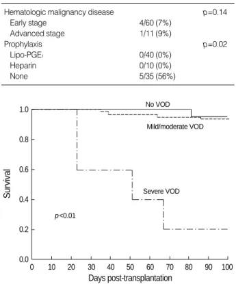
Fig. 1.Survival rate at day +100 relative to VOD development.
*Median and range.
Prophylaxis and VOD severity
Of the 85 patients, 50 received prophylactic low-dose hep- arin or lipo-PGE1treatment for VOD and 35 did not. Al- though the overall incidence of VOD did not differ between treated and non-treated patients (16/50 vs. 9/35, respective- ly), fewer patients receiving prophylaxis developed severe VOD compared to patients who did not receive prophylaxis (0/50 vs. 5/35, respectively, p=0.02) (Table 5). None of patients who received either lipo-PGE1or heparin developed severe VOD.
Survival
All 5 patients with severe VOD died of causes directly att- ributable to VOD, leading to an estimated survival rate of 0%
at 100 days posttransplant. In contrast, the survival rate for patients with mild/moderate VOD was 95%, and for patients without VOD the rate was 92% (p<0.01) (Fig. 1).
DISCUSSION
Hepatic VOD is one of the most common life-threatening complications that can occur during HSCT. The incidence of VOD is reported to range from 5% to 60% in adults (1- 4, 21, 23), and the incidence of post-transplantation VOD in children is reported to range from 18% to 28%, with the ranges probably reflecting the differences in conditioning regimens, type of graft and patient characteristics (5-8, 24, 25). Comparable to those results, the present retrospective study found that the incidence of hepatic VOD in pediatric patients undergoing allogeneic HSCT was 29%.
Pre-HSCT risk factors for VOD development include old age, elevated transaminases prior to conditioning therapy, previous abdominal radiation and advanced disease (2, 9).
Our study found that age at HSCT and pre-transplantation liver function were not risk factors, whereas presence of hema- tologic malignancy was a risk factor. Reported HSCT-relat- ed risk factors for VOD development include conditioning regimen, degree of HLA matching, donor type and positive CMV serology in the recipient (2, 9, 24, 25). In particular, others report a higher incidence of VOD in patients receiving Bu and cyclophosphamide (Cy) compared with those receiv- ing TBI and Cy (26). Similarly, in this study the incidence of VOD in patients using Bu and Cy was 30%, and the inci- dence of VOD in patients using TBI and Cy was 10%. These data indicate that Bu and Cy conditioning was a risk factor for VOD development, although there was no difference in VOD incidence according to the formulation of Bu. There- fore, the higher incidence of VOD in malignancy compared to non-malignant disease might be associated with condition- ing regimens including Bu or TBI that were not used in non- malignant disease. Unlike Carreras et al. (2), the present study found that the degree of HLA matching was not a risk factor.
We also found that the incidence of VOD was not affected by stem cell source or viral serology status (CMV, HSV, HBV, HCV) of either the donor or recipient at transplantation. Pre- vious studies have reported that maximum total bilirubin, maximum BUN, maximum Cr and maximum AST levels were higher in severe VOD compared to mild/moderate VOD patients (23, 25).
A previous retrospective study of pediatric patients found that severe VOD was diagnosed earlier than mild/moderate VOD, and that the former was associated with a high inci- dence of ascites, pulmonary dysfunction and renal dysfunc- tion (25). A study of adults reported that ascites, pleural effu- sion and renal dysfunction were common in severe VOD (23).
In our study, maximum levels of total bilirubin and AST did not differ between mild/moderate and severe VOD patients.
However, it may be difficult to draw firm conclusions from these observations as this study included only a relatively small number of severe VOD patients. Possibly reflecting this, al- though the raw data showed ascites (80%) and renal dysfunc- tion (80%) were observed more frequently in severe VOD patients compared to mild/moderate VOD patients, this dif- ference did not reach statistical significance. We found that pulmonary dysfunction requiring O2 supply or intubation occurred more frequently in severe VOD than in mild/mod- erate VOD patients, as reported elsewhere (23).
Recently, several centers conducted studies examining the use of defibrotide (DF) to treat VOD. A multicenter study found the complete response rate in severe VOD patients was 50% for pediatric patients (27) and 36% in children and adults (28).
While DF is a partially successful therapeutic treatment for severe VOD, administration of prophylactic treatments is generally considered a superior strategy. However, currently there are no effective prophylactic treatments for VOD. Pro- phylactic strategies have included administration of low-dose heparin or PGE1. One randomized study has demonstrated that low-dose heparin reduced the overall incidence of VOD (18). There has been a few trials reporting that PGE1or lipo- PGE1is an effective prophylacxis for VOD (21, 22). The lim- ited number of studies in this area indicates there is a criti- cal lack of data regarding these putative prophylactic appro- aches for VOD.
We found that neither prophylactic low-dose heparin nor lipo-PGE1reduced the incidence of VOD. However, these prophylactic strategies reduced the severity of VOD, with no patients treated with low-dose heparin or lipo-PGE1found to develop severe VOD. However development of severe VOD was not different between patients receiving low-dose hep- arin and those receiving lipo-PGE1. This observation may have particular clinical significance given that no severe VOD patients survived more than 100 days in the present study, whereas the non-severe VOD survival rate was greater than 90%. In regard to mortality rates reported by others, Barker et al. (24) found that the post-transplantation mortality rate
at day 100 was 38.5%, and that severe VOD was associated with a mortality rate of 98% by day 100 after HSCT.
The present study made a number of significant findings.
First, pediatric patients with non-malignant disease developed VOD less frequently than those with malignant disease, and the stage of hematologic malignancy did not affect the inci- dence of VOD. Second, pediatric patients who received Bu- based conditioning regimens developed VOD more frequently than those receiving TBI-based or other regimens. Finally, VOD was less severe in pediatric patients administered pro- phylactic lipo-PGE1or low-dose heparin. These data indicate that administration of prophylactic low-dose heparin or lipo- PGE1may improve outcomes after HSCT in children. The present findings highlight the need for a randomized prospec- tive study using a larger cohort.
REFERENCES
1. McDonald GB, Hinds MS, Fisher LD, Schoch HG, Wolford JL, Ba- naji M Hardin BJ, Shulman HM, Clift RA. Veno-occlusive disease of the liver and multiorgan failure after bone marrow transplanta- tion: a cohort study of 355 patients. Ann Intern Med 1993; 118: 255- 67.
2. Carreras E, Bertz H, Arcese W, Vernant JP, Tomas JF, Hagglund H, Bandini G, Esperou GB, Russell J, Rubia J, Girolamo GD, Demuynck H, Hartmann O, Clausen J, Ruutu T, Leblond V, Iriondo A, Bosi A, Ben-Bassat I, Koza V, Gratwohl A, Apperley JF. Incidence and out- come of hepatic veno-occlusive disease after blood or marrow trans- plantation: a prospective cohort study of the European Group for Blood and Marrow Transplantation. Blood 1998; 92: 3599-604.
3. Jones RJ, Lee KS, Beschorner WE, Vogel VG, Grochow LB, Braine HG, Vogelsang GB, Sensenbrenner LL, Santos GW, Saral R. Venooc- clusive disease of the liver following bone marrow transplantation.
Transplantation 1987; 44: 778-83.
4. Ayash LJ, Hunt M, Antman K, Nadler L, Wheeler C, Takvorian T, Elias A, Antin JH, Greenough T, Eder JP. Hepatic venoocclusive disease in autologous bone marrow transplantation of solid tumors and lymphomas. J Clin Oncol 1990; 8: 1699-706.
5. Hasegawa S, Horibe K, Kawabe T, Kato K, Kojima S, Matsuyama T, Hirabayashi N. Veno-occlusive disease of the liver after allogeneic bone marrow transplantation in children with hematologic malignan- cies: incidence, onset time and risk factors. Bone Marrow Transplant 1998; 22: 1191-7.
6. Ozkaynak MF, Weinberg K, Kohn D, Sender L, Parkman R, Lenarsky C. Hepatic veno-occlusive disease post-bone marrow transplanta- tion in children conditioned with busulfan and cyclophosphamide:
incidence, risk factors, and clinical outcome. Bone Marrow Trans- plant 1991; 7: 467-74.
7. Meresse V, Hartmann O, Vassal G, Benhamou E, Valteau-Couanet D, Brugieres L, Lemerle J. Risk factors for hepatic veno-occlusive disease after high-dose busulfan-containing regimens followed by autologous bone marrow transplantation: a study in 136 children.
Bone Marrow Transplant 1992; 10: 135-41.
8. Rosenthal J, Sender L, Secola R, Killen R, Millerick M, Murphy L, Cairo MS. Phase II trial of heparin prophylaxis for veno-occlusive disease of the liver in children undergoing bone marrow transplan- tation. Bone Marrow Transplant 1996; 18: 185-91.
9. Bearman SI. The syndrome of hepatic veno-occlusive disease after marrow transplantation. Blood 1995; 85: 3005-20.
10. Rozman C, Carreras E, Qian C, Galo RP, Bortin MM, Rowlings PA, Ash RC, Champlin RF, Henslee-Downey PJ, Herzig RH, Hinterberg- er W, Klein JP, Prentice HG, Reiffers J, Zwaan FE, Horowitz MM.
Risk factors for hepatic veno-occlusive disease following HLA-iden- tical sibling bone marrow transplants for leukemia. Bone Marrow Transplant 1996; 17: 75-80.
11. Kami M, Mori S, Tanikawa S, Akiyama H, Onozawa Y, Tanaka T, Okamoto R, Maeda Y, Sasaki T, Kaku H, Matsuura Y, Hiruma K, Sakamaki H. Risk factors for hepatic veno-occlusive disease after bone marrow transplantation: retrospective analysis of 137 cases at a single institution. Bone Marrow Transplant 1997; 20: 397-402.
12. Wadleigh M, Richardson PG, Zahrieh D, Lee SJ, Cutler C, Ho V, Alyea EP, Antin JH, Stone RM, Soiffer RJ, DeAngelo DJ. Prior gem- tuzumab ozogamicin exposure significantly increases the risk of veno- occlusive disease in patients who undergo myeloablative allogeneic stem cell transplantation. Blood 2003; 102: 1578-82.
13. Rajvanshi P, Shulman HM, Sievers EL, McDonald GB. Hepatic sinu- soidal obstruction after gemtuzumab ozogamicin (Mylotarg) thera- py. Blood 2002; 99: 2310-4.
14. Giles FJ, Kantarjian HM, Kornblau SM, Thomas DA, Garcia-Manero G, Waddelow TA, David CL, Phan AT, Colburn DE, Rashid A, Estey EH. Mylotarg (gemtuzumab ozogamicin) therapy is associated with hepatic venoocclusive disease in patients who have not received stem cell transplantation. Cancer 2001; 92: 406-13.
15. Nabhan C, Rundhaugen L, Jatoi M, Riley MB, Boehlke L, Peterson LC, Tallman MS. Gemtuzumab ozogamicin (MylotargTM) is infre- quently associated with sinusoidal obstructive syndrome/veno-occlu- sive disease. Ann Oncol 2004; 15: 1231-6.
16. Tack DK, Letendre L, Kamath PS, Tefferi A. Development of hep- atic veno-occlusive disease after Mylotarg infusion for relapsed acute myeloid leukemia. Bone Marrow Transplant 2001; 28: 895-7.
17. Cohen AD, Luger SM, Sickles C, Mangan PA, Porter DL, Schuster SJ, Tsai DE, Nasta S, Gewirtz AM, Stadtmauer EA. Gemtuzumab ozogamicin (Mylotarg) monotherapy for relapsed AML after hema- topoietic stem cell transplant: efficacy and incidence of hepatic veno- occlusive disease. Bone Marrow Transplant 2002; 30: 23-8.
18. Attal M, Huguet F, Rubie H, Huynh A, Charlet JP, Payen JL, Voigt J, Brousset P, Selves J, Muller C, Pris J, Laurent G. Prevention of hepatic venoocclusive disease after bone marrow transplantation by continuous infusion of low-dose heparin: a prospective, random- ized trial. Blood 1992; 79: 2834-40.
19. Park SH, Lee MH, Lee H, Kim K, Kim WS, Jung CW, Im YH, Yoon SS, Kang WK, Park K, Park CH, Kim SW. A randomized trial of heparin plus ursodiol vs heparin alone to prevent hepatic veno-occlu- sive disease after hematopoietic stem cell transplantation. Bone Mar- row Transplant 2002; 29: 137-43.
20. Vaughan DE, Plavin SR, Schafer AI, Loscalzo J. PGE1accelerates thrombolysis by tissue plasminogen activator. Blood 1989; 73: 1213-7.
21. Gluckman E, Jolivet I, Scrobohaci ML, Devergie A, Traineau R, Bour- deau-Esperou H, Lehn P, Faure P, Drouet L. Use of prostaglandin E1 for prevention of liver veno-occlusive disease in leukaemic patients treated by allogeneic bone marrow transplantation. Br J Haematol 1990; 74: 277-81.
22. Cho B, Sung IK, Kim DW, Lee HJ, Kim SY, Chung NG, Kim CC, Kim HK. A trial use of prophylactic low-dose lipo PGE1(Eglandin) for the prevention of hepatic veno-occlusive disease after hematopoi- etic stem cell transplantation in children with hematologic malignan- cies. Korean J Pediatr Hematol Oncol 2000; 7: 242-8.
23. Litzow MR, Repoussis PD, Schroeder G, Wismayer DS, Batts KP, Anderson PM, Arndt CA, Chen MG, Gastineau DA, Gertz MA, Inwards DJ, Lacy MQ, Tefferi A, Noel P, Solberg LA Jr, Letendre L, Hoagland HC. Veno-occlusive disease of the liver after blood and marrow transplantation: analysis of pre- and post-transplant risk factors associated with severity and results of therapy with tissue plas- minogen activation. Leuk Lymphoma 2002; 43: 2099-107.
24. Barker CC, Butner JD, Anderson RA, Brant R, Sauve RS. Incidence, survival and risk factors for the development of veno-occlusive dis- ease in pediatric hematopoietic stem cell transplant recipients. Bone Marrow Tranaplant 2003; 32: 79-87.
25. Reiss U, Cowan M, McMillian A, Horn B. Hepatic venoocclusive
disease in blood and bone marrow transplantation in children and young adults: incidence, risk factors, and outcome in a cohort of 241 patients. J Pediatr Hematol Oncol 2002; 24: 746-50.
26. Ringden O, Ruutu T, Remberger M, Nikoskelainen J, Volin L, Vin- delev L, Parkkali T, Lenhoff S, Sallerfors B, Ljungman P, Mellander L, Jacobsen N. A randomized trial comparing busulfan with total body irradiation as conditioning in allogeneic marrow transplant recipients with leukemia: a report from the Nordic Bone Marrow Transplantation Group. Blood 1994; 83: 2723-0.
27. Corbacioglu S, Greil J, Peters C, Wulffraat N, Laws HJ, Dilloo D, Strahm B, Gross Wieltsch U, Sykora KU, Ridolfi-Luthy A, Basu O, Gruhn B, Gungor T, Mihatsch W, Schulz AS. Defibrotide in the treat- ment of children with veno-occlusive disease: a retrospective multi- centre study demonstrates therapeutic efficacy upon early interven- tion. Bone Marrow Tranaplant 2004; 33: 189-95.
28. Richardson PG, Murakami C, Jin Z, Warren D, Momtaz P, Hoppen- steadt D, Strahm B, Gross-Wieltsch U, Sykora KW, Ridolfi-Luthy A, Basu O, Gruhn B, Gungor T, Mihatsch W, Schulz AS. Multi-insti- tutional use of defibrotide in 88 patients after stem cell transplanta- tion with severe veno-occlusive disease and multisystem organ fail- ure: response without significant toxicity in a high-risk population and factors predictive of outcome. Blood 2002; 100: 4337-43.
. . . . . .
. . . .
. .