1)
서 론
급성 신손상 (Acute kidney injury, AKI) 은 입 원 중인 환자의 입원 기간은 물론 사망률을 증가시키 고 장기적으로는 만성 신장병의 발생과도 연관이 있 다 [1]. 소아에서도 이러한 급성 신손상이 환아의 장 기적인 예후를 결정하는 중요한 인자로 인식되면서
접수 :2011년 8월 18일 수정, :2011년 9월 5일 승인 :2011년 9월 5일
책임저자 : 조민현 대구광역시 중구 삼덕동, 2가 50번지 경북대학교병원 소아청소년과
Tel : 053)420-5719 Fax : 053)425-6683 E-mail : chomh@knu.ac.kr
그 임상적 중요성이 증가되고 있다 소아에서는 급성 . 신손상의 정의로 주로 두 가지 기준을 사용하고 있는 데, pRIFLE (the pediatric Risk, Injury, Failure, 와 Loss, End-Stage Kidney Disease) criteria AKIN (the Acute Kidney Injury Network) sta-
이 그것이다
ging [2, 3]. 이러한 진단 기준에서는 신 기능을 평가하는 지표로서 혈청 크레아티닌(serum 이 이용되고 있는데 많은 연구에서 혈 creatinine) ,
청 크레아티닌은 사구체 여과율이 현저히 감소된 이 후에 변화를 보여 정확한 예후를 판정하고 조기에 , 예방적 치료를 시행하는 근거로는 부적합하다는 의 견이 지배적이다 [1]. 뿐만 아니라 혈청 크레아티닌 ,
급성 신손상의 생물학적 표지자
경북대학교 의학전문대학원 소아과학교실
조 민 현
= Abstract =
Biomarkers in Acute Kidney Injury
Min Hyun Cho, M.D.
Department of Pediatrics, Kyungpook National University School of Medicine, Daegu, Korea
Acute kidney injury (AKI) can result in mortality or progress to chronic kidney disease in hospitalized patients. Although serum creatinine has long been used as the best biomarker for diagnosis of AKI, it has some clinical limitations, especially in children. New biomarkers are needed for early diagnosis, differential diagnosis, and reliable prediction of prognosis in AKI. Up to the present, candidate AKI biomarkers include neutrophil gelatinase-asso- ciated lipocalin (NGAL), kidney injury molecule-1 (KIM-1), interleukin-18 (IL-18), liver- type fatty acid-binding protein (L-FABP), matrix metalloproteinase-9 (MMP-9), and N- acetyl- -D-glucosaminidase (NAG). However, whether these are superior to serumß creatinine in the confirmation of diagnosis and prediction of prognosis in AKI is unclear.Further studies are needed for clinical application of these new biomarkers in AKI. (J Korean Soc Pediatr Nephrol 2011;15:116-124)
Key Words : Acute kidney injury, Biomarker, Children, NGAL, KIM-1, IL-18, L-FABP
This is an open-access article distributed under the terms of the Creative Commons Attribution Non-Commercial License (http://creativecommons.org/licenses/bync/3.0/) which permits unrestricted non-commercial use, distribu- tion, and reproduction in any medium, provided the original work is properly cited.
은 다양한 신장외적인 요인들 예를 들면 근육량 식 , , 이 약물 등에 영향을 쉽게 받기 때문에 급성 신손상 , 의 생물학적 표지자 이하 표지자 로서 그 중요성은 ( ) 줄어들고 있다 [1]. 동물 실험에서 보면 급성 신손상 의 성공적인 치료 결과를 위해서는 조기 치료가 필수 적이라는 보고가 있다 [4]. 이러한 이유로 인해 급성 신손상의 새로운 표지자를 개발하는 것이 중요시되 면서 다양한 후보 표지자들이 실험적 임상적 근거를 , 가지고 소개되고 있다 여기에서 표지자라 함은 정상 . 생물학적 과정 (normal biological process), 병리 적인 과정 (pathogenic process) 혹은 치료에 대한 약리학적 반응(pharmacological response to a 에 대한 지표로서 객관 therapeutic intervention)
적인 측정이 가능한 물질을 일컫는 용어이다 [5]. 급 성 신손상의 표지자는 크게 가지의 역할을 기대하 3 고 개발되고 있는데 첫 번째는 급성 신손상의 조기 , 진단이고 두 번째는 다양한 신손상의 원인들에 대한 , 감별 진단이며 세 번째는 급성 신손상에 의한 예후 , 판정이다 급성 신손상의 주된 병리 기전이 신세뇨관 . 손상이므로 새로운 표지자는 사구체 여과율을 주로 반영하는 혈청 크레아티닌과는 달리 신세뇨관 손상 , 을 조기에 반영할 필요가 있다 이러한 급성 신손상 . 의 조기 발견은 일반적인 급성 신손상 뿐만 아니라 신장이식 후 급성 거부반응과 원질환의 재발 등에 대 한 조기 발견의 의미를 포함하고 있다 [1]. 급성 신손 상의 감별진단은 다양한 원인을 구별하고 사구체성, 세뇨관성 신후성 요인들을 감별하는 것을 의미한다 ,
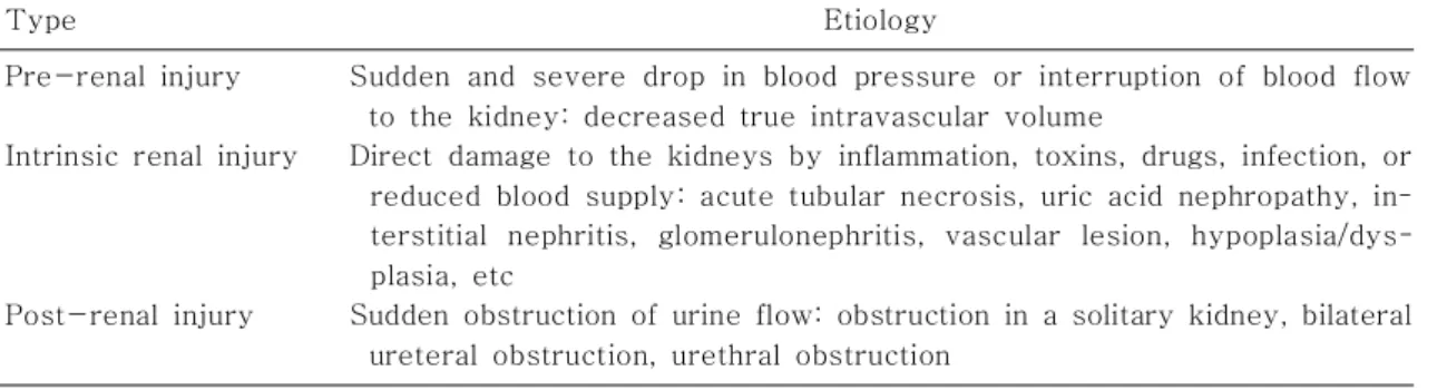
예후 판정은 환자의 생존률 신대체 요
(Table 1). ,
법의 필요성 만성 신질환으로의 진행 등을 예측 , 하는 역할이다 이러한 역할을 수행함에 있어 좋은 표지자 . 라 함은 비침습적인 방법으로 측정이 가능하고 빠른 결과 확인이 가능하면서 그 비용이 합리적인 표지라 라 할 수 있다 [1]. 본 종설에서는 문헌고찰을 통해 현재까지 보고된 급성 신손상의 표지자들을 알아 보 고 특히 소아에서의 임상적 적용 및 그 의의를 알아 보고자 하였다(Table 2).
Neutrophil gelatinase-associated lipocalin (NGAL)
은 활성화된 중성구에서 분비되는
NGAL 25 kDa
의 lipocalin 으로 다양한 세포에서 발현되며 감염 , 악성종양 신세뇨관 손상과 같은 여러 손상 형태에서 , 증가된다 신장에서는 주로 . Henle loop 나 원위 세뇨 관에서 발현하지만 사구체에서 여과 후 근위 세뇨관 에서도 재흡수되어 발현될 수 있다[6, 7]. NGAL 은 심장수술을 받은 소아 환자를 대상으로 연구가 시작 되었는데 심장수술 후 시간이 지나서 혈장 및 소변 , 2 내 NGAL 의 현저한 증가를 보인 환자들이 이후에 급성 신손상을 보였다 [8]. 특히 수술 후 시간째 소 , 2 변 내 NGAL 의 농도의 cutoff 값을 100 ng/mL 로 정한 경우 Area under the curve (AUC) 가 0.95, 민감도 (sensitivity) 가 0.82, 특이도 (specificity) 가 0.90 을 보였고 급성 신손상의 정도 기간 입원기 , ,
Table 1. Etiologies of Acute Kidney Injury
Type Etiology
Pre-renal injury
Intrinsic renal injury
Post-renal injury
Sudden and severe drop in blood pressure or interruption of blood flow to the kidney: decreased true intravascular volume
Direct damage to the kidneys by inflammation, toxins, drugs, infection, or reduced blood supply: acute tubular necrosis, uric acid nephropathy, in- terstitial nephritis, glomerulonephritis, vascular lesion, hypoplasia/dys- plasia, etc
Sudden obstruction of urine flow: obstruction in a solitary kidney, bilateral
ureteral obstruction, urethral obstruction
간 사망률 등과도 의미 있는 상관관계를 보였다 , [9].
유사한 연구로 조영제를 사용하는 심도자술 이후 2 시간이 지나서 혈장 및 소변 내 NGAL 의 증가는 조 영제에 의한 신손상의 예측인자로 사용할 수 있다고 보고되었다 [10]. 그 외 요로감염 , IgA 신병증 용혈 , 요독 증후군 루푸스 신염 만성 신장병과 같은 다른 , , 종류의 신장질환에서도 NGAL 의 증가가 의미 있는 것으로 알려져 있다 [11-13]. Ichino 등은 동물 실 험에서 상부 요로감염의 초기에 소변 내 NGAL 의 농도가 증가하며 이는 신반흔의 생성과 연관이 있다 고 보고하였고 [14], Mitsnefes 등은 혈청 NGAL 이 와 더불어 만성 신장병을 가진 cystatine C (CysC)
소아 환자에서 만성 신장병의 정도를 정량화할 수 있 는 유용한 표자자라고 보고하였다 [15]. 소아의 요로 감염에 대한 연구에서 소변 내 NGAL 및 NGAL/ 크 레아티닌 비는 급성 신손상이나 만성 신질환이 없는 단계에서 요로감염을 조기 발견하는데 도움을 줄 수 있다고 보고하였는데 소변 내 , NGAL 의 경우 cutoff 값을 20 ng/mL 로 하면 민감도는 97%, 특이도는
이었으며 크레아티닌
76% (AUC: 0.979) NGAL/
비의 경우 cutoff 값을 30 ng/mg 으로 하면 민감도 는 98%, 특이도는 76% (AUC: 0.992) 라고 하여 급성 신손상에 비해 요로감염의 cutoff 값은 낮은 경 향이 있음을 보고하였다 [13]. 신생아의 경우 재태연 령이나 출생체중에 따라 소변 내 NGAL 의 정상 농 도는 차이가 있는데 재태연령이 어릴수록 출생체중 ,
이 적을수록 소변 내 NGAL 의 농도가 높다고 알려 져 있다 [16]. 신생아실에서 흔히 신장 기능의 지표 로 사용되는 소변량이나 혈청 크레아티닌은 신장 기 능이 정상이라 하더라도 공급되는 수액의 양이나 , 계열의 항생제의 사용 유무와 같은 aminoglycoside
변수들에 의해 쉽게 달라질 수 있으나 , NGAL 은 이 러한 변수들과는 상관없이 비교적 일관된 정확성을 가진다고 보고하였다 [16]. 사실 신생아나 미숙아는 불완전한 신장 형성 다양한 신독성 약물의 사용 호 , , 흡 곤란 증후군과 같은 급성 신손상이 쉽게 발생할 수 있는 환경 등을 흔히 경험하게 되는데 소변 내 , 의 측정은 비록 신장기능이 정상이라 하더라 NGAL
도 패혈증과 같은 중증질환을 감별하는데도 도움을 줄 수 있다[17].
Kidney injury molecule-1 (KIM-1)
은 의 제 형
KIM-1 104 kDa 1 transmembrane 으로 정상 신장에서는 극히 낮은 정도 glycoprotein
로 발현하고 있지만 허혈성 혹은 독성 신손상 후에는 근위 세뇨관에서 발현이 현저히 증가된다 [1]. 실제 로 급성 신세뇨관 괴사를 가진 환자의 신장조직을 이 용한 연구에서 다른 급성 신부전의 원인보다 허혈성 원인의 경우 근위 세뇨관의 KIM-1 발현이 현저히 나타났다 [18]. 신세뇨관 손상이 발생되면 KIM-1 의 세포 외 부분이 단백분해 효소에 의해 분리되어 Table 2. List of New Biomarkers in Acute Kidney Injury
Name References
Neutrophil gelatinase-associated lipocalin (NGAL) Kidney injury molecule-1 (KIM-1)
Interleukin-18 (IL-18)
Liver-type fatty acid-binding protein (L-FABP) Interleukin-6 & 8 (IL-6 & 8)
Cystatine C (CysC)
N-acetyl- -D-glucosaminidase (NAG) ß Matrix metalloproteinase-9 (MMP-9) Cysteine rich protein 61
Na+/H+ exchanger isoform 3
[6-17]
[18-27]
[28-32]
[33-46]
[47,48]
[49]
[26, 50, 51]
[26, 52, 53]
[54]
[55]
소변 내로 분비되는데[18, 19], 이러한 변화는 다낭 성 신장 독성 신손상 심한 단백뇨 등에서도 나타난 , , 다 [20-22]. 다양한 형태의 급성 신세뇨관 괴사를 가진 중환자를 대상으로 한 연구에서 소변 내 KIM- 의 증가가 사망률 및 투석 치료의 필요성 증가와 연 1
관이 있으며 [23], 심장수술 후의 급성 신손상 과도 연관이 있음이 알려져 있다[24, 25]. 소아에서는 아 직 관련 연구가 부족한 실정이나 , Han 등이 심장 수 술을 받은 소아 40 명을 대상으로 한 연구에서 수술 시간 후 소변 이 이후의 급성 신손상을 예
12 KIM-1
측하는 인자로서 의미가 있다고 보고하였다 [26]. 또 한 폐쇄성 신병증을 가진 소아 환자의 연구에서는 소 변 내 KIM-1 과 NGAL 의 증가가 폐쇄의 악화와 연 관이 있음이 보고되었는데 [27], 수술 시행 개월이 3 지난 이후 소변 내 KIM-1 과 NGAL 의 농도는 현저 한 감소를 보였다 선천성 심장병을 가진 소아환자의 . 심도자술시 사용되는 조영제에 의한 급성 신손상의 연구에서는 조영제 투여 후 48 시간까지 혈청 크레아 티닌은 특별한 변화를 보이지 않은 반면 소변 내 , 은 지속적으로 증가되는 양상을 보였으며 특 KIM-1
히 조영제 투여 , 24 시간 후에는 투여 전에 비해 유의 하게 증가되는 양상을 보였다 (Cho 등 , unpublished data) (Fig. 1).
Interleukin-18 (IL-18)
은 의
IL-18 18 kDa pro-inflammatory cyto- 으로 초기에는 비활성형으로 생성되어 이후 kine
에 의해 활성형으로 변하며 급성 신손상 caspase-1
시 신 세뇨관 상피세포에 분포한다 [28]. 동물실험에 서 IL-18 의 중화 항체를 주입하여 허혈성 신손상을 예방할 수 있었다는 연구에 근거하여 IL-18 은 허혈 성 신손상의 발생에 중요한 역할을 가진 것으로 생각 된다 [29]. Parikh 등은 신장 이식 당일의 소변 내 의 농도로 이후의 혈청 크레아티닌의 경향을 IL-18
예측할 수 있어 delayed graft function 에 대한 의 미 있는 예측인자라고 보고하였다 [30]. 또한 중환 , 자실에 입원 중이거나 급성 호흡 곤란 증후군을 가진 환자의 급성 신손상을 예견하고 사망률을 예측하는 표지자로서 가치가 있다고 보고하였는데 [31], 소변 내 IL-18 의 값이 100 pg/mL 이상인 경우 다음 24 시간 동안 급성 신손상에 대한 odds 값이 6.5 였고 , 이는 사망자와 생존자 간에 유의한 차이를 보였으며, 특히 입원 첫날 측정값은 사망률에 대한 독립적인 예 측인자라고 하였다 실제로 소변 내 . IL-18 은 다른 신장질환 예를 들면 신전성 고질소혈증 요로감염 , , , 만성 신장병 신증후군보다는 급성 신부전에서 현저 , 한 증가를 보였다 [31]. 반면 Washburn 등은 급성 신손상의 정도가 심해질수록 소변 내 IL-18 의 농도 가 증가되는 것으로 알려져 있지만 실제로 급성 신손 상의 발생을 예측하는 인자로서의 가치는 낮다고 보 고하였다[32].
Liver-type fatty acid-binding protein (L-FABP)
는 의 단백으로 근위 세뇨관에 L-FABP 14 kDa
서 발현하며 소변 내 free fatty acid 의 대사와 연관 이 있다 . FABP 는 세포 내 carrier protein 으로 분 , 포하는 장기 간 장관 근육과 심장 지방세포 상피 ( , , , , , Fig. 1. Change of urinary kidney injury mole-
cule-1 after cardiac catheterization with contrast
(
*P<0.05, before infusion of contrast versus 24
hrs after infusion).
회장 뇌 척수 고환 에 따라 총 종류로 구분되며 , , , ) 9 주로 근위 세뇨관에 분포하는 와 원
[33], L-FABP
위 세뇨관에 분포하는 heart-type FABP (H- 로 대표된다
FABP) [34, 35]. H-FABP 의 경우 , 이나
creatinine kinase MB-isoform troponin-T 같은 기존의 급성 심근 경색의 표지자에 비해 현저한 상승을 보여 발병 후 시간 이내에 급성 관상동맥 질 6 환을 진단하는 새로운 표지자로 보고되었다 [36]. 막 성 신증 환자에서는 이 두 가지 FABP 가 모두 소변 내에 증가되며 이는 장기적인 신장의 예후와 연관이 있다고 보고되었다 [37]. 반면 , Wang 등은 in vitro 연구에서 L-FABP 가 허혈 재관류 상황에서 발생 - 된 oxidative stress 를 줄이는 효과가 있다고 발표 하였다 [38]. 허혈성 혹은 독성 신손상의 동물 실험 에서 L-FABP 는 신손상 발생 후 수 분 내지 수시간 내에 증가되며 그 증가 정도는 병리적인 손상 정도와 상관관계를 보이는 것으로 알려져 있으며 조영제에 의한 신손상의 초기 표지자로도 보고되었다[39, 40].
등은 심장 수술을 받은 명의 소아 환자를
Portilla 40
대상으로 한 연구에서 수술 후 시간과 4 12 시간째 소 변 내 L-FABP 의 증가는 이후 발생될 급성 신손상 과 의미 있는 상관관계가 있다고 보고하였다[41].
신장 이식 환자에 대한 연구에서 소변 내 L-FABP 는 허혈 시간 및 세뇨관 주위 모세혈관의 혈류량과 직접적인 연관이 있었으며 이후 입원 기간과도 의미 있는 상관관계를 보였다 [42]. 조영제에 의한 신병증 의 연구에서도 소변 내 L-FABP 가 일시적인 신세 뇨관 손상의 중요한 표지자라고 보고되었다[43].
는 급성 신손상뿐 아니라 만성 신장병의
L-FABP ,
진행기전과 연관이 있는 것으로 보고되었는데 세뇨 , 관 주위의 혈류 장애가 만성적으로 진행되는 경우 의 분비가 증가되고 특히 제 형 당뇨병
L-FABP , 2
성 신장병에서도 이러한 현상이 나타난다고 보고하 였다[44-46].
기타 급성 신손상의 표지자들
과 은 심장 수술을 받은 소아 연구에서 IL-6 IL-8
잠재적인 급성 신손상의 좋은 표지자이며 이는 기계 호흡의 장기적인 의존과도 연관이 있다고 보고되었 다 [47]. 신 이식 환자에서도 허혈 손상을 예측하는 표지자로 소변 내 actin, IL-6, IL-8 이 의미 있는 결과를 보였다[48].
혈청 CysC 또한 급성 신손상의 표지자로 연구되 었는데 이는 직접적인 신세뇨관 손상의 표지자라기 보다는 혈청 크레아티닌과 같은 사구체 여과율의 표 지자로 알려져 있다[49].
그 외에도 다양한 세뇨관 내 효소를 이용한 연구 들도 보고되고 있다 . Westhuyzen 등은 gamma glitamyl transpeptidase, alkaline phosphatase, N-acetyl- -D-glucosaminidase ß (NAG), 와 같 alpha- and pi-glutathione S-transferase 은 세뇨관 효소들이 급성 세뇨관 괴사에서 의미 있는 소변 내 증가를 보였다고 보고하였다 [50]. 특히 ,
는 근위 세뇨관에서 주로 발견되는
NAG lysosomal
으로 다양한 종류의 약제 독성 물질 조영
enzyme , ,
제 허혈성 환경 등에 의한 근위 신세뇨관 손상을 반 , 영하는 민감한 표지자로 알려져 있다[51]. Cardio- 를 시행 받은 소아환자를 대상 pulmonary bypass
으로 한 연구에서 혈청 크레아티닌의 증가가 나타나 기 이전에 소변 내 NAG 는 증가하는 것으로 보고되 었다[26].
는 동 Matrix metalloproteinase-9 (MMP-9) 물실험에서 허혈성 신손상 후 증가되며 NGAL 과 복 합체를 형성하여 심장 수술 후 나타나는 허혈성 신손 상의 의미 있는 표지자로 보고되었다 [52]. 하지만 요 로감염이 있는 환자의 소변에서도 증가되어 급성 신 손상과 감별하는데 한계를 보였다 [26]. 반면 소아 요로감염에 대한 연구에서는 소변 내 MMP-9/
크레아티닌 값이 소변 내 질산염 검사
NGAL/ (nitrite
백혈구 에스테라제 반응
test), (leukocyte esterase
및 신반흔 여부와는 유의한 상관관계를 reaction)
보이나 백혈구 증가증과 C- 반응 단백질과는 그렇지 않았다고 보고하였다[53].
은 양측 신장에 분 Cysteine rich protein 61 30 이상의 허혈을 시행한 동물실험에서 근위 세뇨관 주 변에 그 발현이 증가되는 것으로 알려졌으며 탈수와 같은 다른 조건에서는 이러한 현상이 나타나지 않아 급성 신손상의 표지자로서의 가능성이 보고되었다 [54].
는 신세뇨관에 Na+/H+ exchanger isoform 3
서 가장 풍부한 apical sodium transporter , 로 중 환자실에 입원중인 급성 신부전 환자에서 급성 세뇨 관 괴사가 원인일 경우 가장 현저한 소변 내 증가를 보여 급성 신손상을 진단하는 표지자로 가치가 있다 고 보고되었다[55].
결 론
최근 수 년간 새로운 급성 신손상의 표지자를 발 견하려는 많은 노력이 있었음에도 불구하고 여전히 혈청 크레아티닌만이 유일한 지표로 사용되고 있는 것이 현실이다 서론에서 언급한 것처럼 급성 신손상 . 의 표지자는 조기발견과 감별진단 그리고 정확한 예 , 후를 판정하는 지표로 사용될 수 있어야 하지만 급 , 성 신손상의 발생이 단순히 신장만의 문제로 국한 되 는 것이 아니라 다양한 원인 질환 및 다른 전신장기 와 연관하여 나타나므로 이러한 임상적 요구를 충족 하는 새로운 표지자를 찾기란 여간 어려운 일이 아닐 것이다 또한 연령별로 다양한 특성을 보이는 소아의 . 경우에는 검사의 용이성 비용 효율적인 측면까지 , - 고려되어야 하므로 앞으로 많은 연구와 투자가 필요 할 것으로 생각된다.
References
1) Al-Ismaili Z, Palijan A, Zappitelli M. Bio- markers of acute kidney injury in children:
discovery, evaluation, and clinical application.
Pediatr Nephrol 2011;26:29-40.
2) Akcan-Arikan A, Zappitelli M, Loftis LL, Washburn KK, Jefferson LS, Goldstein SL.
Modified RIFLE criteria in critically ill chil- dren with acute kidney injury. Kidney Int 2007;71:1028-35.
3) Mehta RL, Kellum JA, Shah SV, Molitoris BA, Ronco C, Warnock DG, et al. Acute Kidney Injury Network: report of an initiative to improve outcomes in acute kidney injury.
Crit Care 2007;11:R31.
4) Coca SG, Parikh CR. Urinary biomarkers for acute kidney injury: perspectives on trans- lation. Clin J Am Soc Nephrol 2008;3:481- 90.
5) Biomarkers Definitions Working Group. Bio- markers and surrogate endpoints: preferred definitions and conceptual framework. Clin Pharmacol Ther 2001;69:89-95.
6) Devarajan P. Neutrophil gelatinase-associ- ated lipocalin--an emerging troponin for kidney injury. Nephrol Dial Transplant 2008;
23:3737-43.
7) Mori K, Lee HT, Rapoport D, Drexler IR, Foster K, Yang J, et al. Endocytic delivery of lipocalin-siderophore-iron complex re- scues the kidney from ischemia-reperfusion injury. J Clin Invest 2005;115:610-21.
8) Mishra J, Dent C, Tarabishi R, Mitsnefes MM, Ma Q, Kelly C, et al. Neutrophil gelati- nase-associated lipocalin (NGAL) as a bio- marker for acute renal injury after cardiac surgery. Lancet 2005;365:1231-38.
9) Bennett M, Dent CL, Ma Q, Dastrala S, Grenier F, Workman R, et al. Urine NGAL predicts severity of acute kidney injury after cardiac surgery: a prospective study. Clin J Am Soc Nephrol 2008;3:665-73.
10) Hirsch R, Dent C, Pfriem H, Allen J, Beek- man RH, 3rd, Ma Q, et al. NGAL is an early predictive biomarker of contrast-induced nephropathy in children. Pediatr Nephrol 2007;22:2089-95.
11) Goldstein SL, Devarajan P. Progression from
acute kidney injury to chronic kidney dis- ease: a pediatric perspective. Adv Chronic Kidney Dis 2008;15:278-83.
12) Trachtman H, Christen E, Cnaan A, Patrick J, Mai V, Mishra J, et al. Urinary neutrophil gelatinase-associated lipocalcin in D+HUS:
a novel marker of renal injury. Pediatr Nephrol 2006;21:989-94.
13) Yilmaz A, Sevketoglu E, Gedikbasi A, Kar- yagar S, Kiyak A, Mulazimoglu M, et al. Early prediction of urinary tract infection with urinary neutrophil gelatinase associated li- pocalin. Pediatr Nephrol 2009;24:2387-92.
14) Ichino M, Kuroyanagi Y, Kusaka M, Mori T, Ishikawa K, Shiroki R, et al. Increased uri- nary neutrophil gelatinase associated lipo- calin levels in a rat model of upper urinary tract infection. J Urol 2009;181:2326-31.
15) Mitsnefes MM, Kathman TS, Mishra J, Kartal J, Khoury PR, Nickolas TL, et al. Serum neutrophil gelatinase-associated lipocalin as a marker of renal function in children with chronic kidney disease. Pediatr Nephrol 2007;22:101-8.
16) Lavery AP, Meinzen-Derr JK, Anderson E, Ma Q, Bennett MR, Devarajan P, et al. Uri- nary NGAL in premature infants. Pediatr Res 2008;64:423-8.
17) Parravicini E. The clinical utility of urinary neutrophil gelatinase-associated lipocalin in the neonatal ICU. Curr Opin Pediatr 2010;22:146-50.
18) Han WK, Bailly V, Abichandani R, Thadhani R, Bonventre JV. Kidney Injury Molecule-1 (KIM-1): a novel biomarker for human renal proximal tubule injury. Kidney Int 2002;62:
237-44.
19) Ichimura T, Bonventre JV, Bailly V, Wei H, Hession CA, Cate RL, et al. Kidney injury molecule-1 (KIM-1), a putative epithelial cell adhesion molecule containing a novel immunoglobulin domain, is up-regulated in renal cells after injury. J Biol Chem 1998;
273:4135-42.
20) Ichimura T, Hung CC, Yang SA, Stevens JL,
Bonventre JV. Kidney injury molecule-1: a tissue and urinary biomarker for nephroto- xicant-induced renal injury. Am J Physiol Renal Physiol 2004;286:F552-63.
21) Kuehn EW, Park KM, Somlo S, Bonventre JV. Kidney injury molecule-1 expression in murine polycystic kidney disease. Am J Physiol Renal Physiol 2002;283:F1326-36.
22) van Timmeren MM, Bakker SJ, Vaidya VS, Bailly V, Schuurs TA, Damman J, et al. Tu- bular kidney injury molecule-1 in protein- overload nephropathy. Am J Physiol Renal Physiol 2006;291:F456-64.
23) Liangos O, Perianayagam MC, Vaidya VS, Han WK, Wald R, Tighiouart H, et al.
Urinary N-acetyl-beta-(D)-glucosamini- dase activity and kidney injury molecule-1 level are associated with adverse outcomes in acute renal failure. J Am Soc Nephrol 2007;18:904-12.
24) Liangos O, Tighiouart H, Perianayagam MC, Kolyada A, Han WK, Wald R, et al. Compa- rative analysis of urinary biomarkers for early detection of acute kidney injury fol- lowing cardiopulmonary bypass. Biomarkers 2009;14:423-31.
25) Waikar SS, Bonventre JV. Biomarkers for the diagnosis of acute kidney injury. Nephron Clin Pract 2008;109:c192-7.
26) Han WK, Waikar SS, Johnson A, Betensky RA, Dent CL, Devarajan P, et al. Urinary biomarkers in the early diagnosis of acute kidney injury. Kidney Int 2008;73:863-9.
27) Wasilewska A, Taranta-Janusz K, Debek W, Zoch-Zwierz W, Kuroczycka-Saniutycz E. KIM-1 and NGAL: new markers of ob- structive nephropathy. Pediatr Nephrol 2011;
26:579-86.
28) Leslie JA, Meldrum KK. The role of inter- leukin-18 in renal injury. J Surg Res 2008;
145:170-5.
29) Melnikov VY, Faubel S, Siegmund B, Lucia MS, Ljubanovic D, Edelstein CL. Neutrophil- independent mechanisms of caspase-1- and IL-18-mediated ischemic acute tubular
necrosis in mice. J Clin Invest 2002;110:
1083-91.
30) Parikh CR, Jani A, Mishra J, Ma Q, Kelly C, Barasch J, et al. Urine NGAL and IL-18 are predictive biomarkers for delayed graft function following kidney transplantation.
Am J Transplant 2006;6:1639-45.
31) Parikh CR, Abraham E, Ancukiewicz M, Edelstein CL. Urine IL-18 is an early diag- nostic marker for acute kidney injury and predicts mortality in the intensive care unit.
J Am Soc Nephrol 2005;16:3046-52.
32) Washburn KK, Zappitelli M, Arikan AA, Loftis L, Yalavarthy R, Parikh CR, et al. Uri- nary interleukin-18 is an acute kidney injury biomarker in critically ill children. Nephrol Dial Transplant 2008;23:566-72.
33) Chmurzynska A. The multigene family of fatty acid-binding proteins (FABPs): func- tion, structure and polymorphism. J Appl Genet 2006;47:39-48.
34) Kimura H, Fujii H, Suzuki S, Ono T, Ara- kawa M, Gejyo F. Lipid-binding proteins in rat and human kidney. Kidney Int Suppl 1999;71:S159-62.
35) Maatman RG, Van Kuppevelt TH, Veerkamp JH. Two types of fatty acid-binding protein in human kidney. Isolation, characterization and localization. Biochem J 1991;273:759- 66.
36) Nakata T, Hashimoto A, Hase M, Tsuchi- hashi K, Shimamoto K. Human heart-type fatty acid-binding protein as an early diag- nostic and prognostic marker in acute coro- nary syndrome. Cardiology 2003;99:96- 104.
37) Hofstra JM, Deegens JK, Steenbergen EJ, Wetzels JF. Urinary excretion of fatty acid- binding proteins in idiopathic membranous nephropathy. Nephrol Dial Transplant 2008;
23:3160-5.
38) Wang G, Gong Y, Anderson J, Sun D, Minuk G, Roberts MS, et al. Antioxidative function of L-FABP in L-FABP stably transfected Chang liver cells. Hepatology 2005;42:871-
9.
39) Nakamura T, Sugaya T, Node K, Ueda Y, Koide H. Urinary excretion of liver-type fatty acid-binding protein in contrast me- dium-induced nephropathy. Am J Kidney Dis 2006;47:439-44.
40) Negishi K, Noiri E, Doi K, Maeda-Mamiya R, Sugaya T, Portilla D, et al. Monitoring of urinary L-type fatty acid-binding protein predicts histological severity of acute kidney injury. Am J Pathol 2009;174:1154-9.
41) Portilla D, Dent C, Sugaya T, Nagothu KK, Kundi I, Moore P, et al. Liver fatty acid- binding protein as a biomarker of acute kidney injury after cardiac surgery. Kidney Int 2008;73:465-72.
42) Yamamoto T, Noiri E, Ono Y, Doi K, Negishi K, Kamijo A, et al. Renal L-type fatty acid --binding protein in acute ischemic injury.
J Am Soc Nephrol 2007;18:2894-902.
43) Kato K, Sato N, Yamamoto T, Iwasaki YK, Tanaka K, Mizuno K. Valuable markers for contrast-induced nephropathy in patients undergoing cardiac catheterization. Circ J 2008;72:1499-505.
44) Fine LG, Bandyopadhay D, Norman JT. Is there a common mechanism for the progres- sion of different types of renal diseases other than proteinuria? Towards the unifying theme of chronic hypoxia. Kidney Int Suppl 2000;
75:S22-6.
45) Kang DH, Kanellis J, Hugo C, Truong L, Anderson S, Kerjaschki D, et al. Role of the microvascular endothelium in progressive renal disease. J Am Soc Nephrol 2002;13:
806-16.
46) Noiri E, Tsukahara H. Parameters for mea- surement of oxidative stress in diabetes mellitus: applicability of enzyme-linked im- munosorbent assay for clinical evaluation. J Investig Med 2005;53:167-75.
47) Liu KD, Altmann C, Smits G, Krawczeski CD, Edelstein CL, Devarajan P, et al. Serum in- terleukin-6 and interleukin-8 are early bio- markers of acute kidney injury and predict
prolonged mechanical ventilation in children undergoing cardiac surgery: a case-control study. Crit Care 2009;13:R104.
48) Kwon O, Molitoris BA, Pescovitz M, Kelly KJ. Urinary actin, interleukin-6, and inter- leukin-8 may predict sustained ARF after ischemic injury in renal allografts. Am J Kidney Dis 2003;41:1074-87.
49) Schwartz GJ, Work DF. Measurement and estimation of GFR in children and adoles- cents. Clin J Am Soc Nephrol 2009;4:1832- 43.
50) Westhuyzen J, Endre ZH, Reece G, Reith DM, Saltissi D, Morgan TJ. Measurement of tubular enzymuria facilitates early detection of acute renal impairment in the intensive care unit. Nephrol Dial Transplant 2003;18:
543-51.
51) Gibey R, Dupond JL, Alber D, Leconte des Floris R, Henry JC. Predictive value of urinary N-acetyl-beta-D-glucosaminidase (NAG), alanine-aminopeptidase (AAP) and beta-2-microglobulin (beta 2M) in evalua-
ting nephrotoxicity of gentamicin. Clin Chim Acta 1981;116:25-34.
52) Basile DP, Fredrich K, Weihrauch D, Hattan N, Chilian WM. Angiostatin and matrix me- talloprotease expression following ischemic acute renal failure. Am J Physiol Renal Phy- siol 2004;286:F893-902.
53) Hatipoglu S, Sevketoglu E, Gedikbasi A, Yilmaz A, Kiyak A, Mulazimoglu M, et al.
Urinary MMP-9/NGAL complex in children with acute cystitis. Pediatr Nephrol 2011;
26:1263-8.
54) Muramatsu Y, Tsujie M, Kohda Y, Pham B, Perantoni AO, Zhao H, et al. Early detection of cysteine rich protein 61 (CYR61, CCN1) in urine following renal ischemic reperfusion injury. Kidney Int 2002;62:1601-10.
55) du Cheyron D, Daubin C, Poggioli J, Rama- kers M, Houillier P, Charbonneau P, et al.
Urinary measurement of Na+/H+ exchan- ger isoform 3 (NHE3) protein as new marker of tubule injury in critically ill patients with ARF. Am J Kidney Dis 2003;42:497-506.