Introduction
Isolated posterior cruciate ligament (PCL) injuries are uncom
mon and often go undiagnosed in acutely injured knees1). The PCL, being a strong ligament, presents more commonly with avulsion fractures of its tibial attachment2) unlike the anterior cruciate ligament whose tears commonly present as isolated le
sions. Avulsion injuries differ from other PCL injuries in that they are easily diagnosed on standard radiographs where a bony fragment may be visible. Traditionally, surgery of PCL injuries was deferred due to an apprehension that the approach to the posterior part of the knee is difficult3). However, surgical fixation of avulsion injuries is recommended to avoid morbidity associ
ated with nonunion of the fracture4).
Repair of avulsion injuries can be done with an open or ar
throscopic technique. Biomechanical studies have also dem
onstrated comparable results of screw fixation using open or arthroscopic means5). However, arthroscopic repair is technically more challenging, requires specialized equipment and has a long learning curve. Thus, it unsuitable to perform in primary cen
ters. Open reduction and internal fixation using screws has been considered a favorable method to manage PCL avulsion injuries producing satisfactory results610). In addition, a simplified pos
teromedial approach described by Burks and Schaffer11), which is
Open Reduction and Internal Fixation of Isolated Posterior Cruciate Ligament Avulsion Fractures:
Clinical and Functional Outcome
Sachin Joshi, MD
1, Chirag Bhatia, MD
1,2, Ashwin Gondane, MD
1, Avinash Rai, MD
1, Sumer Singh, MD
1,3, and Shobhit Gupta, MD
11Department of Orthopaedics, Government Medical College, Kota, Rajasthan; 2Department of Orthopaedics, Cygnus Orthocare Hospital, New Delhi; 3Department of Orthopaedics, American International Institute of Medical Sciences, Udaiour, Rajasthan, India
Purpose: The posterior cruciate ligament (PCL) presents commonly with avulsion fractures of its tibial attachment. An avulsion fracture of the PCL, if not surgically fixed, may lead to secondary changes in the knee joint. Various fixation techniques have been explored with use of lag screws, steel wires, absorbable screws, suture anchors and straddle nails. The purpose of this study was to evaluate the clinical and functional outcome of open reduction and internal fixation of tibial avulsion injuries of the PCL using cannulated cancellous screws.
Materials and Methods: We performed open reduction and internal fixation using cannulated cancellous screws in 14 patients (mean age, 33.9 years) with isolated PCL avulsion injuries. Patients with a duration of injury more than 12 weeks were excluded. The minimum followup period was 12 months. Results were assessed radiologically and clinically. Final functional outcome was assessed using the Lysholm knee scoring system.
Results: The average followup period was 13.5 months. At the final followup, all 14 patients achieved fracture union. The average flexion was 121.7°±9.2° with full extension achieved in all patients. Mild instability (1+) was noted in 4 patients. The Lysholm functional score was excellent in 11 patients, good in 2 patients and fair in 1 patient with an average score of 97±7.6.
Conclusions: Stable early fixation for PCL avulsion injuries with early controlled mobilization provided excellent to good results. However, fixation should not be discouraged for latepresenting patients because good to fair results can be achieved in such cases.
Keywords: Knee, Posterior cruciate ligament, Fraucture, Avulsion, Fixation pISSN 2234-0726 · eISSN 2234-2451
Knee Surgery & Related Research
Received March 12, 2017; Revised June 8, 2017; Accepted June 12, 2017 Correspondence to: Chirag Bhatia, MD
Department of Orthopaedics, Cygnus Orthocare Hospital, C5/29, Safdarjung Development Area, Hauz Khas, Opp. IIT Main Gate, New Delhi 110016, India
Tel: +919968849269, Fax: +919968849269 Email: [email protected]
210
This is an Open Access article distributed under the terms of the Creative Commons Attribution NonCommercial License (http://creativecommons.org/licenses/bync/4.0/) which permits unrestricted noncommercial use, distribution, and reproduction in any medium, provided the original work is properly cited.
Copyright © 2017 KOREAN KNEE SOCIETY www.jksrr.org
useful for avoiding difficulties associated with previous posterior approaches to the knee, has become the standard approach to the PCL.
There is currently a wide variety of materials available for inter
nal fixation, including lag screws, steel wires, absorbable screws, suture anchors and straddle nails12,13). Fixation with screws has shown favorable results; however, no one technique has been considered a gold standard for avulsion fractures of the PCL. In this study, we present our experience with fixation using cannu
lated cancellous screws, which is a good implant to achieve com
pression needed to fix avulsion fractures and is easily available.
The approach we used was also simple and helpful in avoiding the neurovascular bundles unlike other posterior approaches.
The purpose of our study was to evaluate the clinical and func
tional outcome after open reduction and internal fixation of tibial avulsion injuries of the PCL using cannulated cancellous screws.
We hypothesized that the technique would provide improved clinical and functional outcome with use of an easily available implant and a simple approach.
Materials and Methods
Twentyone patients with PCL avulsion fractures were enrolled for the study from December, 2012 to November, 2015. Patients aged between 18 and 60 years with a duration of injury less than 12 weeks were included in the study. Patients with any other as
sociated ligament injury of the same joint, any other bony injury of the same limb or any medical comorbidity precluding the sur
gery were excluded from the study. Patients in whom the avulsed fragment was too small (<20 mm2) to be fixed using a screw were also excluded. After excluding 7 patients based on the above criteria, 14 patients having isolated PCL avulsion fracture were ultimately included in the study.
All patients underwent thorough clinical examination. All the cases were clinically assessed by the senior author using the drawer test to confirm PCL injury. Anteroposterior and lateral ra
diographs including the posterior stress view (Fig. 1) of the knee were taken to confirm the injury. A computed tomography scan
Fig. 1. Lateral radiograph of the knee (nonstress view) showing the avulsed fragment from the tibia (case no. 3).
Fig. 2. Threedimensional computed tomography scan showing the avulsed fragment of the tibia (case no. 2).
Fig. 3. Magnetic resonance imaging scan showing a lax posterior cruci
ate ligament avulsed from its tibial attachment (case no. 2).
(Fig. 2) was done for patients in whom the injury could not be defined well on radiographs. Magnetic resonance imaging (MRI) (Fig. 3) was done for every patient in order to confirm associated ligament injuries of the knee.
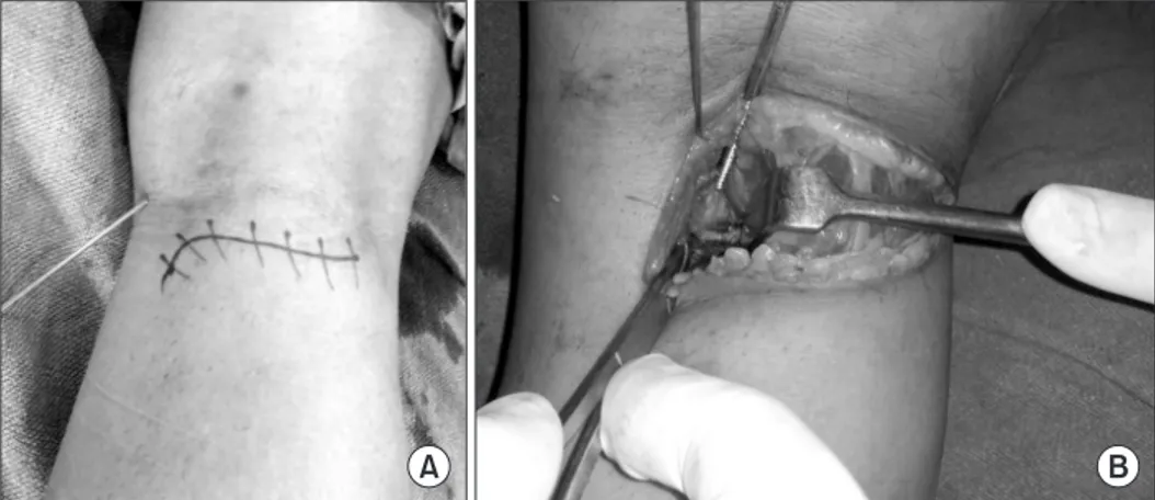
After informed consent was obtained, patients were operated using the modified posterior approach described by Burks and Schaffer11). The approach (Fig. 4) uses an inverted ‘L’ incision starting on the medial border of the gastrocnemius and curv
ing along the flexor crease of the joint towards the lateral side.
The fascia is incised in the line of skin incision. The capsule is exposed using the interval between the semimembranosus and gastrocnemius, which is then incised longitudinally to expose the joint at the site of the avulsed fragment. The avulsed fragment
was debrided if needed and reduced using a clamp. It is of note that the reduction was achieved by slight flexion of the joint. The avulsed fragment was fixed using one or two 4 mm cannulated cancellous screws according to the size of the fragment (Fig. 5).
A washer was used when compression was not achieved with the screw alone.
Postoperatively, the limb was kept immobilized using a long knee brace for 6 weeks. Quadriceps strengthening was started from the 2nd postoperative day. Passive knee bending was started after suture removal (10–12 days after surgery) and active knee mobilization was started 3 weeks after surgery. Partial weight bearing was allowed after 6 weeks and full weight bearing after 12 weeks when the brace was also discarded. Return to heavy activities, such as running and sports, were allowed only after 6–9 months.
Patients were regularly followed up every week for a month, then every month for the first 3 months and every 3 months thereafter for a minimum of 12 months. At every followup, pa
tients were assessed clinically and radiographically. Outcomes were assessed in terms of stability and range of motion (ROM).
Stability was assessed clinically by the drawer test and radiologi
cally by lateral stress Xray (Fig. 6). Final functional outcome was assessed using the Lysholm knee scoring system.
Statistical analysis was done using the Wilcoxon nonparamet
ric paired test to determine the significance of postoperative improvement with respect to the Lysholm score as well as joint stability.
Results
Of the 14 patients, 12 were male and 2 were female. The mean age of the patients was 33.9 years, ranging from 22 to 54 years (Table 1). The most common mode of injury was road traffic accident (n=9) with a majority involving motorcycle accident fol
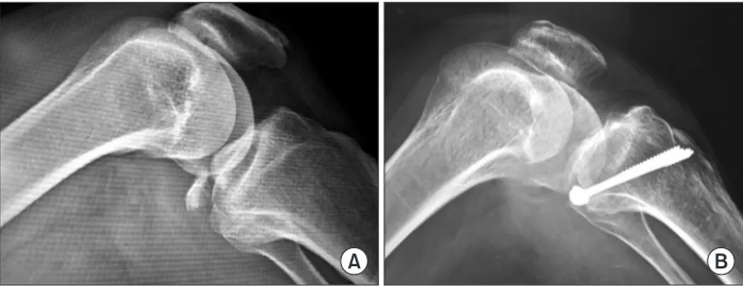
Fig. 5. Lateral radiographs of the knee showing cannulated cancellous screws in situ after fixation of the tibial avulsion of the posterior cruciate ligament in case no. 4 (A) and case no.10 (B). Note the different orienta
tion of screws, which was decided according to the orientation of the fracture line.
A B
Fig. 4. Intraoperative photograph showing the skin incision and fracture site with the screw insertion technique.
lowed by either sportsrelated injury or fall. Eleven patients pre
sented to the clinic within 3 weeks of injury while three patients presented between 3 to 12 weeks after injury. The average period of followup was 13.5 months.
At the final followup, all 14 patients had achieved fracture union. At 6 weeks after surgery, the ROM was more than 90° in 9 patients. Of the remaining 5 patients, 3 patients who had pre
sented later than 3 weeks after injury regained more than 90° of flexion after more than 6 postoperative weeks. At the final follow
up, the average flexion was 121.7°±9.18° with full extension pos
sible in all the patients. The functional outcome assessed by the Lysholm scoring system was excellent in 11 patients, good in 2 patients and fair in one patient. The average Lysholm score was
97±7.6.
Two patients complained of residual pain in the joint while one complained of residual swelling in the joint. Two patients had superficial wound infection, which was treated with intravenous antibiotics and regular dressings. No other complications were observed.
Instability tested clinically using the drawer test and radiologi
cally by stress Xray was found to be mild (1+) in 4 of the patients while the rest of the patients had no residual instability.
The statistical analysis showed highly significant improvement (p<0.001) in functional outcome assessed by the Lysholm score and joint stability (p<0.001) (Table 2).
A B Fig. 6. Preoperative (A) and postoperative
(B) posterior tibial sagging (case no. 3).
Table 1. Details of the 14 Cases Case Age
(yr) Mechanism
of injury Duration of
injury (day) Followup (mo)
Lyscholm score
Clinincal
outcome (°) Postop
instability Complication
Preop Postop
(functional outcome)
1 29 Motorcycle 2 15 0 100 (excellent) 130 Nil
2 34 Fall 10 12 6 99 (excellent) 110 Nil Superficial infection
3 26 Motorcycle 4 12 0 99 (excellent) 113 Nil
4 48 Motorcycle 3 13 0 84 (good) 110 Nil Residual pain
5 27 Road traffic 1 16 0 95 (excellent) 120 Mild (1+)
6 39 Motorcycle 5 13 0 100 (excellent) 130 Nil
7 24 Sports related 8 12 2 95 (excellent) 128 Mild (1+) Superficial infection
8 41 Fall 46 15 20 89 (good) 121 Nil Residual swelling
9 43 Motorcycle 7 12 4 95 (excellent) 127 Mild (1+)
10 22 Sports related 28 17 15 99 (excellent) 110 Nil
11 31 Motorcycle 1 14 0 100 (excellent) 135 Nil
12 29 Road traffic 2 13 0 95 (excellent) 130 Mild (1+)
13 54 Fall 38 12 13 74 (fair) 110 Nil Residual pain
14 28 Motorcycle 5 14 7 100 (excellent) 130 Nil
Mean 33.9 4.8 94.6 121.7
Preop: preoperative, Postop: postoperative.
Discussion
PCL injuries account for approximately 20% of total ligament injuries of the knee14). The incidence is especially high in cases of highenergy trauma (motorcycle and car accidents), and athletic population involved in contact sports is especially vulnerable to PCL injuries4,15). The most common mechanism underlying PCL avulsion fractures of the tibia in road traffic accidents is dash
board collision in which a direct force is applied to the proximal part of the tibia in an anteriortoposterior direction, with the knee in flexion16,17). In our series, most of the injuries resulted from motorcycle accidents because the majority of people com
mute by twowheelers in India.
Though the necessity of surgical treatment of isolated PCL inju
ries is still controversial, fixation methods for avulsion fractures of the PCL at the tibial insertion have been suggested in different series1823). If left untreated, the injury leads to secondary joint changes resulting in osteoarthritis24). Open fixation through the posteromedial approach seems to be a relatively easy procedure that can be performed at any center by any orthopedic surgeon.
The posteromedial approach used in our patients was devised by Burks and Schaffer11), which is a simple approach exposing the desired surgical area without having to disturb the neurovascular bundles as with other approaches described by Abbott, Trickey, Ogata, and McCormick3,20,25,26).
In this study, we performed open reduction and internal fixa
tion of isolated PCL avulsion injuries using cannulated cancellous screws, assuming to attain good functional and clinical outcome.
The most important findings of our study include that excellent to good functional outcome was obtained in the majority of pa
tients (92.9%) and fairly good results were achieved in terms of ROM as well as stability.
In our series, we treated 14 patients with isolated PCL avulsion injuries by internal fixation using cannulated cancellous screws and the results were excellent in 78.6%, good in 14.3% and fair in 7.1% of the patients with an average postoperative Lysholm score of 97±7.6. These results were superior to those reported in previ
ous studies. In a study by Attia and Zanfaly27) among 12 patients treated using navicular screws, excellent results were obtained in 33.3%, good results in 58.3% and fair results in 8.3%. Similarly,
Piedade and Mischan28) showed excellent results in 53% and good in 47% of their cases where screws or Polyester no. 5 were used to fix the fracture. Khatri et al.29) used the same approach and technique, which resulted in a postoperative Lysholm score of 90.85±5.58. In the present study, all patients attained fair to good ROM with an average flexion of 121.7°±9.2° and full exten
sion. Khatri et al.29) achieved normal to nearly normal ROM in 96% of the patients where as 64.3% of the patients achieved re
sults similar to our study. Postoperative knee flexion was inferior in patients who had presented to the clinic relatively late and who did not adhere to the prescribed physiotherapy. Hence early fixa
tion and strict adherence to physiotherapy are recommended.
Four of the patients who had mild (1+) residual instability had an intrasubstance PCL injury determined on MRI. However, this instability did not affect the functional outcome. Bali et al.10) reported that intrasubstance occult injuries were present in 15 out of 42 patients included in their study and 6 of them required a PCL reconstruction at a later stage. By contrast, none of the patients in our study had gross instability that necessitated PCL reconstruction at a later stage.
Although the technique we used in this study resulted in sig
nificant improvement in functional and clinical outcome, it is difficult to conclude that it should be the go to technique for PCL avulsion fractures due to the small sample size. Further studies with a larger sample size or casecontrol studies comparing dif
ferent techniques are necessary to confirm our results. Neverthe
less, the significance of this study is that we explored a novel and simple technique and added to the available literature on the management of PCL avulsion fractures.
Conclusions
Avulsion injuries of the PCL, though rare, should not be ig
nored. These injuries can be easily approached using a modified posterior approach and cancellous screw fixation with early con
trolled knee mobilization provides excellent to good functional results. Early diagnosis and fixation is preferred; however, fixa
tion should not be discouraged in patients presenting late since comparably good to fair results can be obtained in such cases as well.
Table 2. Statistical Analysis
Parameter Preoperative Postoperative pvalue Inference
Lysholm score 3±6.7 97±7.6 <0.001 Significant
Instability 2.5±0.5 0.3±0.5 <0.001 Significant
Values are presented as mean±standard deviation.
Conflict of Interest
No potential conflict of interest relevant to this article was re
ported.
References
1. Allen CR, Kaplan LD, Fluhme DJ, Harner CD. Posterior cru
ciate ligament injuries. Curr Opin Rheumatol. 2002;14:1429.
2. Sonin AH, Fitzgerald SW, Hoff FL, Friedman H, Bresler ME. MR imaging of the posterior cruciate ligament: normal, abnormal, and associated injury patterns. Radiographics.
1995;15:55161.
3. Abbott LC, Carpenter WF. Surgical approaches to the knee joint. J Bone Joint Surgery. 1945;27:277310.
4. Griffith JF, Antonio GE, Tong CW, Ming CK. Cruciate liga
ment avulsion fractures. Arthroscopy. 2004;20:80312.
5. Sasaki SU, da Mota e Albuquerque RF, Amatuzzi MM, Pereira CA. Open screw fixation versus arthroscopic suture fixation of tibial posterior cruciate ligament avulsion inju
ries: a mechanical comparison. Arthroscopy. 2007;23:1226
30.
6. Chen CH, Chen WJ, Shih CH. Fixation of small tibial avul
sion fracture of the posterior cruciate ligament using the double bundles pullthrough suture method. J Trauma.
1999;46:10368.
7. Chiu FY, Wu JJ, Hsu HC, Lin L, Lo WH. Management of avul
sion injury of the PCL with reattachment. Injury. 1994;25:293
5.
8. Jazayeri SM, Esmaili Jah AA, Karami M. A safe postero
medial approach to posterior cruciate ligament avulsion fracture. Knee Surg Sports Traumatol Arthrosc. 2009;17:2447.
9. Nicandri GT, Klineberg EO, Wahl CJ, Mills WJ. Treat
ment of posterior cruciate ligament tibial avulsion fractures through a modified open posterior approach: operative technique and 12 to 48month outcomes. J Orthop Trauma.
2008;22:31724.
10. Bali K, Prabhakar S, Saini U, Dhillon MS. Open reduction and internal fixation of isolated PCL fossa avulsion fractures.
Knee Surg Sports Traumatol Arthrosc. 2012;20:31521.
11. Burks RT, Schaffer JJ. A simplified approach to the tibial at
tachment of the posterior cruciate ligament. Clin Orthop Relat Res. 1990;(254):2169.
12. Sun HQ, Ren YJ, Zhang YQ. Treatment of posterior cruciate ligament avulsion fracture with pressurized staples. Chi J Orthop Trauma. 2011;13:118990.
13. Fu YP, Hang CM, Fam HQ. Treatment of posterior cruciate ligament avulsion fracture using anchor system combined with cannulated screw. J Pract Orthop. 2011;17:734.
14. Deehan DJ, Pinczewski LA. Arthroscopic reattachment of an avulsion fracture of the tibial insertion of the posterior cruciate ligament. Arthroscopy. 2001;17:4225.
15. Wind WM Jr, Bergfeld JA, Parker RD. Evaluation and treat
ment of posterior cruciate ligament injuries: revisited. Am J Sports Med. 2004;32:176575.
16. Janousek AT, Jones DG, Clatworthy M, Higgins LD, Fu FH.
Posterior cruciate ligament injuries of the knee joint. Sports Med. 1999;28:42941.
17. Schulz MS, Russe K, Weiler A, Eichhorn HJ, Strobel MJ.
Epidemiology of posterior cruciate ligament injuries. Arch Orthop Trauma Surg. 2003;123:18691.
18. Torisu T. Isolated avulsion fracture of the tibial attachment of the posterior cruciate ligament. J Bone Joint Surg Am.
1977;59:6872.
19. Seitz H, Schlenz I, Pajenda G, Vecsei V. Tibial avulsion fracture of the posterior cruciate ligament: Kwire or screw fixation? A retrospective study of 26 patients. Arch Orthop Trauma Surg. 1997;116:2758.
20. Trickey EL. Rupture of the posterior cruciate ligament of the knee. J Bone Joint Surg Br. 1968;50:33441.
21. Lee HG. Avulsion fracture of the tibial attachments of the cruciate ligaments: treatment by operative reduction. J Bone Joint Surg. 1937;19:4608.
22. Kim SJ, Shin SJ, Choi NH, Cho SK. Arthroscopically assisted treatment of avulsion fractures of the posterior cruciate liga
ment from the tibia. J Bone Joint Surg Am. 2001;83:698708.
23. Kim SJ, Shin SJ, Cho SK, Kim HK. Arthroscopic suture fixa
tion for bony avulsion of the posterior cruciate ligament.
Arthroscopy. 2001;17:77680.
24. Strobel MJ, Weiler A, Schulz MS, Russe K, Eichhorn HJ.
Arthroscopic evaluation of articular cartilage lesions in posteriorcruciateligamentdeficient knees. Arthroscopy.
2003;19:2628.
25. Ogata K. Posterior cruciate reconstruction using iliotibial band: preliminary report of a new procedure. Arch Orthop Trauma Surg. 1983;101:10710.
26. McCormick WC, Bagg RJ, Kennedy CW Jr, Leukens CA.
Reconstruction of the posterior cruciate ligament: prelimi
nary report of a new procedure. Clin Orthop Relat Res.
1976;(118):301.
27. Attia ME, Zanfaly AI. Fixation of tibial bony avulsion of the posterior cruciate ligament using the posteromedial ap
proach. Egypt Orthop J 2014;49:815.
28. Piedade SR, Mischan MM. Surgical treatment of avulsion fractures of the knee PCL tibial insertion: experience with 21 cases. Acta Ortop Bras. 2007;15:2725.
29. Khatri K, Sharma V, Lakhotia D, Bhalla R, Farooque K.
Posterior cruciate ligament tibial avulsion treated with open reduction and internal fixation through the burks and schaf
fer approach. Malays Orthop J. 2015;9:28.