Available at http://www.formulastudy.com
HFS
Original Article / 원저
카드뮴으로 유발된 산화 스트레스에 대한 진피의 간세포 보호 및 항산화 효과
노규표
1, 변성희
1, 정대화
2, 이종록
3, 박숙자
3*, 김상찬
1**1
대구한의대학교 한의과대학,
2디앤비바이오,
3대구한의대학교 제약공학과
Hepatocyte protection and antioxidant effect of Citri Unshius Pericarpium against cadmium-induced oxidative stress
Gyu Pyo Noh
1, Sung Hui Byun
1, Dae Hwa Jung
2, Jong Rok Lee
3, Sook Jahr Park
3*, Sang Chan Kim
1**1
College of Korean Medicine, Daegu Haany University,
2D&B Bio,
3
Department of Pharmaceutical Engineering, Daegu Haany University
ABSTRACT
Objective : Citri Unshius Pericarpium is the dried peel of mature fruit of Citrus unshiu Markovich and has been used to treat indigestion, vomiting, and removal of phlegm. This study investigated the hepatoprotective and antioxidant effect of CEE (Ethanol extract of Citri Unshius Pericarpium) in cadmium (CdCl2)-treated HepG2 cells.
Methods : Component analysis of Citri Unshius Pericarpium was analyzed by UPLC with C18 column. Cell viability was determined by MTT assay. The enzyme activity of superoxide dismutase (SOD) and the level of reactive oxygen species (ROS) and reduced glutathione (GSH) were analyzed using commercially available kits.
Results : Cadmium caused severe HepG2 cell death. Cadmium also increased ROS production, consistent with depletion of GSH and inhibition of the SOD enzyme. However, CEE treatment reduced cell death and relieved oxidative stress caused by cadmium toxicity. CEE lowered ROS levels and improved depletion of GSH levels. CEE also enhanced the enzymatic activity of SOD. In component analysis, hesperidin was the most abundant of the five marker compounds (Narigenin, Narigin, Narirutin, Hesperidin and Hesperidin), which assumes that hesperidin partially contributed to the antioxidant activity of CEE.
Conclusion : These results suggested that CEE could be a potential substance to solve heavy metal-related health problems. In particular, inhibition of oxidative stress by CEE can be a way to treat liver damage caused by cadmium.
ⓒ 2020 The Korean Medicine Society For The Herbal Formula Study
This paper is available at http://www.formulastudy.com which permits unrestricted non-commercial use, distribution, and reproduction in any medium, provided the original work is properly cited.
Key words : Citri Unshius Pericarpium; antioxidant; reactive oxygen species; glutathione; superoxide dismutase; hesperidin.
Ⅰ. 서론
1)귤피는 운향과(Rutaceae)에 속한 상록 소교목인 귤 (Citrus unshiu Markovich)의 성숙한 과실의 껍질을 건조한 것으로서 귤피의 시간이 경과된 것을 진피(陳 皮)라 하며 이기화중(理氣和中), 조습화담(燥濕化痰) 하는 효능이 있다. 약리학 측면에서는 위장관 평활근 이완작용, 항 위궤양작용, 위액분비 촉진작용, 거담 및 감기 치료효과, 소염 및 항 알러지 효과, 강압 및 혈관확장 작용, 자궁수축억제 작용, 콜레스테롤 저해 작용 등이 있다고 알려져 있다1,2).
陳皮는 vitamin류, limonoid류 등의 화합물을 함 유하고 있다. 감귤류 과피에는 페놀성 화합물 농도가 높아 flavonoid류를 다량 함유하고 있으며 주요 성분 으로는 hesperidin, naringin, naringenin, narirutin 등이 있다. 陳皮 내 hesperidin의 90% 이상이 과피 의 흰 부위에 함유되어 있다3). 陳皮의 주요 성분 중 하나인 hesperidin은 산화적 스트레스에 대한 억제효 과가 보고되었으며 hesperidin의 aglycone 형태인 hesperitin은 카드뮴으로 유도되는 간 기능 장애와 관련된 산화적 스트레스로부터의 보호 효과가 있다고 연구되어진 바 있다3,4).
카드뮴은 환경에 널리 분포하는 위험한 중금속으로 전기 도금을 비롯한 산업현장 뿐만 아니라 음식을 통 해서도 인체에 노출될 수 있다. 또한 습관적인 담배 흡연은 카드뮴에 대한 만성 노출의 주요한 원인으로 알려져 있다5,6). 이러한 카드뮴에 대한 환경적 노출은 신장, 골격, 심혈관계를 악화시키고7) 암 발생을 초래 하며8) 낮은 수준의 노출에도 간 손상이 유발될 수 있 다고 보고되었다9).
산화 스트레스는 카드뮴으로 유도된 간 손상에서 독성 작용의 주요 메커니즘 중 하나로 인식되어 왔으 며7,10), in vitro 및 in vivo 연구에서 얻은 결과는
카드뮴이 free radical과 reactive oxygen species (ROS)의 생성에 관여함을 보여주고 있다10-12). 카드 뮴에 의한 ROS의 과도한 증가는 간세포 구조의 정상 기능을 손상 시키거나 억제하여 결과적으로 세포 사 멸을 초래할 수 있다12). 또한 카드뮴은 효소 및 비효 소적 항산화 장벽을 약화시킴으로써 간 손상을 유발 하며 카드뮴에 의한 세포 사멸 효과는 적어도 부분적 으로 항산화 효소의 억제와 세포 내 대표적 항산화 물질인 glutathione (GSH)의 수준 변화에 의한 산화 스트레스 유도를 통해 매개된다12,13).
카드뮴이 인체를 비롯한 다양한 생물체에 노출됨으 로써 나타나는 악영향을 방지하고 치료하기 위해 항 산화 잠재력을 가진 천연물 또는 phytochemical을 사용하고자 하는 가능성에 과학계의 관심이 증가하고
있다7,14). 본 연구에서는 陳皮 에탄올 추출물을 사용
하여 간 세포주에서 카드뮴으로 유도된 산화 스트레 스에 대한 세포 보호 효과를 확인하고자 하였으며 陳 皮의 주요 성분 화합물에 대한 분석을 실시하였다.
Ⅱ. 재료 및 방법
1. 시약
Rhodamine 123 (Rh123)은 Calbiochem (San Diego, CA, USA)에서 구입하였고, superoxide dismutase (SOD) assay kit는 Cayman Chemical (Ann Arbor, MI, USA)에서 구입하였다. GSH determination kit 는 Oxis International Inc (Tampa, FL, USA)에서 구입하였고, narigenin, narigin, narirutin, hesperitin, hesperidin, 3-(4,5-dimethylthiazol-2-yl)-2,5- diphenyltetrazolium bromide (MTT), 2,7’-dichlorofluorescein diacetate (DCFH-DA)는 Sigma (St. Louis, MO, USA)에서 구입하였다. Penicillin-streptomycin, fetal bovine serum (FBS), Dulbecco’s modified Eagle’s
*Corresponding author: Sook Jahr Park. Department of Pharmaceutical Engineering, Daegu Haany University, 1, Hanuidae-ro, Gyeongsan-si, Gyeongsangbuk-do, 38610, Republic of Korea
Tel : +82-53-819-1298, Fax : +82-53-819-1406, E-mail : [email protected]
**Corresponding author: Sang Chan Kim. College of Korean Medicine, Daegu Haany University, 1, Hanuidae-ro, Gyeongsan-si, Gyeongsangbuk-do, 38610, Republic of Korea
Tel : +82-53-819-1862, Fax : +82-53-819-1860, E-mail : [email protected]
∙Received : November 6, 2020 / Revised : November 23, 2020 / Accepted : November 25, 2020
medium (DMEM)은 Gibco BRL (Grand Island, NY, USA)에서 구입하였다.
2. 陳皮 에탄올 추출물(CEE)의 제조 및 UPLC 분석 陳皮는 품질 검증이 확인된 제조품(제조번호 KJP 140603-02)으로 미륭생약(경기도 포천)에서 구입하 였다. 건조 중량으로 300 g의 陳皮를 정량한 후, 100% ethanol 3 L에 침지하여 24시간 동안 상온에 서 추출하였으며, 陳皮 에탄올 추출물(CEE)의 최종 수율은 17.58%였다. UPLC 분석에 사용된 陳皮 표준 물질은 narigenin, narigin, narirutin, hesperidin 및 hesperitin으로 각각 1 ㎍을 1 ㎖의 methanol (HPLC용, JT BAKER)에 녹여 표준원액을 만들고, 각각의 표준원액을 ㎖ 당 1, 5, 10 ng이 함유되도록 methanol로 희석하여, 표준액으로 사용하였다.
UPLC 정량용 검액은 陳皮 시료를 분쇄한 후, 균질하 게 혼합하여 1 g에 70% methanol 10 ㎖에 첨가하여
1시간 동안 sonification(화신테크사, power sonic 405)하여 용출하였고 0.2 ㎛이하의 멤브레인 필터로 여과하여 검액을 제조하였다. 고성능 액체크로마토그 래피(Ultra performance liquid chromatography, UPLC)는 BEH C18 column (1.7 ㎛, 2.1×100 ㎜)과 photodiode array detector가 연결된 Waters ACQUITYTM UPLC (Milford, MA, USA)을 사용하 였다. Column의 온도는 실온에서 분석하였다. Hesperidin 과 hesperitin은 280 ㎚, narigenin, narigin, narirutin 은 380 ㎚에서 분석하였다. 이동상으로는 0.1% formic acid (FA)를 함유한 acetonitrile (JT BAKER)과 물 의 혼합액을 사용하였으며, Table 1의 조건으로 분석 하였다. 시료는 2 ㎕를 주입하였으며, 유속은 0.4 ㎖ /min이었다. 머무름 시간에 의한 정성분석과 피크 면적법을 이용하여 陳皮 내 활성성분의 함량을 정량 하였다.
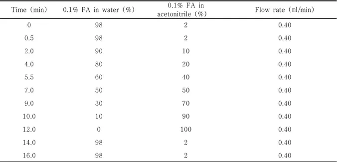
Table 1. Gradient profile for the analysis of UPLC
Time (min) 0.1% FA in water (%) 0.1% FA in
acetonitrile (%) Flow rate (㎖/min)
0 98 2 0.40
0.5 98 2 0.40
2.0 90 10 0.40
4.0 80 20 0.40
5.5 60 40 0.40
7.0 50 50 0.40
9.0 30 70 0.40
10.0 10 90 0.40
12.0 0 100 0.40
14.0 98 2 0.40
16.0 98 2 0.40
3. 세포배양
HepG2 cell은 American Type Culture Collection (ATCC, Rockville, MD, USA)에서 구입하였으며 10% FBS, 100 units/㎖ penicillin이 포함된 DMEM 배지를 이용하여 37℃와 5% CO2 조건에서 배양하였다.
세포는 100 ㎜의 배양접시에서 80% 이상의 confluence 에 도달하도록 배양하여 실험에 이용하였다.
4. MTT assay
세포 생존율은 MTT assay 법을 통해 조사하였다.
HepG2 cell을 24 well plate에 1×10⁵ cells/well로 분주하여 24 시간 배양한 후, FBS가 없는 배지에 100 ㎍/㎖의 CEE를 24 시간 전 처치하였다. CEE를 전 처치한 세포는 카드뮴(CdCl2, 100 μM)에 3시간 동안 노출시킨 후에 배양 배지는 걷어내고 phosphate- buffered saline (PBS)로 세척하였다. 세척한 세포에
0.1 g/㎖ MTT 용액을 첨가하여 37℃에서 2 시간 반 응시킨 후 생성된 formazan을 DMSO로 녹여 microplate reader (Tecan, Männedorf, Switzerland)를 사용 하여 570 ㎚의 파장에서 흡광도를 측정하였다. 세포 생존률은 대조세포에 대한 백분율로 다음과 같은 수 식에 의해 계산하였다 [cell viability (%) = 100 × (absorbance of treated sample)/(absorbance of control)].
4. Reactive oxygen species (ROS) 측정
세포 내 ROS 측정은 활성산소 및 과산화수소와 반 응하여 dichlorofluorescein 형광을 생성하는 DCFH-DA 를 이용하였다. 처치가 완료된 세포에 10 μM DCFH-DA 를 30분 동안 반응시키고, trypsin으로 세포를 분리 하여 black plate에 100 ㎕씩 옮겨준 다음, excitation 485 ㎚, emission 530 ㎚ 파장에서 형광강도를 측정 하였다.
5. Glutathione (GSH) 함량 측정
처치가 완료된 세포에서 GSH의 함량을 측정하기 위해서 500 ㎕의 metaphosphoric acid를 첨가하여 용해한 후, 원심분리(3,000 ×g, 4℃, 10 min)하여 단백질을 포함하는 상층액을 얻었다. 상층액은 GSH determination kit를 이용하여 405 ㎚ 파장에서 측 정하였다.
6. Superoxide dismutase (SOD) assay
CEE로 전 처치가 완료된 세포를 PBS로 2회 세척 후, radioimmunoprecipitation assay (RIPA) buffer (1% NP-40, 1% sodium deoxycholate, 0.1% SDS, 25 mM Tris-HCl pH 7.6, 150 mM NaCl)와 halt protease and phosphatase inhibitor cocktail을 100:1로 혼합한 lysis buffer를 첨가하여 4℃에서 10
분간 반응시킨 후, 초음파(Branson Ultrasonics Sonifier S-450 Digital Ultrasonic Cell Disruptor/
Homogenizer)로 파쇄한 후, 원심분리(1,500 ×g, 4℃, 5 min)하여 상층액을 회수하였다. 회수한 상층 액은 다시 원심분리(10,000 ×g, 4℃, 15 min)하여 단백질을 포함하고 있는 상층액을 취하였다. 상층액 은 SOD assay kit를 사용하여 460 ㎚ 파장에서 흡 광도를 측정하여 효소 활성을 측정하였다.
7. 통계적 검정
모든 실험은 3회 이상 반복 실시하였으며 실험 결 과는 mean ± S.D.로 나타내었다. 유의성 검정은 윈 도우용 SPSS ver. 23 프로그램을 사용하여 one way analysis of variance (ANOVA) 분석을 실시하였다.
사후 검증은 Tukey HSD를 사용하였으며, p값이 0.05 미만일 때 통계적으로 유의하다고 판정하였다.
Ⅲ. 실험결과
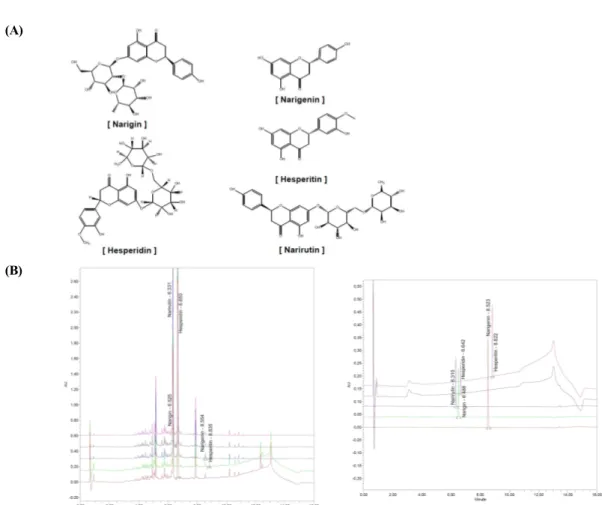
1. CEE에 포함된 활성성분 분석
陳皮에는 narigenin, narigin, narirutin, hesperidin 및 hesperitin 등이 함유되어 있는 것으로 보고되었 다3,4). 陳皮 추출물에 포함되어 있는 narigenin, narigin, narirutin, hesperidin 및 hesperitin의 함유 여부 및 함유량을 UPLC를 통해 분석하였다. 본 연구에 서 이용한 陳皮에는 narigenin이 0.42 ± 0.040 ppm, narigin이 0.34 ± 0.045 ppm, narirutin이 105.27
± 6.283 ppm, hesperidin이 530.84 ± 4.738 ppm, hesperitin이 0.18 ± 0.006 ppm 포함되어 있 었다 (Fig. 1, Table 2). Hesperidin은 진피에 다량 함유되어진 주요 성분 화합물로 알려져 있으며15) 본 실험의 결과에서도 5개의 화합물 중에서 가장 많은 량으로 확인되었다.
(A)
(B)
Fig. 1. Chemical structures and UPLC chromatogram of five marker compounds in CEE
(A) Chemical structures of five marker compounds, narigenin, narigin, narirutin, hesperidin and hesperitin. (B) UPLC chromatograms of standard compounds (left) and five marker compounds in CEE (right).
Table 2. UPLC retention time and content of 5 marker compounds in CEE
Compounds Retention time (min) Content (ppm)
Narigenin 8.523 0.42 ± 0.040
Narigin 6.486 0.34 ± 0.045
Narirutin 6.310 105.27 ± 6.283
Hesperidin 6.642 530.84 ± 4.738
Hesperitin 8.822 0.18 ± 0.006
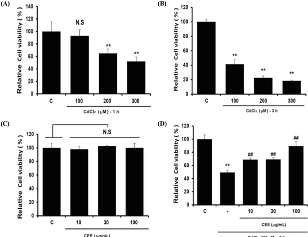
2. 카드뮴으로 유도된 간세포 사멸에 대한 CEE의 보 호 효과
카드뮴으로 유도된 간세포 사멸에 대한 CEE의 보 호 효과를 규명하기 위해, HepG2 cell에서 카드뮴의 농도 (100, 200, 300 μM)와 처리 시간 (1, 3 h)에
따른 세포독성 조건을 MTT assay를 통해 결정하였 다. 그 결과 카드뮴 100 μM을 3시간 동안 처치를 하였을 때 효과적으로 세포 독성이 유도되었다 (Fig.
2A, B). 또한 CEE가 HepG2 cell에서 세포독성을 나 타내는지를 확인하기 위하여 10-100 ㎍/㎖ CEE를
24시간 동안 처치하여 MTT assay로 분석하였다. 그 결과, CEE의 단독 처치에 의한 세포 독성은 확인되 지 않았다 (Fig. 2C). CEE가 카드뮴으로 유도한 세 포 독성에 미치는 영향은 CEE를 24 시간 전 처치 후, 카드뮴을 3시간 처치하여 살펴보았다. 카드뮴을 단독 처치한 군에서는 대조세포 대비 49.4 ± 2.9%
로 세포 생존율이 감소하여 세포 독성이 확인되었다.
하지만 10, 30 및 100 ㎍/㎖의 CEE와 카드뮴을 처 치한 군에서는 농도 의존적으로 대조세포 대비 각각 68.8 ± 2.4%, 69.1 ± 2.7%, 89.4 ± 6.9%로 세포 생존율이 각각 증가하였다 (Fig. 2D). 이 결과를 통 해 CEE는 카드뮴에 의한 간세포 독성에 대해 세포 보호 효과가 있음을 확인하였다.
(A) (B)
(C) (D)
Fig. 2. Effect of CEE on cadmium-induced cytotoxicity in HepG2 cells
Various concentrations (100, 200, 300 μM) of cadmium were treated into the cells for 1 h (A) or 3 h (B) to determine the optimum experimental condition of cadmium in HepG2 cells. (C) HepG2 cells were treated with CEE (10, 30, 100 ㎍/㎖) alone for 24 h. (D) HepG2 cells were pre-treated with CEE (10, 30, 100 ㎍/㎖) for 24 h and then exposed to 100 μM of cadmium for 3 h. (significant as compared to control, **p < 0.01; significant as compared to cadmium alone, ##p < 0.01; N.S, not significant; C, control).
3. CEE가 카드뮴으로 과다 생성된 ROS에 미치는 영향 세포 내 ROS는 HepG2 cell에 CEE를 24시간 전 처치 후, 카드뮴을 1 시간 동안 처치한 다음 DCFH-DA 로 염색하여 측정하였다. 카드뮴을 단독으로 처치하
였을 때, 대조세포와 비교하여 ROS가 276.74 ± 15.14% 증가하였다. CEE와 카드뮴을 같이 처치하였 을 때 대조세포 대비 92.26 ± 6.38%로 카드뮴 단독 처치 대비 통계적으로 유의하게 감소하였고, CEE를
단독 처치하였을 때 대조세포 대비 89.88 ± 3.19%
였다 (Fig. 3). 따라서 CEE에 의해 카드뮴으로 증가
된 ROS가 감소함을 확인하였다.
Fig. 3. Effect of CEE on cadmium-induced ROS production
Cells were exposed with cadmium for 1 h after CEE treatment for 24 h. ROS level was measured by DCFH-DA staining and the fluorescence intensity was expressed as percentage of control. (significant as compared to control, **p < 0.01; significant as compared to cadmium alone, ##p < 0.01; C, control).
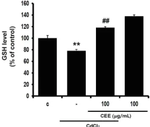
4. CEE가 카드뮴으로 유도된 GSH 감소에 미치는 영향 카드뮴 3 시간 처치는 대조군 대비 GSH 함량을 78.24 ± 2.29%로 감소하였으며, CEE 전처치에 의 해 GSH는 118.32 ± 1.46%로 통계적으로 유의하게
증가하였다. CEE의 단독 처치에 의해 GSH는 137.89
± 2.45%로 대조군 대비 증가하였다 (Fig. 4). 이 결과를 통해 CEE는 카드뮴으로 인한 GSH 함량의 감 소를 회복시킬 수 있음을 확인하였다.
Fig. 4. Effect of CEE on GSH levels altered by cadmium treatment
Cells were exposed with cadmium for 3 h after CEE treatment for 24 h. GSH contents were measured in cell homogenates as described in material and method section. Data were expressed as percentage of control. (significant as compared to control, **p < 0.01;
significant as compared to cadmium alone, ##p < 0.01; C, control).
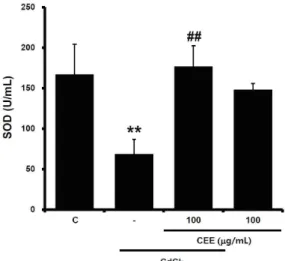
5. CEE가 SOD 활성에 미치는 영향
대조군 세포에서 SOD 활성은 167.18 ± 37.04 U/
㎖이었고, 카드뮴 3 시간 처치에 의해 SOD 활성이 68.83 ± 18.06 U/㎖으로 감소하였다. CEE 전처치 는 카드뮴에 의한 SOD 활성을 176.97 ± 25.5 U/㎖
으로 증가시켰다. CEE 단독 처치는 148.38 ± 7.35
U/㎖으로 SOD 활성이 감소하는 경향을 나타내었으 나 통계적으로 유의하지 않았다 (Fig. 5). 이 결과를 통해 카드뮴으로 인한 SOD의 활성 감소가 CEE에 의 해 회복됨을 확인하였다.
Fig. 5. Effect of CEE on SOD activity altered by cadmium treatment
Cells were exposed with cadmium for 3 h after CEE treatment for 24 h. SOD activity was measured as described in material and method section. (significant as compared to control,
**p < 0.01; significant as compared to cadmium alone, ##p < 0.01; C, control).
Ⅳ. 고찰
카드뮴은 인간에게 해를 입히는 대표적인 중금속 중의 하나로 환경, 식품 등으로부터 오염되어 인체의 여러 장기에 손상을 입힌다7,8). 특히 간은 해독작용을 담당하는 곳으로 이러한 중금속에 오염되어 간세포 독성을 나타낼 경우 인체에 치명적인 결과를 초래할 수 있다9). 陳皮는 우리나라에서 과수로 재배하고 있 는 상록 소교목인 귤(Citrus unshiu Markovich)의 잘 익은 열매껍질로 이기(理氣), 조습(燥濕), 조중(調 中), 화담(化痰), 지해(止咳) 등의 효능을 얻고자 할 때 약재로 사용되어 왔다1,2). 약리학 측면에서는 항궤 양16) 및 항알러지17) 효과 등이 보고되었다. 본 연구 에서는 간세포를 활용하여 카드뮴으로 세포 손상을 유발한 후에 陳皮의 세포 보호 작용을 확인하고자 하 였으며, 효소적 및 비효소적인 항산화 활성을 통해 陳皮가 간세포 보호 작용을 나타냄을 확인하였다.
카드뮴에 의해 유도되는 세포 독성의 핵심적인 기 전으로 산화 스트레스(oxidative stress)가 알려져 있 으며 간에서 카드뮴이 염증과 산화 스트레스를 통해 세포자멸사를 유발하는 것으로 보고되었다12,13). 산화 스트레스는 항산화 시스템과 ROS 사이의 균형이 깨 질 때 발생한다. ROS는 산소의 정상적인 대사의 부 산물이며 신호전달과 항상성 유지에 중요한 역할을 하지만 비정상적으로 축적되면 산화 스트레스가 나타 나게 된다18,19). ROS에 의한 산화 스트레스는 자외선, 전리 방사선 또는 중금속 등에 노출된 결과로 나타날 수 있으며 지질, 단백질, DNA에 대한 산화적 손상, 항산화 효소 억제 등을 통해 노화 및 세포 사멸이 일 어나게 된다20). 본 연구에서는 카드뮴이 간세포의 사 멸과 세포 내 ROS의 과다 증가를 유도하였으나 CEE 의 전 처치로 인해 이러한 세포 손상과 ROS 수준의 변화가 억제됨을 확인하였다.
GSH는 세포 내에서 비효소적으로 항산화 시스템을 유지하는 강력한 항산화 물질로 자유라디칼, 과산화
물, 과산화 지질, 중금속 반응, ROS에 의한 세포 성 분의 손상을 방지할 수 있다21). GSH의 산화된 형태 인 glutathione disulfide (GSSG)는 NADPH에 의해 환원되어 다시 GSH가 되며, 세포 내 GSH와 GSSG 의 산화 환원 비율은 산화적 스트레스의 척도로 사용
된다22,23). 간에는 카드뮴에 대해 높은 친화성을 갖는
대부분의 금속 결합 단백질(metallothionein)이 포함 되어 있기 때문에 하고 있기 때문에 주로 카드뮴 축 적이 간에서 발생하며24) 고농도의 카드뮴에 노출되었 을 때 metallothionein에 결합되지 않은 카드뮴은 GSH 수준을 상당히 감소시킬 수 있는 것으로 보고되 었다25). 본 연구에서는 세포 내 GSH의 함량을 측정 한 결과 카드뮴에 의해 감소한 GSH의 함량이 CEE에 의해 증가함을 확인하였다.
SOD는 초과산화이온을 산소와 과산화수소로 바꿔 주는 불균등화 반응을 촉매하는 효소이며, 항산화작 용을 하는 것으로 알려져 있다19,26). 초과산화 이온은 세포에 해로운 영향을 주는 자유 라디칼 음이온을 가 지고 있으며, SOD는 초과산화이온을 산소와 과산화 수소로 바꿔, 산화적 스트레스로부터 세포방어 작용 을 나타낸다26,27). 본 연구에서는 카드뮴으로 인해 SOD 활성이 감소되었으나 CEE에 의해 회복됨을 확 인하였다.
감귤의 주요성분인 hesperidin은 혈액 내 카드뮴 농도를 감소시키고 뇨와 변을 통한 카드뮴 배설효과 가 뛰어나며28) 지질과산화물의 형성을 억제하는 항산 화 작용이 있다고 알려져 있다4,29). 본 연구에 사용한 陳皮는 메탄올로 추출한 것으로 성분 분석에서 hesperidin이 가장 많이 포함되어 있음을 확인하였 다. 따라서 본 실험에서 나타난 CEE의 세포 보호효 과와 항산화 활성에 hesperidin이 기여하였을 것으로 사료되지만 추후 실험을 통해 hesperidin에 의한 효 과를 살펴봐야 할 것이다.
이상의 결과로 카드뮴에 의해 유도된 산화적 스트 레스 매개 세포 사멸에 대한 CEE의 세포 보호 효과 를 확인하였다. CEE는 ROS의 증가와 GSH의 감소를 개선시키고 SOD의 효소 활성을 회복시킴으로써 카드 뮴에 의한 간세포 사멸을 억제하였다.
Ⅴ. 결론
본 연구에서는 카드뮴을 처리한 간세포에서 진피추
출물(CEE)이 항산화 작용과 세포 보호 활성을 나타 냄을 확인하였다. 카드뮴에 의해 과다 생산된 ROS는 GSH의 고갈을 유발하였으나 진피에 의해 이러한 산 화 스트레스가 억제되었다. 또한 진피는 항산화 효소 인 SOD의 활성도 증가시켰다. 진피 성분 분석에서는 항산화 활성이 보고되어진 hesperidin이 가장 많은 양으로 함유되어 있었다. 이러한 결과들은 중금속에 의한 간세포 독성에서 진피의 비효소적 및 효소적 항 산화 작용을 이해하게 함으로써 중금속 독성에 대한 진피의 활용 가능성을 제시하고 있다.
감사의 글
이 논문은 2020년도 정부(과학기술정보통신부)의 재 원으로 한국연구재단의 지원을 받아 수행된 연구임 (No. 2018R1A5A2025272).
References
1. Oriental Pharmacology Committee. Oriental Pharmacology. 2nd ed. Seoul:Shinil. 2006:670-5.
2. Hung KC. The Pharmacology of Chinese herbs.
2nd ed. Cleveland:CRC Press. 1998:285-8.
3. Hyon JS, Kang SM, Mahinda S, Koh WJ, Yang TS, Oh MC, Oh CK, Jeon YJ, Kim SH. Antioxidative activities of extracts from dried Citrus sunki and C. unshiu peels. J Korean Soc Food Sci Nutr.
2010;39(1):1-7.
4. Leelavinothan P, Kalist S. Hesperetin protects against oxidative stress related hepatic dysfunction by cadmium in rats. Exp Toxicol Pathol. 2012;64(5):513-20.
5. Nawrot TS, Staessen JA, Roels HA. Cadmium exposure in the population: From health risks to strategies of prevention. Biometals.
2010;23:769-82.
6. Satarug S, Vesey DA, Gobe GC. Health risk assessment of dietary cadmium intake: Do current guidelines indicate how much is safe? Environ Health Perspect. 2017;125:284-8.
7. Mezynska M, Brzóska MM. Environmental exposure to cadmium—A risk for health of the
general population in industrialized countries and preventive strategies. Environ Sci Pollut Res.
2018;25:3211-32.
8. Lin J, Zhang F, Lei Y. Dietary intake and urinary level of cadmium and breast cancer risk: A meta-analysis. Cancer Epidemiol.
2016;42:101-7.
9. Kang MY, Cho SH, Lim YH, Seo JC, Hong YC.
Effects of environmental cadmium exposure on liver function in adults. Occup Environ Med.
2013;70:268-73.
10. Ramakrishnan R, Elangovan P, Pari L. Protective role of tetrahydrocurcumin: An active polyphenolic curcuminoid on cadmium- induced oxidative damage in rats. Appl Biochem Biotechnol. 2017;183:51-69.
11. Brzóska MM, Rogalska J, Roszczenko A, Galazyn-Sidorczuk M, Tomczyk M. The mechanism of the osteoprotective action of a polyphenol-rich Aronia melanocarpa extract during chronic exposure to cadmium is mediated by the oxidative defense system. Planta Med.
2016;82:621-31.
12. Skipper A, Sims JN, Yedjou CG, Tchounwou PB.
Cadmium chloride induces DNA damage and apoptosis of human liver carcinoma cells via oxidative stress. Int J Environ Res Public Health. 2016;13(1):88.
13. Rani A, Kumar A, Lal A, Pant M. Cellular mechanisms of cadmium-induced toxicity: A review. Int J Environ Health Res. 2013;
24:378-99.
14. Brzóska MM, Borowska S, Tomczyk M.
Antioxidants as a potential preventive and therapeutic strategy for cadmium. Curr Drug Targets. 2016;17:1350-84.
15. Cha BJ. Changes in the constituents and antioxidant activity in accordance with the processing conditions of Citrus unshiu Markovich. Kor J Pharmacogn. 2015;46(1):
23-30.
16. Borrelli F, Izzo AA. The plant Kingdom as a
source of anti ulcer remedies. Phytother Res.
2000;14(8):581-91.
17. Kim DK, Lee KT, Eun JS, Zee OP, Lim JP, Eum SS, Kim SH, Shin TY. Anti-allergic components from the peels of Citrus unshiu.
Arch Pharm Res. 1999;22(6):642-5.
18. Lee HI, Park AR, Chun SW. Effects of mitochondrial reactive oxygen species on neuronal excitability in rat spinal substantia gelatinosa neurons. Int J Oral Biol.
2012;37(1):17-23.
19. Kwon GR, Moon IS, Lee WC. Neuroprotective effects of Pinelliae rhizoma water-extract by suppression of reactive oxygen species and mitochondrial membrane potential loss in a hypoxic model of cultured rat cortical cells. J Life Sci. 2009;19(5):598-606.
20. Cichoż-Lach H, Michalak A. Oxidative stress as a crucial factor in liver diseases. World J Gastroenterol. 2014;20(25):8082-91.
21. Wang L, Yan B, Liu N, Li Y, Wang Q. Effects of cadmium on glutathione synthesis in hepatopancreas of freshwater crab, Sinopotamon yangtsekiense. Chemosphere.
2008;74(1):51-6.
22. Noctor G, Foyer CH. Ascorbate and glutathione: keeping active oxygen under control. Annu Rev Plant Physiol Plant Mol Biol. 1998;49:249-79.
23. Romero MJ, Canada AT. The evaluation of Escherichia coli as a model for oxidant stress in mammalian hepatocytes: role of glutathione.
Toxicol Appl Pharmacol. 1991;111: 485-95.
24. Haouem S, Hmad N, Najjar MF, Hani AE, Sakly R. Accumulation of cadmium and its effects on liver and kidney functions in rats given diet containing cadmium-polluted radish bulb. Exp Toxicol Pathol. 2007;59:77-80.
25. Dkhil MA, Marwa SM, Diab MSM, Lokman MS, El-Sayed H, Aljawdah HMA, Al-Quraishy S, Bauomy AA. Hepato-protective effect of Pleurotus ostreatus extracts in cadmium-
intoxicated rats. J King Saud Univ Sci.
2020;32(8):3432-6.
26. Fridovich I. Superoxide dismutases. Annu Rev Biochem. 1975;44:147-59.
27. Imlay JA, Fridovich I. Assay of metabolic superoxide production in Escherichia coli. J Biol Chem. 1991;266(11):6957-65.
28. Choi JH, Rhee SJ. Effects of green tea catechin on cadmium accumulation in chronic cadmium poisoned rats. Kor J Nutr.
2001;34(4):384-92.
29. Cho EJ, Li L, Noriko Y, Kim HY.
Antioxidative effects of hesperidin and hesperetin under cellular system. CNU J Agric Sci. 2011;38(4):717-22.