․ 접수 : 2008년 9월 1일 ․ 논문심사 : 2008년 10월 30일 ․ 채택 : 2008년 12월 5일
* Corresponding author : Seung Hwa Baek, Department of Herbal Resources, Professional Graduate School of Oriental Medicine, Wonkwang University, Iksan 570-749, Korea. Tel : +82-63-850-6225.
Fax : +82-63-841-4893. Email : [email protected] Kor. J. Oriental Preventive Medical Society 2008;12(3):91-98
흑미 추출물에 대한 항동맥경화 효과(Notes)
임치환1)․김종민1)․백승화2)*
1)충남대학교 농업생명과학대학 생물환경화학전공,
2)원광대학교 한의학전문대학원 한약자원개발학과
Antiarteriosclerosis Effects of Black Rices Extract
Chi Hwan Lim,1) Jong Min Kim1) & Seung Hwa Baek2)*
1)Department of Bioenvironmental Chemistry, College of Agricultural & Life Sciences, Chungnam National University, Daejeon, 305-764, Korea.
2)Department of Herbal Resources, Professional Graduate School of Oriental Medicine, Wonkwang University, Iksan 570-749, Korea.
Abstract
80% MeOH extract of black rices fractionated with n-hexane, EtOAc, and n-BuOH. Copper-induced LDL oxidation inhibition assay and ACAT inhibition assay examined with fractions. n-Hexane and EtOAc fractions showed high inhibition activity. We divided n-hexane fraction into three sub-layers(H1 - H3) and EtOAc into eight sub-layers(E1 - E8). H1, H2, E6, and E7 are showed higher inhibition activity than standard in Lp-PLA2 inhibition assay and H1 and E6 are showed higher ACAT inhibition activity than standard.
Key words:Black rices, Copper-induced LDL Ooxidation inhibition assay, ACAT inhibition assay, Lp-PLA2 inhibition assay.
I. 서 론
흑미는 화본과의 한해살이풀로 한국, 일본, 중국 그리고 남부 아시아에서 재배되는 진한 보라색을 띄는 특용 작물로서 예로부터 그 의 학적 효과가 널리 알려졌으며 떡, 빵, 아이스크 림 그리고 술의 착색제로 많이 사용되고 있 다.1) 흑미 중의 안토시아닌의 항염증 활성,2) 환원 활성3) 그리고 항산화 활성4)이 보고 되면 서 안토시아닌이 생리활성 물질로서 간주되고 있으며, 최근에 적포도주의 페놀성 물질이 동 맥 경화의 주요인이라고 알려진 HLDL(human low density lipoprotein)의 산화를 억제한다고 보고되어 있다.5)
동맥경화증은 한국인의 사망원인 중 높은 비 중을 차지하는 질병으로, 뇌경색, 협심증, 심근 경색증 등 허혈성 심질환과 말초혈관 폐쇄를 유발하는 원인 질환으로, 활성산소에 의한 산화 적 스트레스의 일례로 들 수 있다.6) Cholesterol 에 의해 간에서 합성되는 LDL-cholesterol이 활성산소에 의해 산화되면, 허혈성 심장 질환 및 동맥경화증, 뇌혈관 질환 등을 유발하는 고 지혈증의 주요 인자가 되는 것이다.7, 8) 동맥경 화의 발병 초기 메카니즘으로서 혈관벽 평활 근 세포 증식에 의한 내막의 비대화와 내막으 로의 LDL의 과다 유입을 들 수 있다. 혈중 LDL은 receptor에 의해 세포내에서 분해되지 만, 콜레스테롤 다량 섭취로 receptor가 부족하 거나 결핍되면 혈중에 많아지게 되고, 결국 축 적되어 oxidized LDL이 생성된다.9-11) 또한 LDL 은 내피세포의 lipoxygenase, 대식세포에서 생 성되는 superoxide radical, 그리고 식품을 통하 여 들어온 과산화지질에 의하여, oxidized LDL 로 산화되는 것으로 밝혀졌다.12) 산화적 스트 레스에 의해 생성된 oxidized LDL은 높은 세 포 독성이 있는 지질과산화물을 가지고 있어
서, 세포 조직에 확산되어 독성을 나타내고 내 피세포에 염증을 일으키며, 동맥내에서 지방선 조를 일으키고, 섬유소의 용해를 억제하여 조 직인자의 발현을 증가시켜 응혈원의 활성도를 촉진시키는 데, 이러한 작용기작에 의해서 동 맥이 탄성을 잃게 되는 것이다.13) 이와 같이 LDL 산화는 초기 동맥 경화성 병변의 형성과 진전에 주요한 역할을 하므로, 동맥경화의 원 인을 예방하기 위해서는 항산화제에 의해 LDL 의 산화를 방지해야 하는 것이다.14-17)
본 연구에서는 흑미의 ACAT와 Lp-PLA2
inhibition assay, 동맥경화 발생저해(LDL Oxi- dation inhibition)에 대한 활성검정 결과를 보 고하고자 한다.
II. 실험방법 및 재료
1. 시약 및 기기
본 실험에서 사용된 n-hexane, chloroform, EtOAc, EtOH, MeOH, n-BuOH, 용매는 국내 삼전순약 제품, 기타 시약은 특급시약을 사용하 였다. Medium pressure liquid chromatography (MPLC)용 MeOH는 공업용 용매를 정제하여 사용하였으며, Silica gel(70 - 230mesh, Merk, Germany), TLC plate(20 × 20cm, Merck, Kie- selgel 60 F254)를 사용하였다. 생리활성물질의 정 제를 위해 Combi Flash(ISCO, U.S.A)의 MPLC 를 이용하였고, UV spectrum은 Carry 100 conc (Varian)로 측정하였다.
2. 재료
흑미는 cyanidin-3-glucoside를 약 4.7mg/g, peonidin-3-glucose 약 0.23mg/g을 함유하고 있는 수원 415호를 사용하였다.
3. LDL oxidation inhibition assay18-20) LDL 분리는 신선한 human plasma에 0.1%
EDTA와 0.02% NaN3를 가하고, 교반한 후, KBr(d = 1.006 - 1.025)를 가하고 1차 원심분리 (40,000 rpm, 5℃, 24hrs)를 한다. 이때 분리된 VLDL을 제거하고, LDL이 포함된 fraction을 취한 후, KBr(d = 1.026 - 1.055)을 가하여 2차 원심분리(40,000rpm, 5℃, 24hrs)하여, density 1.025 - 1.055g/mL의 신선한 human LDL을 얻 었다. 분리한 LDL은 pH 7.4 buffer Tris. 0.05M NaCl, 0.02% EDTA로 투석시키고, 냉동 건조 하여 사용하였다. LDL 산화는 LDL(400μg/mL) 과 CuSO4(16μM)에 전체부피가 1mL가 되도록 phosphate buffer(pH 7.4)를 섞어 37℃에서 배 양시키고, 1mM EDTA와 1mM BHT(20μL)를 첨가하여 산화를 중지시켰다.
산화된 LDL에 trichloroacetic acid를 넣어 단백질을 침전시키고 원심분리 후, 산화 지질 을 포함한 상층액을 분리하였다. 분리된 상층 액에 시료 200μg 및 1mL의 thiobarbituric acid (TBA 25%)를 첨가하여, 95℃에서 10분간 가 열 후 얼음으로 냉각시키고, 생성된 MDA의 양을 532nm에서 spectrophtometer를 이용하여 측정하였다.
4. Lp-PLA2 inhibition assay21)
신선한 혈액에 0.04% EDTA, 0.05% NaN3, 0.015% phenylmethylsulfonyl flouride(PMSF) 를 넣어 혈액에 있는 지단백질의 변성을 막는 다. 100,000 × g, 4℃에서 20시간 동안 초원심 분리하여 VLDL과 chylomicron을 제거하고, 나머지 하층을 다시 100,000 × g, 4℃에서 20시간 동안 초원심 분리하여 상층의 LDL을 분리하였 다. 10μL의 [3H] PAF(250μCi, 21.50Ci/mmole)와 12.5μM PAF(2.5μL)를 질소가스 하에서 완전 농 축시킨 후, 2.7mM EDTA를 포함하는 10mM
PBS(3.2mL)를 첨가하여 micellar 형태의 기질 을 준비한다. (A)시험관에 희석한 LDL(20μL) 와 (A)용액(160 μL)을 DMSO에 녹인 시료 20 μL를 첨가하여, 37℃에서 15분간 반응시킨 후, chloroform/methanol(2 : 1) 용액 600μL를 첨가 하여 반응을 중지시켰다. 무처리군는 시료 대 신 DMSO를 사용하였고, 대조군는 Lp-PLA2
활성저해 표준물질인 SB 381320을 사용하였 다. 1,500 × g에서 3분간 원심분리하여 250μL의 물층을 취하고, chloroform(250μL)을 첨가하여 1,500 × g에서 3분간 원심분리하여, 다시 물층 을 취하였다. 최종 물층(500μL) 중 100μL를 취해 서 scintillation cocktail(3mL)를 첨가하여, liquid scintillation counter를 이용하여, 1-O-hexadecyl- [acetyl-3H(N)] phosphatidyl-choline으로부터 생성된 [3H] acetate를 측정하였으며, 아래와 같 은 식으로 저해율을 계산하였다.
Inhibition r ates(%)
= 100 ×[1- Sample(cpm)-Backgr ound (cpm)Control (cpm)-Background (cpm) ]
5. ACAT inhibition assay22-24)
ACAT 효소 저해 활성은 [14C]-oleoyl-CoA를 기질로 하여 사용하였다. 4μL Liver microsome, 20μL assay buffer(1M KH2PO4, 10mM DTT, pH 7.4), water(41μL), BSA(15μL, 40mg/mL), cholesterol(2μL, 20mg/mL) 그리고 inhibitor(10 μL, 시료)를 37℃에서 20분간 예비 반응 시키고, 이 반응액에 8μL의 [14C]-oleoyl-CoA(0.05 μCi) 를 가하여 37℃에서 25분간 반응 시켰다. 1mL 의 isopropanol-heptaneL(4 : 1, v/v)를 가해 반 응을 정지시켰다. 0.6mL의 heptane과 0.4mL의 assay buffer를 첨가한 후 vortex하고 원심 분 리했다. 원심분리하여 얻은 상층액 100μL에 lipo- lumn 3mL를 가하여, scintillation counter를 이 용하여 radioactivity를 측정하였다. ACAT 저해 활성은 아래와 같은 식을 이용하여 계산하였다.
Inhibition Rate(%)=(1- CPMCPMac))-CPM-CPMdb)))×100
CPMa): 시료와 효소를 넣었을 때 CPM*
CPMb): 시료는 넣고 효소는 넣지 않았을 때 CPM CPMc): 시료는 넣지 않고 효소는 넣었을 때 CPM CPMd): 효소와 시료 모두 넣지 않았을 때 CPM CPM*: Counter per minute
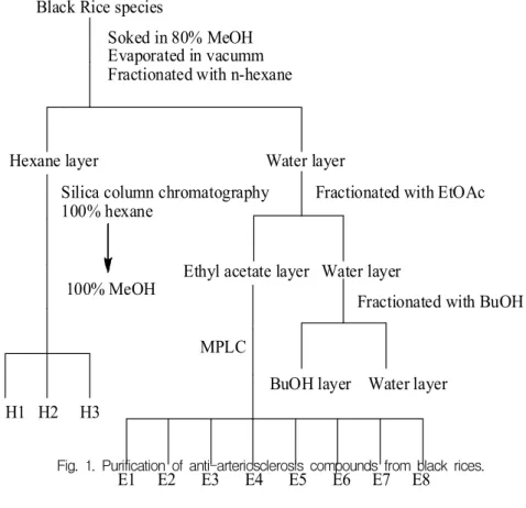
6. 흑미 추출물에 대한 항동맥경화 활성
흑미 10kg을 80% MeOH(H2O : MeOH, v/v) 로 상온에서 3회 추출하여 감압 농축하고, n- hexane, EtOAc, 그리고 n-BuOH의 순으로 분 획하여 41g(0.41%), 8g(0.08) 그리고 28g(0.28%)
의 분획물을 얻었다. 이 분획들을 이용하여 LDL-oxidation inhibition assay 그리고 Lp- PLA2 inhibition assay를 수행하여, n-hexane 분획과 EtOAc 분획에 활성이 있음을 확인하였 다. n-hexane 분획을 이용하여, silica column chromatography(100% n-hexane →100% MeOH) 를 수행하여, 3개의 소분획(H1 - H3)을 얻었으며, EtOAc 분획을 이용하여 MPLC(silica packed column, 100% chlorofrom → 100% MeOH)를 수행하여 8개의 소분획을 얻었다. n-Hexane 분획과 EtOAc 분획에서 얻어진 11개의 소분 획을 이용해, Lp-PLA2 inhibition assay 그리 고 ACAT-2 inhibition assay를 수행하여 활성 을 확인하였다(Fig. 1).
Black Rice species
Soked in 80% MeOH Evaporated in vacumm Fractionated with n-hexane
Hexane layer Water layer
Fractionated with EtOAc
Ethyl acetate layer Water layer
Fractionated with BuOH
BuOH layer Water layer Silica column chromatography
100% hexane
100% MeOH
H1 H2 H3
MPLC
E1 E2 E3 E4 E5 E6 E7 E8
Fig. 1. Purification of anti-arteriosclerosis compounds from black rices.
Fig. 2. LDL Oxidation inhibition rates of solvent extract of black rices.
III. 결과 및 고찰
1. 흑미의 항동맥경화 활성
Copper-induced LDL oxidation inhibition assay에서는 standard로서 probucol을 사용하 였으며, 0.5, 1, 2, 4, 10μM의 농도를 만들어 생 성된 MDA의 농도를 비교하여, 저해율을 확인 하였다. 각 용매 분획물은 4μg/mL와 40μg/mL 의 농도를 만들어 저해율을 확인한 결과, 40μg/
mL에서는 MeOH(M) 추출물의 저해율이 80.9%
로 확인되었으며, EtOAc(E) 분획은 99%의 저 해율을 보여 높은 활성을 확인할 수 있었다.
다른 n-hexane(H), n-BuOH(B) 그리고 water (W) 층은 89.3, 73.8 그리고 71.8%의 저해율을 관찰 할 수가 있었다. 4μg/mL에서는 EtOAc 분 획이 84.5%의 저해율이 관찰되어, 낮은 농도에 서도 활성을 유지하고 있음을 확인하였으며, 나 머지 분획의 저해율은 현저히 떨어졌다(Fig. 2).
Lp-PLA2 inhibition assay 그리고 ACAT-2 inhibotion assay에서는 copper-induced LDL oxidation inhibition assay 결과를 토대로, LDL
항상화 활성이 큰 hexane층과 EtOAc층을 silica column chromatography를 수행하여, 얻은 총 11개의 소분획을 이용하여 표준물질의 저해율 과 비교하여, 표준물질보다 높은 저해율을 보 인 소분획은 활성이 관찰되었다. 그 결과 Lp- PLA2 inhibition assay에서는 H2 분획이 90%
의 저해율을 관찰되어, 높은 활성이 있음을 확 인하였으며, 표준 항산화제(SB)보다 높은 활성 을 보인 분획은 H1, E6, E7, 그리고 E8 분획 이었다(Fig. 3).
ACAT inhibition assay에서는 H1과 E6 분 획이 표준물질(KSS)보다 높은 활성을 보였으 며, 나머지 분획은 낮은 활성을 보였다. 전체적 인 활성의 크기는 H1 = E6 > KSS = E3 > E7 >
E2의 순으로 관찰되었다(Fig. 4). EtOAc 분획 의 4μg/mL에서 84.5%의 높은 활성이 관찰되 어, 흑미 추출물에 대한 항동맥경화에 효능이 있는 생리활성물질을 분리 및 분석하는 연구 를 진행중에 있다.
IV. 결 론
흑미를 이용한 항동맥경화 물질 탐색에서는
Fig. 3. Lp-PLA2 Inhibition rates of each fraction of black rices.
Fig. 4. ACAT Inhibition rates of each fraction of black rice.
80% MeOH 추출물 그리고 각 용매 분획물을 이용하여, LDL oxidation inhibition assay, Lp- PLA2 inhibition assay, 그리고 ACAT-2 inhi- bition assay를 확인하였다. LDL oxidation inhi- bition assay에서 40μg/mL의 농도에서, 모든 추출물 및 분획물의 활성이 70~90%를 보였으 나, EtOAc 분획만이 4μg/mL의 농도에서도 84.5%의 높은 활성을 유지하였으며, 나머지는 활성이 현저히 떨어졌다. n-Hexane과 EtOAc 분획의 소분획을 이용한 Lp-PLA2 inhibition
assay에서는, H2 > E6 > H1 > E7 > E8 > SB >
E2 > E1 > H3의 순으로 활성이 관찰되었으며, ACAT inhibition assay에서는 H1 > E6 > KKS
= E3 > E7 > E2 > H2 > E1 > E5 > E8 > H3의 순으로 활성을 나타내었다.
참고문헌
1) Cho, M.H., Yoon, H.H., Han, T.R. Thermal
stability of the major color component, cyanidin 3-glucoside, from a Korean pig- mented rice variety in aqueous solution.
Agri. Chem. Biotech. 39 : 245-248, 1996.
2) Valskovska, M., Drenska, D., Ovcharov, R. Effect of antioxidant, alone and in combination, on the inflammatory process.
Probl. Vutr. Med. 18 : 13-19, 1990.
3) Gabar, E. Possible biological role of some anthocyanins in foods. Bull. Liaison Group Polyphenols 14 : 130-133, 1988.
4) Drenska, D., Bantutova, I., Ovcharov, R.
Anticonvulsant effect of anthocyanins and antioxidants. Farmatsiya 39 : 33-40, 1989.
5) Meunier, M. T., Duroux, E., Bastide, P.
Antioxidant activity of procyanidol oli- gomers and anthocyanins with regard to superoxide anion and lipid peroxidation.
Plant Med. Phytother. 23 : 236-247, 1989.
6) Caroline, K. H. Pharmacognosy Phyto- chemistry Medicinal Plants ; 1. Compounds of Toxic Plants, Lavoisier Publishing Inc.
pp 275-499, 1999.
7) Aruoma, O. I. Nutrition and health aspects of free radicals and antioxidants. Food Chem. Toxicol. 32 : 671-683, 1994.
8) Davies, K. J. A., Goldberg, A. L. Proteins damaged by oxygen radicals are rapidly degraded in extracts of red blood cells.
J. Biol. Chem. 262(17) : 8227-8234, 1987.
9) Esterbauer, H., Gebicki, J., Pohl, H., Jurgens, G. The role of lipid peroxidation and antioxidants in oxidative modification of LDL. Free Radic. Biol. Med. 13 : 341- 390, 1992.
10) Ishikawa, Y, Inadera, H., Shirai, K., Hashimoto, H., Fukamachi, I., Saito, Y., Yoshida, S. Moderate oxidation of hyper-
triglyceridemic LDL causes apolipoprotein- Bepitope change and enhances its uptake by macrophages. Biochim Biophys. Acta 1129 : 60-64, 1992.
11) Ross, R. The pathogenesis of atheros- clerosis, perspective for the 1990's. Nature 29 : 801-809, 1993.
12) Fogelman, A.M., Shechter, I., Sieger, J., Hook, M., Child, J. S., Edward, P.A.
Malondialdehyde alteration of low density lipoprotein leads to cholesteryl ester accu- mulation in human monocyte-macrophages.
Proc. Natl. Acad. Sci. USA, 77 : 2214- 2218, 1980.
13) Morel, D.W., Hessler, J.R., Chisolm, G.M.
Low density lipoprotein cytotoxicity induced by free radical peroxidation of lipids. J.
Lipid Res. 24 : 1070-1076, 1983.
14) Akio, K., Yoshinori, O. Antioxidative acti- vities of bioactive substances. Fragrance J. 88 : 53, 1988.
15) Bakatani, N. Recent advances in the study on natural antioxidants. Nippon Sho- kuhin Kogyo Gakkaishi. 37 : 569, 1990.
16) Yang, K.S., Jeon, C.M. Effect of Tara- xacum coreanum Nakai on LDL oxidation.
Kor. J. Pharmacogn. 27(3) : 267-273, 1996.
17) Chen. G. C., Hardman. D. A., Hamilton.
R. L., Mendel. C. M., Chhilling. J. M., Kan. J. P. Distribution of lipid binding regions in human apo B-100. Biochem.
28 : 2477-2488, 1989.
18) Lue. G., Fruchart. J. C. Oxidation of lipo- proteins and artherosclerosis. Am. J. Clin.
Nutr. 53 : 206-210, 1988.
19) Hanfang. Z., Yuzhou. Y., Urs. P. S.
Structural requirements for the binding of modified proteins to the scavenger
receptor of macrophages. J. Biol. Chem.
268 : 5535-5542, 1993
20) Wulf. P., Michael. E. R., Seppo. Y. H., Geoff. C. G., Steve. S. S. LDL undergoes oxydative modification in vivo. Proe. Natl.
Acad. Sci. USA 86 : 1372-1376, 1989.
21) Wu, C.A., Tsujita, M., Hayashi, M., Yo- koyama, S. Probucol inactivates ABCA1 in the plasma membrane with respect to Its mediation of apolipoporotein binding and high density lipoprotein assembly and to its proteolytic degradation, J. Biol.
Chem. 279(29) : 30168-30174, 2004.
22) Asami, Y., Yamagishi, I., Murakami, S., Araki, H., Tsuchida, K., Higuchi, S. HL- 004, the ACAT inhibitor, prevents the progression of atherosclerosis in cholesterol- fed rabbits. Life Sci. 62(12) : 1055-1063, 1998.
23) Bang, M.H., Jang, T.O., Song, M.C., Kim, D.H., Kwon, B.M., Kim, Y.K., Lee, H.S., Chung, I.S., Kim, D.K., Kim, S.H., Park, M.H., Baek, N.I. Screening of biologically active compound from edible plant sources. IX. Isolation and iden- tification of sesquiterpene lactones isolated from the root of Ixeris dentata forma albiflora ; inhibition effects on ACAT, DGAT and FPTase activity. Han'guk Eungyong Sangmyong Hwahakhoeji, 47 (2) : 251-257, 2004.
24) Gill, P.J., Robinson, C., Rathgeb, K. A.
Regulation of ACAT activity by a chole- sterol substrate pool during the prog- ression and regression phases of athero- sclerosis implications for drug discovery.
Atherosclerosis. 83 : 177-185, 1990.