107
Broden photographing method for Calcaneus Subtentaculum tali observation of Ankle plaster patient
Byeongju An
Dept. of Radiology, Nambu University
발목부상을 당한 석고환자의 발꿈치뼈·목말받침돌기 관찰을 위한 Broden 촬영법 연구
안병주 남부대학교 방사선학과
Abstract
There is an Ankle Oblique(Broden’ low) as an examination for the patient whose ankle was encased in orthopedic plaster. Some types of Ankle Joint coalition - Calcaneonavicular coalition, Talocalcaneal coalition and Naviculocuneiform coalition. This study is focused on the relation between Ankle Joints and the structural change of soft tissues, also finding the most proper angle to obtain good images of Ankle Joint from the patient who wore a plaster on his ankle, when we x-ray with Harris-Beath View(30°∼55°) - for observing Subtalar joint, Calcaneus Fracture, Subtentaculum, Tali Fracture and Talocalcaneal coalition. We intend to get the angle which makes us achieve the good image that shows Calcaneus Fracture, Subtentaculum, and Tali Fracture by changing internal angles of the patient’s ankle. We evaluated the images obtained from 51 patients with PACS monitor. The result of the evaluation, subtalar joint was not seperated but opened, and Subtentaculum Tali Fracture was seen overlaid. at the angle 30, we could observe Calcaneus Fracture, Subtentaculum Tali Frature and the front part of behind side of subtalar joint well. And Calcaneo Navicularcoalition, Talocal Canealcoalition, Naviculo Cuneiform coalition condition were clearly seen at that angle. At the angle 35, we could achieve the clear images of subtalar comminuted fracture, talus, the behind joint of heel bone and get the high definition image on the degree of talocalcaneal joint separation. In addition to, We could obtain the good wide image of Sinus Tarsi. At the area of 45, We can distinguish the soft tissues from gyps separation. The outer-talus and density of the bone were definitely seen and Calcaneus is more separated than that of at the angle of 35, but this image is distorted. Calcaneus , Subtentaculum Tali show 1.20±0.414 at the angle 25, 2.47±0.516 at the angle 30, 2.27±0.458 at the angle 45. This difference is statistically meaningful. (p<0.05).
Including the degree of distortion, The distortion appears less at the area of 30° but at the area of 40, there is heavy distortion. So, We could get the best image for making a diagnosis. At the 30∼35° degree for X-raying ankle. and at the 30∼40° for Calcaneus Fracture, Subtentaculum Tali Fracture.
Key word : Ankle Joint cast photography, X-ray angle and patients potion, Calcaneus Fracture, Subtentaculum Tali Frature , Calcaneus Fracture.
Corresponding Author: Byeongju An, E-mail:[email protected], Tel: +82-010-6744-7575 Add. 506-7064, NamBu Univ., Wolgye-dong, Gwangsan-gu, Gwangju, Korea
요약
발목 병변을 관찰하는 단순 방사선촬영은 여러 가지가 있다. 그 중에서 Ankle Broden법은 발꿈치뼈(Calcaneus)및 발목뼈(Ankle Joint)의 결합상태와 발목뼈의 발꿈치뼈 골절(Calcaneus Fracture), Subtentaculum Tali Frature를 관찰 하기 위하여 촬영을 한다. 이 촬영법은 전·후면과 측면뿐 아니라, 사면 상(oblique view)이나 축 상(axial view)의 촬 영을 하며 잦은 부상이 발생하는 목말뼈· 발꿈치뼈간, 발꿈치뼈·발배뼈간 결합상을 보기 위해 주로 촬영을 한다. 본 연 구는 석고처치 환자들은 발목관절이 영상에 잘 묘출되기가 어렵기 때문에 발목뼈 사이의 관계 및 연부조직의 구조적 변화와 Subtalar joint, Calcaneus Fracture, Subtentaculum Tali Frature에 따른 목말뼈·발꿈치뼈의 골 결합 (talocalcaneal coalition)을 관찰하기 위한 Harris-Beath View(30°∼55°) 촬영 시 평가에 유용한 발목뼈의 결합상을 얻을 수 있는 각도를 알아보고자 하였다. 연구의 진행은 40명의 환자에게서 획득한 영상을 평가하였으며, 평가결과 25°의 촬영상은 목말밑 관절(subtalar joint)이 열려져 분리되지 않았고 또한, Subtentaculum Tali Frature에 중복되어 보였다. 30°의 촬영상은 발꿈치뼈 골절(Calcaneus Fracture), Subtentaculum Tali Frature, 목말밑관절(subtalar joint) 과 후방 관절면의 앞부분이 가장 잘 나타나고 Calcaneo Navicularcoalition, Talocal Canealcoalition, Naviculo Cuneiform coalition의 결합상태가 명확하게 보였다. 35°의 촬영상은 목말밑관절(subtalar joimt), 목말뼈(talus), 발뒤 꿈치뼈의 뒤쪽관절면이 명확하고 발목발꿈치관절(talocalcaneal joint)의 분리정도가 좋고 발목뼈굴(sinus tarsi)이 넓 게 나타나며 좋은 평가가 되었다. 45°영역에서는 연부조직과 석고 분리를 판별할 수 있었고 가쪽복사와 뼈 밀도가 명 확하게 보였다. 35°에 비해 발꿈치뼈(Calcaneus)이 분리되었지만 영상이 왜곡되어 보였다. Calcaneus, Subtentaculum Tali Frature는 25°에서 1.20±0.414, 30°에서는 2.47±0.516, 35°에서는 2.87±0.352, 45°에서는 2.27±0.458로 나타 났고 통계적으로 유의한 차이가 나타났다(p<0.05). 왜곡의 정도를 포함시키면 30°영역의 왜곡이 적게 나타났고, 40°
영역은 심한 왜곡현상을 보였다. 따라서 Ankle(Broden)촬영에서 30∼35°상이 가장 좋은 영상이었으며, 30∼40°상에 서는 Calcaneus Fracture, Subtentaculum Tali Frature의 진단에 가장 좋은 영상을 묘출하였다.
중심단어: 석고처치환자, Broden촬영, Harris-Beath View(30°∼55°) 촬영
Ⅰ. 서 론
현대사회에서 진정으로 인생을 복되게 살고 싶어 하며 추구하고 있는 Well Being Mood는 자신의 육체가 가지고 있는 제한과 장애가 무엇이든지간에 가능한 생동하며, 정기적인 자기 관리를 통해서 자신이 생각 하고 있는 것보다도 더 훌륭한 건강을 유지하고자 노 력하고 있다[1]. 요즘은 주 5일제와 Well Being 시대에 접어들어 자신의 몸을 위해 레저, 스포츠 등을 많이 하고 있으며, 이러한 과정에서 불의의 사고나 사전 준 비운동 부족으로 인하여 사고도 많이 발생한다고 한 다[2]. 영상 정보기술의 눈부신 발전으로 인하여 많은 방사선 분야가 세분화되어 지고 전문화 되어졌다. 방 사선촬영 중 일반촬영은 점점 비중이 낮아지고 있으 나 부인할수 없는 사실은 일반촬영은 정형외과 환자 의 진단에 있어서 지금도 그 중요성이 매우 크고 가장 많이 이용되고 있다는 점이다.
인간의 신체의 다리(하지) 중 발(족부)는 발목뼈 (ankle), 발가락마디뼈(phalanges) 및 발허리뼈(metatarsal) 의 3쌍으로 구성되며, 이중에서 발목뼈는 7개의 뼈로 구성된다. 발목뼈 중 발꿈치뼈(calcaneus)는 가장 크고 목말뼈로 부터 체중 부하를 흡수하며 경고한 지주역 할을 수행하는 뼈로 목말뼈 및 입방뼈와 관절면을 이 루며 그중 목말뼈와는 3개의 관절면을 가지고 있다[3]. 외상환자 및 인대손상 환자들의 진단 및 치료하는 과정에서 방사선검사를 실시한다. 발목손상 환자는 일 반촬영 방법 중 broden상을 얻기 위한 Harris-Beath View (30° ~ 55°) 촬영을 실시한다.
연구자는 정형외과 환자 중 발목부상으로 석고 처 치를 한 환자를 대상으로 X선각도, 환자의 position 등 의 변화를 주어 발꿈치뼈 골절(Calcaneus Fracture) 및 발목결합(talocalcaneal coalition), Subtentaculum Tali Frature, 발목뼈굴(sinus tarsi), 목말밑관절(subtalar joint) 이 최적으로 표현될 수 있도록 하는 Harris-Beath View
를 알아보고자 한다.
Ⅱ. 대상 및 방법
1. 연구대상
한 2차 병원 정형외과에 내원 또는 입원한 발목 부 상 환자 중 20세 이상의 성인 40명(남 25명, 여 15명)을 대상으로 하였다. 직접촬영 한 환자(남 3, 여2)와 (2012 년 3월 1일부터 6월 31일) PACS에 저장된 영상으로 구 성 하였다. 환자들은 모두 발목 관절(ankle Joint)에 석 고처치를 한 환자였다. 환자들의 결과는 4명(정형외과 전문의 2명, 영상의학과 전문의 2명)을 통하여 판정하 였다.
2. 검사방법
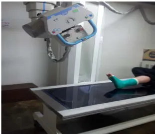
환자는 누운 자세 또는 앉은 자세로 한다. 종아리 (하퇴)부와 발꿈치 뒤에 detecter가 오도록 하고, 다리와 detecter의 중앙선이 일치하도록 한다. 발꿈치가 필름 아래모서리(하연)에서 2cm 정도 떨어지게 놓는다.
Ankle과 Foot이 90°가 되도록 Dorsiflexion시키고, Ankle 을 Medial쪽으로 45° Invert되도록 한다. Central Ray는 Ankle을 향해 입사하고 Tube angle은 caphalad로 25°, 30°, 35°, 45°로 각각 촬영하여 총 4장을 촬영한다 [Fig.
1].
Fig. 1. Inspection methods
3. 실험기기 및 실험조건
3.1 실험장비
1) X-ray발생장치: COMED EVA-HF525 2) Detecter: COMED KDR1000 (CCD Type)
3.2 촬영조건 70Kv, 8mAs
3.3 FFD 100cm
4. 영상 평가기준 및 평가방법
4.1 평가기준
1) 목말밑관절(subtalar joint)의 Posterior Facet Fracture, 발목발꿈치관절(talocalcaneal joint)을 포함하여 발꿈치가 보이는가?
2) 분쇄골절(comminuted fracture)의 발목관절의 관련 여부 및 목말뼈(talus)와 발뒤꿈치뼈의 뒤쪽 관절면 영 상에서 발꿈치뼈(Calcaneus)는 회전되지 않았는가?
3) 발꿈치뼈 골절(Calcaneus Fracture), Subtentaculum Tali Frature, 목말밑관절(subtalar joimt)이 열려져 보여야 하며, 발목뼈굴(sinus tarsi)이 넓게 나타나고 가쪽복사 와 뼈 밀도가 명확하게 나타날 수 있도록 적정조건에 서 촬영되었는가?
4) 검사에서는 후방 관절면의 앞부분이 가장 잘 나 타나고 Calcaneo Navicularcoalition, Talocal Canealcoalition, Naviculo Cuneiform coalition의 결합상태 와 발꿈치관절사이가 명확한가?
Fig. 2. Evaluation Criteria
4.2 평가방법
평가는 전문가 4명(정형외과 전문의 2명, 영상의학 과 전문의 2명)으로 구성하였으며, Broden법으로 setting한 자세에서 검사하여 영상을 획득하였다.
획득된 영상을 통하여 발목뼈굴(sinus tarsi)이 열린 정도를 판독하여 평가하였다 [Fig. 3]. 발꿈치뼈 골절 (Calcaneus Fracture), Subtentaculum Tali Frature , 목말밑 관절(subtalar joimt)과 발목발꿈치관절(talocalcaneal joint) 가 열려져 분리되는 정도를 각각 0점(분리안됨), 1점 (나쁨), 2점(보통), 3점(좋음)으로 평가하고 SPSS 12.0를 사용하여 분산분석(ANOVA)을 시행하였다 [Table 1.].
Fig. 3. degree of sinus tarsi open
Ⅲ. 결 과
한 2차 병원의 정형외과에 내원 또는 입원한 발목 부상 환자 중 석고처치를 받은 환자 40명의 남, 여 환 자에게서 획득한 영상을 PACS 모니터에서 평가하였 다. 25°의 영역에서는 목말밑관절(subtalar joimt)이 열려 져 분리 되지 않았고 또한 Subtentaculum Tali Frature에 걸쳐서 보였다. 나머지 평가는 양호하게 나타났다 [Fig.4].
30°영역에서는 발꿈치뼈 골절(Calcaneus Fracture), Subtentaculum Tali Frature, 목말밑관절(subtalar joimt)이 후방 관절면의 앞부분이 가장 잘 나타나고 Calcaneo Navicularcoalition, Talocal Canealcoalition, Naviculo Cuneiform coalition의 결합상태가 명확하게 보였다 [Fig. 5].
35°에서는 목말밑관절(subtalar joimt)분쇄골절, 목말뼈 (talus), 발뒤꿈치뼈의 뒤쪽 관절면이 명확하고 발목발 꿈치관절(talocalcaneal joint)분리정도가 양호하고 발목 뼈굴(sinus tarsi)이 넓게 관찰되는 영상을 얻었다 [Fig.
6].
45°영역에서는 연부조직이나 석고 분리를 판별할 수 있었고 가쪽복사와 뼈밀도가 명확한 영상이었으며 35°에 비해 발꿈치뼈(Calcaneus)가 왜곡되어 보였다 [Fig. 7].
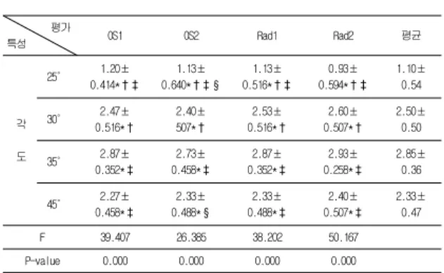
25°는 1.10±0.54, 30°는 2.50±0.50, 35°는 2.85±0.36, 45°
는 2.33±0.47 로 나타났고 통계적으로 유의한 차이를 보였다 (P<0.05, [Table 1.]). 35°영역에서 목말밑관절 (subtalar joimt)의 분쇄골절이 잘 분리되고 30°와 45° 영 역은 비슷한 점수를 나타냈지만 왜곡의 정도를 포함
시키면 30°영역의 왜곡이 적게 나타났고 40°영역은 심 한 왜곡현상을 보였다. 따라서 30~35°상이 가장 좋은 발(foot) Broden 촬영법에 해당하였다. 또한 30~35°상 이 발뒤꿈치 뼈뒤쪽 관절면, 발목뼈굴(sinus tarsi), 목말 밑관절(subtalar joimt)을 명확하게 묘출하였다.
Fig. 4. Angle 25° Fig. 5. Angle 30.°
Fig. 6. Angle 35° Fig. 7. Angle 45°
Table 1. Angle changes according to the observer Anova results
평가
특성 OS1 OS2 Rad1 Rad2 평균
각
도
25° 1.20±
0.414*†‡
1.13±
0.640*†‡§
1.13±
0.516*†‡
0.93±
0.594*†‡
1.10±
0.54
30° 2.47±
0.516*†
2.40±
507*†
2.53±
0.516*†
2.60±
0.507*†
2.50±
0.50
35° 2.87±
0.352*‡
2.73±
0.458*‡
2.87±
0.352*‡
2.93±
0.258*‡
2.85±
0.36
45° 2.27±
0.458*‡
2.33±
0.488*§
2.33±
0.488*‡
2.40±
0.507*‡
2.33±
0.47
F 39.407 26.385 38.202 50.167
P-value 0.000 0.000 0.000 0.000
* †‡§ by tukey comparisons test(p<0.05)
N 평균 표준
편차 표준 오차
95%신뢰구간 최
소 값
최 대
하한값 상한값 값
각도 25도
OS1 51 1.14 0.601 0.084 0.97 1.31 0 2
OS2 51 0.69 0.678 0.095 0.5 0.88 0 2
RD1 51 0.73 0.777 0.109 0.51 0.94 0 2
합계 153 0.85 0.714 0.058 0.74 0.96 0 2
각도 30도
OS1 51 1.65 0.658 0.092 1.46 1.83 1 3
OS2 51 1.57 0.755 0.106 1.36 1.78 0 3
RD1 51 1.8 0.775 0.109 1.59 2.02 0 3
합계 153 1.67 0.733 0.059 1.56 1.79 0 3
각도 35도
OS1 51 2.43 0.5 0.07 2.29 2.57 2 3
OS2 51 2.49 0.505 0.071 2.35 2.63 2 3
RD1 51 2.69 0.469 0.066 2.55 2.82 2 3
합계 153 2.54 0.5 0.04 2.46 2.62 2 3
각도 45도
OS1 51 2.12 0.621 0.087 1.94 2.29 1 3
OS2 51 1.75 0.659 0.092 1.56 1.93 1 3
RD1 51 1.78 0.757 0.106 1.57 2 1 3
합계 153 1.88 0.697 0.056 1.77 1.99 1 3
Table 2. Angle changes according to the observer scores
Ⅳ. 고 찰
발목의 병변을 관찰하는 단순 방사선촬영은 여러 가지가 있다. 그 중에서 Ankle Broden 검사는 발목에 대하여 대략 전방 40° 정도 기울어진 사방향으로 보이 게 한다. 이 검사에서는 정상적으로 Talocal Canealcoalition, Naviculo Cuneiform coalition결합과 관절 와연이 렌즈모양으로 겹치게 된다. 주위 연부조직을 포함하여 발의 구조를 보여주며, 섬세함과 우수한 시 각화를 제공하여 유용하게 이용된다. 또한 ankle broden 검사는 다리를 중립위치, 내전 또는 내전한 상 태에서 촬영할 수 있다. 발목의 내전 시 Subtentaculum Tali Frature , 목말밑관절(subtalar joimt)이 잘 보인다 [4].
또 다른 검사로 Grashey법으로 앞에서 기술한 ankle Broden 검사에서 정강뼈(Tibia)에 대해 측방으로 약 45°
및 다른 관절의 이상들이 쉽게 평가되어 진다. 그러나 사방향으로 촬영하는 것은 밀도의 급격한 변화뿐만 아니라 발목을 덮는 연부조직 밀도를 더 높아지게 만 든다. 따라서 Ankle AP 검사에 비해 골의 섬세함은 감 소한다. 내측 검사의 다른 단점은 전부 및 후부 관절 이 겹치는 것이다. 그래서 전위가 없는 Fracture병변 같 은 이상을 놓칠 수 있다. 추가로 정강뼈(Tibia)의 겹침 은 ankle AP 검사보다 더 평가하기 어려워진다[5]. 불안 정성이나 외상의 병력을 가진 환자의 경우, Hill sachs 변형이나 Trough defect, 관절와연 골절 등을 발견하기 좋은 촬영술을 선택하여야한다. 발꿈치뼈(Calcaneus)의 후측방이 손상되는 Hill Sachs defect, 발꿈치뼈 (Calcaneus)의 전면부의 Trough defect는 Shoulde AP(내 전한 상태), Garth view[6] , axillary view[6], Stryker notch view[7]에서 잘 보이며 전후 관절와연은 Axillary view나 그 변법촬영에서 잘 보인다. 전면 아래쪽 관절와는 어 깨촬영(West Point view)처럼 잘 보인다[8].
V. 결 론
CT나 MRI 영상의 관상면에서 발목발꿈치관절의 상 태를 증명할 수 있지만 본 연구는 발목 부상으로 석고 처치를 받은 성인 환자를 대상으로 일반촬영을 실시 함에 있어서 엑스선 각도 및 환자 position에 변화를 주어 발목 결합 및 발꿈치뼈 골절(Calcaneus Fracture), Subtentaculum Tali Frature , 목말밑관절, 발목뼈굴 영상 이 잘 표현될 수 있는 적합한 Broden 각도를 연구 하 였다. 본 연구를 통해 환자는 누운 자세 또는 앉은 자 세에서 발목뼈를 내측 (Lateral - Medio) 방향으로 45°로 하며, Tube angle은 caphalad로 30°~35° 주어 검사하는 것이 석고처치를 한 환자의 발목관절 영상을 얻는 데 가장 적합하다는 결론을 얻었다.
참 고 문 헌
[1] Munchen, Lehman, Grashey R, Atlas Typischer Rontgenfilder, R. Grashey.
[2] 최진석, " 주 5일제와 여가활동 변화에 따른 환경영향, 한국환경정책․평가 연구원, pp.20-24, 2002.
[4] Sanders et al, Imaging Techniques for the Evaluation of Glenohumeral Instability, Am J sports Vol. 28, Med,, p414~434, 2000.
[5] Munchen, Lehman, Grashey R, Atlas Typischer Rontgenfilder, R. Grashey 1923.
[6] Lawrence WS, A new position in radiographing the shoulder joint. Am J Roentgenol, Vol.2, p728~730.
[7] Hall RH, Isaac F, Booth CR, Dislocations of the shoulder with special reference to accompanying small fractures. J Bone Joint Surg, Vol.41A, 1959.
[8] Rokous JR, Feagin JA, Abbott HG, Modified axillary roentgenogram; A useful adjunct in the diagnosis of recurrent instability of the shoulder, Clin Orthop, Vol.82, pp84~86, 1972.