159
"J. Korean Soc. Radiol., Vol. 13, No. 2, April 2019"
The Effect of Advanced Modeling Iterative Reconstruction(ADMIRE) on the Quality of CT Images : Non-contrast CT in Chest
SangHeon Lee,1 HyoYeong Lee1,2*
1Department of Biomedical Science, Graduate School, Dong-Eui University
2Department of Radiological Science, Dong-Eui University
Received: February 18, 2019. Revised: April 26, 2019. Accepted: April 30, 2019
ABSTRACT
We examined the effect of Siemens ADMIRE (Advanced Modeled Iterative Reconstruction) on image quality by measuring changes in HU, noise, and SNR of background air, fat, muscle, and background signals on a chest CT scan. Experimental results show that as the ADMIRE Strength increases, the noise decreases and the signal increases, consequently the signal-to-noise ratio increases. ADMIRE can reduce noise by 28 ~ 61% compared to FBP, which is a conventional image reconstruction algorithm, and improves SNR by 16 ~ 100%.
Keywords: ADMIRE, HU, Noise, SNR
Ⅰ. INTRODUCTION
필터보정 역 투영법(filtered back projection, FBP) 은 CT에서 영상 재구성의 표준 알고리즘이었지만, 줄무늬 인공물(streak artifact)을 생성하고 낮은 선량 에서 영상의 잡음(noise)이 현저하게 증가하는 단점 이 있다.[1] 최근 컴퓨터 기술의 진보로 영상의 인공 물(artifact) 및 잡음을 줄이는 목적으로 고안된 반복 적 재구성법(Iterative Reconstruction, IR)이 임상에 적용되었다. 이 재구성 방법은 data를 한 번만 처리 하는 FBP와 달리 영상의 잡음을 줄이기 위해 raw data 또는 reconstructed data를 반복적으로 처리하는 데 사용되는 알고리즘(algorithm)을 총칭하며, 다양 한 알고리즘이 임상에 도입되고 있으며 상당한 선 량 감소의 가능성을 보여준다.[2] IR은 새로운 것이 아니며 1970년대 CT 도입 초기에 데이터 재구성을 위해 처음 제안된 방법이었으나, CT의 많은 양의 데이터와 수학적으로 계산해 낼 수 있는 기술의 한 계로 임상에 적용할 수 없었다.[3] 제조사별로 IR 알 고리즘은 다르지만, 영상의 잡음을 줄여 영상의 질
을 개선한다는 목적은 동일하다.[4] 그리고 IR을 적 절히 이용하면 기존의 FBP와 비교해서 영상의 질 을 유지하면서 환자 선량을 줄이거나, 영상의 질이 향상된다는 많은 연구가 있었다.[5-19] 이후 개발된 Siemens 사의 3세대 IR 인 고급 모델링 반복 재구 성법(ADMIRE, Siemens Medical Solutions, Forchheim, Germany)은 현재 임상에서 이용 가능하며, ADMIRE 알고리즘은 원시 프로젝션 데이터(raw projection data)와 이미지 데이터(image data)에 대한 통계 모델(statistical model)뿐만 아니라, 앞으로 투영 (projection)을 위한 시스템 모델(system model)을 구현 하므로 소위 모델 기반 반복 재구성(model - based iterative reconstruction, MBIR) 알고리즘의 범주에 포함된다.[20]
본 연구는 Siemens 사의 ADMIRE가 영상의 화질 에 미치는 영향을 알아보기 위하여 흉부 비조영 CT 검사에서 ADMIRE Strength에 의한 Signal, Noise 그리고 SNR(signal-to-noise ratio)의 변화를 정 량적으로 비교 분석하여 어떠한 차이가 있는지 알 아보고자 한다.
* Corresponding Author: HyoYeong Lee E-mail: [email protected] Tel: +82-51-890-2679 https://doi.org/10.7742/jksr.2019.13.2.159
Ⅱ. MATERIAL AND METHODS
2018년 12월 1일부터 2019년 1월 31일까지 양산 P 병원 건강증진센터(health promotion center, HPC) 에서 질병의 조기발견(early finding)을 목적으로 흉 부 비조영 CT 검사를 받은 48명의 환자를 대상으 로 하였다. 평균 나이 60.8±11.2세, 평균 키 168.6±
8.8 cm, 평균 몸무게 67.9±10.7 kg 그리고 성별은 남자 27명, 여자 21명이었다. 위 대상 환자는 대한 비만학회 비만 치료지침(2012) 기준의 따라 신체 질량지수(body mass index, BMI)가 정상인 환자를 대상으로 선별하였다.
CT 검사에 사용된 장비는 192-slice dual energy CT (Somatom Force, Siemens Medical Solutions, Forchheim, Germany)이며, 흉부 비조영 CT 검사를 대상으로 하였다. 관전압은 120 kVp, 관전류 40 Eff. mAs로 고정 관전류 기법을 사용하였으며, CTDIvol(32 cm)은 2.67 mGy로 설정하였다. 본 연구 의 대상자 48명의 평균 DLP (dose length product)는 108.2±15.8 mGy·cm였다. 그리고 갠트리(gantry) 회 전속도 0.5 s/rotation, 절편 두께(slice thickness) 1.5 mm, 피치(pitch) 1.2, 콜리메이션(collimation) 192 × 0.6 mm, 관측시야(field of view, FOV) 400 mm로 하 였다.
획득한 영상은 mediastinum Window, Br54 Kernel 으로 재구성하였다. IR은 Siemens 사의 ADMIRE를 사용하였으며, ADMIRE의 ADMIRE Strength는 0부 터 5까지 조절할 수 있다. 본 연구에서는 ADMIRE 0, 1, 3, 5로 재구성하였다. ADMIRE 0은 ADMIRE 를 적용하지 않은 기존의 재구성 방법인 FBP이다.
ADMIRE가 CT 영상의 화질에 미치는 영향을 알 아보기 위하여 ADMIRE Strength에 따른 Signal, Noise 그리고 SNR을 비교하였다. 관심영역(region of interest, ROI)은 공기(Air), 지방(Fat), 근육(Muscle) 그리고 Background (BG)로 하였다. Signal은 관심영 역 안의 하운스필드 유닛(Hounsfield unit, HU)의 평 균값(mean), Nosie는 관심영역 안의 HU의 표준편차 (standard deviation, SD) 그리고 SNR은 각 관심영역 의 HU의 평균값(Signal)을 관심영역의 표준편차 (Noise)로 나눈 값으로 정의하였으며[21] Eq. (1)로
나타냈다.
SNR =
(1)
정량적인 평가를 하기 위하여 CT Workstation (Syngo.via, Siemens Medical Solutions, Forchheim, Germany)과, Segmentation Tools의 자동 컨투어링 사용(enable automatic contouring) 기능을 적용하여 환자의 몸 전체를 관심 영역으로 설정하였다. 관심 영역 안의 각 픽셀(pixel)의 HU의 평균값과 HU의 표 준편차를 구하기 위해서 Region growing 기능을 이 용하여 공기, 지방, 근육의 문턱 값(threshold value) 을 설정하였다. 공기의 문턱 값은 -1024 ~ -200 HU, 지방의 문턱 값은 -200 ~ -40 HU, 근육의 문턱값 -50 ~ 142 HU 로 Workstation에 입력된 값으로 측정 하였으며 Fig. 1, 2와 같다.
통계분석은 PASW (PASW statistics, ver. 18.0, SPSS, Chicago, USA)를 이용하여 일원 배치 분산분 석(one-way ANOVA)을 실시하였으며, 유의한 차이 는 Tukey의 사후분석(post hoc analysis)으로 확인하 였다. 또한 신뢰구간은 95%로 p-value는 0.05 미만 일 때 통계적으로 유의한 차이가 있다고 하였다.
Ⅲ. RESULT
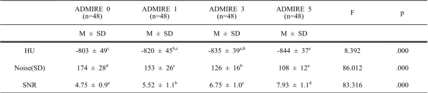
1. Air의 HU(Hounsfield unit)와 Noise 그리고 SNR ADMIRE Strength에 따른 Air의 HU와 Noise 그리 고 SNR을 분석하였다. 먼저 HU를 분석한 결과 ADMIRE Strength 간에 유의한 차이가 있는 것으로 나타났다(F=8.392, p<0.001). 추가로 사후분석을 한 결과 ADMIRE 0과 ADMIRE 1은 차이가 없었다 (p=0.199).
그러나 ADMIRE 0 (M=-803) 은 ADMIRE 3 (M=
-835), 5 (M=-844) 보다 HU가 통계적으로 유의하게 높게 나타났으며(p=0.002, p=0.000), Table 1과 같다.
Noise와 ADMIRE Strength 간에는 유의한 차이가 있는 것으로 나타났다(F=83.012, p<0.001). 추가로 사후분석을 한 결과 ADMIRE 0에서 5로 갈수록 Noise는 통계적으로 유의하게 나타났고 Table 1과
"J. Korean Soc. Radiol., Vol. 13, No. 2, April 2019"
같다. SNR과 ADMIRE Strength 간에는 유의한 차이 가 있는 것으로 나타났다(F=83.316, p<0.001). 추가 로 사후분석을 한 결과 ADMIRE 0에서 5로 갈수록 Noise는 통계적으로 유의하게 나타났고 Table 1과 같다.
Air의 HU는 ADMIRE 0과 비교하였을 때 ADMIRE 1은 차이가 없지만, ADMIRE 3은 3%, ADMIRE 5 는 5% 감소하였다. Air의 Noise는 ADMIRE Strength가 증가할수록 13%, 38%, 61% 감 소하였다. Air의 SNR은 ADMIRE Strength가 증가할 수록 6%, 42%, 66% 증가하였다.
2. Fat의 HU(Hounsfield unit)와 Noise 그리고 SNR ADM IRE Strength에 따 른 Fat의 HU와 Noise 그 리 고 SNR을 분석하였다. HU를 분석한 결과 ADMIRE Strength 간에 유의한 차이가 있는 것으로 나타났다(F=20.166, p<0.001). 추가로 사후분석을 한 결과 ADMIRE 0과 ADMIRE 1에는 차이가 없었다 (p=0.701). 그러나 ADMIRE 0 (M=-103) 은 ADMIRE 3 (M=-98), 5 (M=-94) 보다 HU가 통계적으로 유의 하게 낮게 나타났고(p=0.000, p=0.000) Table 2와 같 다. Noise와 ADMIRE Strength 간에는 유의한 차이 가 있는 것으로 나타났다(F=472.729, p<0.001). 추가 로 사후분석을 한 결과 ADMIRE 0에서 5로 갈수록 Noise는 통계적으로 유의하게 낮게 나타났고 Table 2와 같다. SNR과 ADMIRE Strength 간에는 유의한 차이가 있는 것으로 나타났다(F=75.064, p<0.001).
추가로 사후분석을 한 결과 ADMIRE 0에서 5로 갈 수록 Noise는 통계적으로 유의하게 높게 나타났고 Table 2와 같다.
결과적으로 Fat의 HU는 ADMIRE 0과 비교하였 을 때 ADMIRE 1은 차이가 없지만, ADMIRE 3 은 5%, ADMIRE 5는 9% 증가하였다. Fat의 Noise는 ADMIRE Strength가 증가할수록 4%, 17%, 41% 감 소하였다. Fat의 SNR은 ADMIRE Strength가 증가할 수록 25%, 50%, 100% 증가하였다.
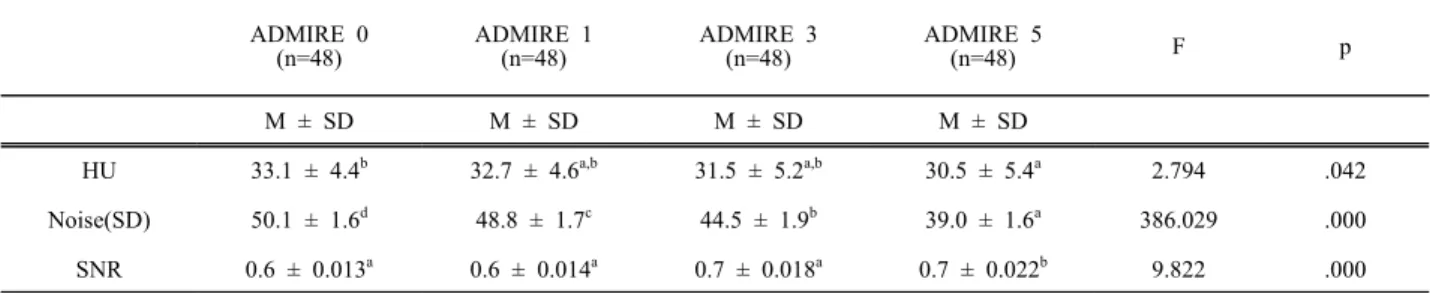
3. Muscle의 HU(Hounsfield unit)와 Noise 그리고 SNR
ADMIRE Strength에 따른 Muscle의 HU와 Noise
그리고 SNR을 분석하였다. 먼저 HU를 분석한 결 과 ADMIRE Strength 간에 유의한 차이가 있는 것 으로 나타났다(F=2.794, p=0.042). 추가로 사후분석 을 한 결과 ADMIRE 0과 ADMIRE 1, 3 에는 차이 가 없었다(p=0.976, p=365). 그러나 ADMIRE 0 (M=33.1)은 ADMIRE 5 (M=30.5)보다 HU가 통계적 으로 유의하게 낮게 나타났고(p=0.049) Table 3과 같다.
Noise와 ADMIRE Strength 간에는 유의한 차이가 있는 것으로 나타났다(F=386.029, p<0.001). 추가로 사후분석을 한 결과 ADMIRE 0에서 5로 갈수록 Noise는 통계적으로 유의하게 낮게 나타났고 Table 3과 같다.
SNR과 ADMIRE Strength 간에는 유의한 차이가 있는 것으로 나타났다(F=9.822, p<0.001). 추가로 사 후분석을 한 결과 ADMIRE 0과 ADMIRE 1, 3에는 차이가 없었다(p=0.983, p=237). 그러나 ADMIRE 0 (M=0.6)은 ADMIRE 5 (M=0.7) 보다 HU가 통계적 으로 유의하게 낮게 나타났고(p=0.000) Table 3과 같다.
결과적으로 Muscle의 HU는 ADMIRE 0과 비교하 였을 때 ADMIRE 1, 3은 차이가 없지만, ADMIRE 5 는 8% 감소하였다. Muscle의 Noise는 ADMIRE Strength가 증가할수록 3%, 13%, 28% 감소하였다.
Muscle의 SNR은 ADMIRE 0과 비교하였을 때 ADMIRE 1, 3은 차이가 없지만, ADMIRE 5는 16%
증가하였다.
4. Background(BG)의 HU(Hounsfield unit)와 Noise 그리고 SNR
ADMIRE Strength에 따른 Background의 HU와 Noise 그리고 SNR을 분석하였다. 먼저 HU를 분석 한 결과 ADMIRE Strength 간에 유의한 차이가 있 는 것으로 나타났다(F=116.208, p<0.001). 추가로 사 후분석을 한 결과 ADMIRE 0에서 ADMIRE 5로 갈 수록 HU는 통계적으로 유의하게 낮게 나타났고 Table 5와 같다.
Noise와 ADMIRE Strength 간에는 유의한 차이가 있는 것으로 나타났다(F=24.971, p<0.001). 추가로 사후분석을 한 결과 ADMIRE 0에서 5로 갈수록
Noise는 통계적으로 유의하게 낮게 나타났다. 그러 나 ADMIRE 0과1은 차이가 없었고(p=0.412) Table 4와 같다.
SNR과 ADMIRE Strength 간에는 유의한 차이가 있는 것으로 나타났다(F=92.592, p<0.001). 추가로 사후분석을 한 결과 ADMIRE 0에서 5로 갈수록 Noise는 통계적으로 유의하게 높게 나타났고 Table 4와 같다. 그러나 ADMIRE 0과 1은 차이가 없었고
(p=0.509) Table 4와 같다. 결과적으로 Background의 HU는 ADMIRE Strength가 증가할수록 0.3%, 1.1%, 1.7% 감소하였다. Background의 Noise는 ADMIRE Strength가 증가할수록 11%, 46%, 109% 감소하였다.
Background의 SNR은 ADMIRE 0과 비교하였을 때 ADMIRE 1은 차이가 없지만, ADMIRE 3은 51%, ADMIRE 5는 234% 증가하였다.
(A) (B) (C) (D)
Fig. 1. By applying the automatic contouring function of Syngovia workstation Segmentation Tools, the entire body of the patient is set as an area of interest(A). Refining segmentation objects by adjusting thresholds (B) Air, (C) Fat, (D) Muscle.
(A) Air (B) Fat (C) Muscle
Fig. 2. Quantitative measurement using threshold value in ROI.
Table 1. Comparison of HU, noise and SNR difference of air by ADMIRE (N=192)
ADMIRE 0 (n=48)
ADMIRE 1 (n=48)
ADMIRE 3 (n=48)
ADMIRE 5
(n=48) F p
M ± SD M ± SD M ± SD M ± SD
HU -803 ± 49c -820 ± 45b,c -835 ± 39a,b -844 ± 37a 8.392 .000
Noise(SD) 174 ± 28d 153 ± 26c 126 ± 16b 108 ± 12a 86.012 .000
SNR 4.75 ± 0.9a 5.52 ± 1.1b 6.75 ± 1.0c 7.93 ± 1.1d 83.316 .000
* a<b<c<d : Turkey's multiple comparison
"J. Korean Soc. Radiol., Vol. 13, No. 2, April 2019"
(A) HU (B) Noise (C) SNR
Fig. 3. Average chart of air by ADMIRE Strengh.
Table 2. Comparison of HU, noise and SNR difference of fat by ADMIRE (N=192)
ADMIRE 0 (n=48)
ADMIRE 1 (n=48)
ADMIRE 3 (n=48)
ADMIRE 5
(n=48) F p
M ± SD M ± SD M ± SD M ± SD
HU -103 ± 5.3a -102 ± 5.5a -98 ± 6.0b -94 ± 6.6c 20.166 .000
Noise(SD) 41.3 ± 1.4d 39.7 ± 1.6c 35.2 ± 1.9b 29.2 ± 1.8a 472.729 .000 SNR 0.04 ± 0.01a 0.05 ± 0.01b 0.06 ± 0.01c 0.08 ± 0.01d 75.064 .000
* a<b<c<d : Turkey's multiple comparison
(A) HU (B) Noise (C) SNR
Fig. 4. Average chart of fat by ADMIRE Strengh.
Table 3. Comparison of HU, noise and SNR difference of muscle by ADMIRE (N=192)
ADMIRE 0 (n=48)
ADMIRE 1 (n=48)
ADMIRE 3 (n=48)
ADMIRE 5
(n=48) F p
M ± SD M ± SD M ± SD M ± SD
HU 33.1 ± 4.4b 32.7 ± 4.6a,b 31.5 ± 5.2a,b 30.5 ± 5.4a 2.794 .042
Noise(SD) 50.1 ± 1.6d 48.8 ± 1.7c 44.5 ± 1.9b 39.0 ± 1.6a 386.029 .000
SNR 0.6 ± 0.013a 0.6 ± 0.014a 0.7 ± 0.018a 0.7 ± 0.022b 9.822 .000
* a<b<c<d : Turkey's multiple comparison
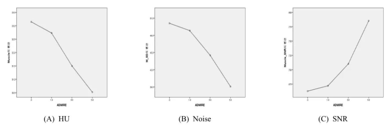
(A) HU (B) Noise (C) SNR Fig. 5. Average Chart of Muscle by ADMIRE Strengh.
Table 4. Comparison of HU, noise and SNR difference of BG by ADMIRE (N=192)
ADMIRE 0 (n=48)
ADMIRE 1 (n=48)
ADMIRE 3 (n=48)
ADMIRE 5
(n=48) F p
M ± SD M ± SD M ± SD M ± SD
HU -981 ± 6.3d -984 ± 5.5c -992 ± 3.9b -998 ± 2.9a 116.208 .000
Noise(SD) -21.4 ± 6.8c -19.2 ± 6.6c -14.7 ± 6.7b -10.2 ± 7.1a 24.971 .000
SNR 49 ± 12.4a 55 ± 14.0a 74 ± 19.0b 115 ± 33.4c 92.592 .000
* a<b<c<d : Turkey's multiple comparison
(A) HU (B) Noise (C) SNR
Fig. 6. Average chart of BG by ADMIRE Strengh.
Ⅳ. DISCUSSION
CT 장비 제조사별로 IR 알고리즘은 다르지만, 영 상의 잡음을 줄여 영상의 질을 개선한다는 목적은 동일하다.[4] 흉부 CT 검사에서 IR을 적절히 이용하 면 기존의 FBP와 비교해서 영상의 질을 보존하면 서 환자 선량이 감소 가능하다는 것은 이미 선행
연구에서 보고되었다.[22-29] 이러한 연구들은 Phantom 또는 커대버(cadaver)를 이용하거나, 임상연구의 경 우는 어떤 질환이나 특정 관심영역(ROI)을 그려 IR 적용시 영상에 미치는 영향을 주관적으로 평가하 는 것이 대부분이다.
본 연구는 Siemens 사의 고급 모델링 반복 재구 성법(Advanced Modeled Iterative Reconstruction,
"J. Korean Soc. Radiol., Vol. 13, No. 2, April 2019"
ADMIRE)이 영상의 화질에 미치는 영향을 알아보 기 위하여 흉부 비조영 CT 검사에서 ADMIRE Strength에 의한 Air, Fat, Muscle, Background의 Signal, Noise 그리고 SNR의 변화를 정량적으로 비 교 분석하여 어떠한 차이가 있는지 알아보고자 하 였다.
연구 결과 영상의 잡음은 FBP(ADMIRE 0)를 기 준으로 ADMIRE 1, 3, 5를 비교하였을 때, ADMIRE Strength가 증가할수록 Air의 Noise는 13%, 38%, 61%, Fat의 Noise는 4%, 17%, 41%, Muscle의 Noise 는 3%, 13%, 28% 그리고 Background의 Noise는 11%, 46%, 109% 감소하였다. 이는 흉부 CT에서 IR 의 적용은 영상의 잡음을 크게 감소시킨다는 선행 연구와 결과가 일치하였다.[24,30,31]
영상의 SNR은 FBP(ADMIRE 0)를 기준으로 ADMIRE 1, 3, 5를 비교하였을 때, ADMIRE Strength가 증가할수록 Air의 SNR은 6%, 42%, 66%, Fat의 SNR은 25%, 50%, 100% 증가하였다. 하지만 Muscle의 SNR은 ADMIRE 0과 비교하였을 때 ADMIRE 1, 3은 차이가 없지만, ADMIRE 5는 16%
증가하였다. 그리고 Background의 SNR은 ADMIRE 0과 비교하였을 때 ADMIRE 1은 차이가 없지만, ADMIRE 3 은 51%, ADMIRE 5 는 234% 증가하였 다. Hu 등은 흉부 비조영 CT 검사에서 IR을 적용하 면 40% 방사선량을 감소시킬 수 있으며, Siemens 사의 IR 인 IRIS (Iterative Reconstruction in Image Space)를 적용하면 기존의 FBP와 비교해서 영상의 잡음, SNR, 대조도 대 잡음비(contrast to noise ratio, CNR)가 크게 개선된다고 보고한 것과 일치하는 결 과이다.[22]
영상의 HU는 FBP를 기준으로 ADMIRE 1, 3, 5를 비교하였을 때, ADMIRE Strength가 증가할수록 Air 의 HU는 ADMIRE 0과 비교하였을 때 ADMIRE 1은 차이가 없지만, ADMIRE 3은 3%, ADMIRE 5는 5%
감소하였다. Fat의 HU는 ADMIRE 0과 비교하였을 때 ADMIRE 1은 차이가 없지만, ADMIRE 3은 5%, ADMIRE 5는 9% 증가하였다. Muscle의 HU는 ADMIRE 0과 비교하였을 때 ADMIRE 1, 3은 차이 가 없지만, ADMIRE 5는 8% 감소하였다.
Background의 HU는 ADMIRE Strength가 증가할수
록 0.3%, 1.1%, 1.7% 감소하였다. 여기서 특이한 점 은 ADMIRE Strength가 증가할수록 Air, Muscle, Background의 HU는 증가하였으나, Fat의 HU는 감 소했다는 점이다.
Omoumi Patrick 등은 경추(cervical spine) CT 검사 에서 저 선량으로 검사를 하여 영상의 질을 기존의 검사와 유사하게 하기 위한 Siemens 사의 SAFIRE (Sinogram Affirmed Iterative Reconstruction) Strength 수준을 연구하였는데, SAFIRE Strength가 증가할수 록 신호 대 잡음비와 대조도 대 잡음비는 증가하였 지만, 연한 조직(soft tissue)은 SAFIRE Strength가 증 가할수록 감소하였다는 연구와 관련이 있다고 생 각된다.[32]
본 연구에서 Air, Muscle, Background는 ADMIRE Strength가 높아질수록 잡음은 감소하고 신호는 증 가하여, 결론적으로 신호 대 잡음비는 증가하는 것 으로 나타났다. 하지만 Fat은 ADMIRE Strength가 높아질수록 잡음은 감소하고 신호도 감소하지만, 신호 대 잡음비는 증가하는 결과를 나타낸다. 이는 신호감소의 폭 보다 영상잡음의 감소가 더 크기 때 문에 결론적으로 신호 대 잡음비가 증가한다고 볼 수 있다.
Ⅴ. CONCLUSION
ADMIRE는 기존의 영상 재구성 알고리즘인 FBP 와 비교하여 Noise를 28~61%까지 줄일 수 있으며, SNR은 16~100%까지 향상시킨다. 이러한 연구를 기반으로 ADMIRE 적절한 사용은 낮은 선량으로 영상의 잡음이 적은 질 좋은 영상 획득이 가능하 며, CT 검사에서 환자가 받는 방사선량을 최소화하 는데 도움이 될 것이다.
Reference
[1] Hsieh, Jiang, et al., "Recent advances in CT image reconstruction," Current Radiology Reports, Vol. 1, No. 1, pp. 39-51, 2013.
[2] Beister, Marcel, Daniel Kolditz, Willi A. Kalender.,
"Iterative reconstruction methods in X-ray CT,"
Physica medica, Vol. 28, No. 2, pp. 94-108, 2012.
[3] Hounsfield, Godfrey N., "Computerized transverse axial scanning (tomography): Part 1. Description of
system," The British journal of radiology, Vol. 4, No. 6, pp. 552, 1016-1022, 1973.
[4] Geyer, Lucas L., et al. "State of the art: iterative CT reconstruction techniques." Radiology, Vol. 276, No.
2, pp. 339-357, 2015.
[5] Willemink, Martin J., et al., "Iterative reconstruction techniques for computed tomography Part 1: technical principles," European radiology, Vol. 23, No. 6, pp.
1623-1631, 2013.
[6] Vardhanabhuti, Varut, et al., "Image comparative assessment using iterative reconstructions: clinical comparison of low-dose abdominal/pelvic computed tomography between adaptive statistical, model-based iterative reconstructions and traditional filtered back projection in 65 patients," Investigative radiology, Vol. 49, No. 4, pp. 209-216, 2014.
[7] Pickhardt, Perry J., et al., "Abdominal CT with model-based iterative reconstruction (MBIR): initial results of a prospective trial comparing ultralow-dose with standard-dose imaging." American journal of roentgenology, Vol. 199, No. 6, pp. 1266-1274, 2012.
[8] Ploussi, Agapi, et al., "Patient radiation exposure and image quality evaluation with the use of iDose4 iterative reconstruction algorithm in chest–abdomen–
pelvis CT examinations," Radiation protection dosimetry, Vol. 158, No. 4, pp. 399-405, 2013.
[9] Fontarensky, Mikael, et al., "Reduced radiation dose with model-based iterative reconstruction versus standard dose with adaptive statistical iterative reconstruction in abdominal CT for diagnosis of acute renal colic," Radiology, Vol. 276, No. 1, pp.
156-166, 2015.
[10] Gervaise, Alban, et al. "Low-dose CT with
automatic tube current modulation, adaptive statistical iterative reconstruction, and low tube voltage for the diagnosis of renal colic: impact of body mass index," American Journal of Roentgenology, Vol.
202, No. 3, pp. 553-560, 2014.
[11] Prakash, Priyanka, et al., "Reducing abdominal CT radiation dose with adaptive statistical iterative reconstruction technique," Investigative radiology, Vol. 45, No. 4, pp. 202-210, 2010.
[12] Padole, Atul, et al., "CT radiation dose and iterative reconstruction techniques." American Journal of
Roentgenology, Vol. 204, No. 4, pp. 384-392, 2015.
[13] Singh, Sarabjeet, et al., "Abdominal CT: comparison of adaptive statistical iterative and filtered back projection reconstruction techniques," Radiology, Vol.
257, No. 2, pp. 373-383, 2010.
[14] May, Matthias S., et al., "Dose reduction in abdominal computed tomography: intraindividual comparison of image quality of full-dose standard and half-dose iterative reconstructions with dual-source computed tomography," Investigative radiology, Vol. 46, No. 7, pp. 465-470, 2011.
[15] Schabel, Christoph, et al., "Clinical evaluation and potential radiation dose reduction of the novel sinogram-affirmed iterative reconstruction technique (SAFIRE) in abdominal computed tomography angiography," Academic radiology, Vol. 20, No. 2, pp. 165-172, 2013.
[16] Kilic, K., et al., "Lowering the dose in head CT using adaptive statistical iterative reconstruction,"
American journal of neuroradiology, Vol. 32, No. 9, pp. 578-1582, 2011.
[17] Vorona, G. A., et al., "The use of adaptive statistical iterative reconstruction in pediatric head CT: a feasibility study," American Journal of Neuroradiology, Vol. 34, No. 1, pp. 205-211, 2013.
[18] Kaza, Ravi K., et al. "CT enterography at 80 kVp with adaptive statistical iterative reconstruction versus at 120 kVp with standard reconstruction: image quality, diagnostic adequacy, and dose reduction,"
American Journal of Roentgenology, Vol. 198. No.
5, pp. 1084-1092, 2012.
[19] Goldman, Alice R., Pierre D. Maldjian, "Reducing radiation dose in body CT: a practical approach to optimizing CT protocols," American Journal of Roentgenology, Vol. 200, No. 4, pp. 48-754, 2013.
[20] Solomon, Justin, et al., "Diagnostic performance of an advanced modeled iterative reconstruction algorithm for low-contrast detectability with a third-generation dual-source multidetector CT scanner: potential for radiation dose reduction in a multireader study," Radiology, Vol. 275, No. 3, pp.
735-745, 2015.
[21] Greffier, Joël, et al., "CT dose reduction using automatic exposure control and iterative
"J. Korean Soc. Radiol., Vol. 13, No. 2, April 2019"
reconstruction: a chest paediatric phantoms study,"
Physica Medica, Vol. 32, No. 4, pp. 582-589, 2016.
[22] Hu, X. H., et al., "Radiation dose of non-enhanced chest CT can be reduced 40% by using iterative reconstruction in image space," Clinical radiology, Vol. 66, No. 11, pp. 1023-1029, 2011.
[23] Katsura, Masaki, et al., "Model-based iterative reconstruction technique for radiation dose reduction in chest CT: comparison with the adaptive statistical iterative reconstruction technique," European
radiology, Vol. 22, No. 8, pp. 1613-1623, 2012.
[24] Katsura, Masaki, et al., "Model-based iterative reconstruction technique for ultralow-dose chest CT:
comparison of pulmonary nodule detectability with the adaptive statistical iterative reconstruction technique," Investigative radiology, Vol. 48, No. 4, pp. 206-212, 2013.
[25] Leipsic, Jonathon, et al., "A prospective evaluation of dose reduction and image quality in chest CT using adaptive statistical iterative reconstruction,"
American Journal of Roentgenology, Vol. 195, No.
5, pp. 1095-1099, 2010.
[26] Pontana, François, et al., "Chest computed
tomography using iterative reconstruction vs filtered back projection (Part 1): evaluation of image noise reduction in 32 patients," European radiology, Vol.
21, No. 3, pp. 627-635, 2011.
[27] Prakash, Priyanka, et al., "Diffuse lung disease: CT of the chest with adaptive statistical iterative reconstruction technique," Radiology, Vol. 256, No.
1, pp. 261-269, 2010.
[28] Singh, Sarabjeet, et al., "Adaptive statistical iterative reconstruction technique for radiation dose reduction in chest CT: a pilot study," Radiology, Vol. 258, No. 2, pp. 565-573, 2011.
[29] Vardhanabhuti, Varut, et al., "Image quality
assessment of standard-and low-dose chest CT using filtered back projection, adaptive statistical iterative reconstruction, and novel model-based iterative reconstruction algorithms," American Journal of Roentgenology, Vol. 200, No. 3, pp. 545-552, 2013.
[30] Yuki, Hideaki, et al., "Clinical impact of
model-based type iterative reconstruction with fast reconstruction time on image quality of low-dose
screening chest CT," Acta Radiologica, Vol. 57, No.
3, pp. 295-302, 2016.
[31] Jensen, Kristin, et al., "Image quality in oncologic chest computerized tomography with iterative reconstruction: a phantom study," Journal of computer assisted tomography, Vol. 40, No. 3, pp.
351-356, 2016.
[32] Omoumi, Patrick, et al., "Low-dose multidetector computed tomography of the cervical spine:
optimization of iterative reconstruction strength levels," Acta Radiologica, Vol. 55, No. 3, pp.
335-344, 2014.
[33] Ott, Julien G., et al., "Update on the non-prewhitening model observer in computed tomography for the assessment of the adaptive statistical and model-based iterative reconstruction algorithms," Physics in Medicine & Biology, Vol.
59, No. 15, pp. 4047-4064, 2014.
[34] Verdun, F. R., et al., "Image quality in CT: From physical measurements to model observers," Physica Medica, Vol. 31, No. 8, pp. 823-843, 2015.
[35] Qiu, D., E. Seeram., "Does iterative reconstruction improve image qual-ity and reduce dose in computed tomography," Radiology, Vol. 1, No. 2, pp. 42-54, 2016.
고급 모델링 반복 재구성법(ADMIRE)이 CT 영상의 화질에 미 치는 영향: 흉부 비조영 CT에서
이상헌,1 이효영1,2,*
1동의대학교 대학원 보건의과학과
2동의대학교 방사선학과
흉부 CT스캔에서 공기, 지방, 근육, Background의 HU, 잡음 그리고 신호 대 잡음비의 변화를 측정해 영 상 화질에 미치는 지멘스 ADMIRE(Advanced Modeled Iterative Reconstruction)의 영향을 조사했다. 실험 결 과에 따르면 ADMIRE Strength가 커질수록 잡음은 감소하고 신호는 증가하여, 결론적으로 신호 대 잡음비 는 증가하는 것으로 나타났다. ADMIRE는 기존 영상 재구성 알고리즘인 FBP와 비교하여 잡음을 28~61%까 지 줄일 수 있으며, 신호 대 잡음비는 16~100%까지 향상시킨다.
중심단어: 고급모델링 반복적 재구성, Hounsfield unit, 잡음, 신호 대 잡음비
성명 소속 직위
(제1저자) 이상헌 동의대학교 대학원 보건의과학과 대학원생(박사과정)
(교신저자) 이효영 동의대학교 방사선학과 교수
연구자 정보 이력