Total Phenolic Contents, Radical Scavenging Capacities and Inhibitory Effects on Lipid Peroxidation and LDL Oxidation of Prunus persica Branch
Hyo Seung Yi, Won Hwan Park, Sun Hee Lim
1, Jin Young Moon
1*
Medical Science Research Center for Cardiovascular Disease, 1 : College of Oriental Medicine, Dongguk University
This study was undertaken to elucidate the antioxidant activity of the ethanol (EEPB) and water (WEPB) extracts of Prunus persica branches. The extracts contained a high phenolic content and revealed a potent hydrogen donating activity in DPPH scavenging assay. Compared to α-tocopherol, EEPB (p < 0.001) and WEPB (p < 0.05) significantly inhibited FeCl
2-ascorbic acid-induced lipid peroxidation, and also exhibited potent antiradical activities against hydroxyl radical, superoxide anion, nitric oxide and peroxynitrite. In copper- and AAPH-mediated human low-density lipoprotein (LDL) oxidation systems, the extracts demonstrated a strong antioxidant function by metal chelating, rather than direct scavenging, action. Furthermore, EEPB at 5 μg/mL concentration showed 80.77% inhibition of the electrophoretic mobility of LDL, compared to 77.69% for ascorbic acid and 76.92% for BHT. These results suggest that PB branch extracts may protect against oxidative stress-induced diseases
Key words : Prunus persica antioxidant; lipid peroxidation low-density lipoprotein (LDL)
* To whom correspondence should be addressed at : Jin-Young Moon, College of Oriental Medicine, Dongguk University, Sukjang-Dong 707, Kyungju 780-714, Korea.
․E-mail : [email protected], ․Tel : 054-770-2665
․Received : 2008/07/22 ․Revised : 2008/08/06 ․Accepted : 2008/08/29
Introduction
In biological systems, the overproduction of reactive oxygen species (ROS) and reactive nitrogen species (RNS) induces oxidative damage to various biomolecules, including phospholipids, proteins and DNA, which areimplicated in the pathogenesis and progression of cardiovascular diseases
1). Several cell culture studies and animal model have shown that oxidative modification of phospholipids, especially low-density lipoprotein (LDL), can damage vascular endothelial cells, induce cell adhesion molecules, and form foam cells, which play a central role in initiating and promoting atherosclerosis
2-4). On the other hand, it has been suggested that natural antioxidants derived from plant and food sources are beneficial in protecting against oxidative damage, and play a versatile role in minimizing the risk of atherosclerosis and myocardial infarction
5-6). Accordingly, growing attention has been focused on the role of natural antioxidants in the management of atherosclerosis.
The peach tree, Prunus persica L. Batsch (PB), is commonly cultivated in Korea and other parts of the world. It
has been reported that parts of the peach tree, particularly the seed and fruit have antiradical
7), anticholinesterase
8-9)and antitumor
10)activities. The PB branch, called Dojee in Korea, is used as a natural dye and has also been used as a medicinal herb in the treatment of cardiovascular diseases such as paroxysmalcardiodynia and cardialgia in traditional medicine
11). However, the phytochemical and pharmacological activities of the PB branch extracts have not yet been demonstrated. In the present study, we evaluated the antioxidant activity of PB branch extracts using free radical-induced, lipid peroxidation and LDL oxidation systems.
Moreover, we determined their total phenolic contents for preliminary chemical studies.
Materials and Methods
1. Chemicals
2,2-Diphenyl-1-picryl-hydrazyl (DPPH), butylated
hydroxytoluene (BHT), dimethyl sulfoxide (DMSO),
ethylenediaminetetraacetic acid (EDTA), L-ascorbic acid, α
-tocopherol, hydrogen peroxide (H
2O
2), nitro blue tetrazolium
(NBT), trichloroacetic acid (TCA), 2-thiobarbituric acid (TBA),
hypoxanthine, xanthine oxidase, bicinchoninic acid kit,
DL-penicillamine, diethylenetriaminepentaacetic acid (DTPA),
4,5-diaminofluorescein (DAF-2), 3-morpholinosydnonimine
hydrochloride (SIN-1), copper(II) sulfate (CuSO
4), human LDL,
gallic acidand caffeic acid were purchased from Sigma Chemical Co. (St. Louis, MO). Folin-Ciocalteu phenol reagent was obtained from Merck (Merck KGaA, Germany), dihydrorhodamine 123 (DHR 123) from Molecular Probes (Eugene, OR), peroxynitrite from Cayman Chemical Co. (Ann Arbor, MI), agarose (LE, analytical grade) from Promega (Madison, WI), and ferrous chloride (FeCl
2) and 2,2'-azobis(2-methylpropionamidine) dihydrochloride (AAPH) from Wako Pure Chemical Industries Ltd. (Osaka, Japan). All other chemicals used were analytical grade.
2. Plant material
The PB branches were collected from Cheongdo, Kyungbuk Province in Korea and authenticated by Dr.
Sundong Park, Division of Medical Botany, Dongguk University. A voucher specimen (No MRC-42) was deposited at our institute.
3. Extraction procedure
For preparation of the ethanol extract (EEPB), the dried PB branches were finely cut and ground in an electric blender, and then extracted three times with 70% ethanol for 3 days.
The obtained extract was filtered to remove debris, and the filtrate was evaporated under vacuum condition and dried using a Freeze Drier (Freezone 6. Labconco). For preparation of the water extract (WEPB), the ground PB branches were mixed with boiling water for 3 h. After filtration to remove insoluble debris, the filtrate was evaporated and then frozen.
4. Determination of total phenolics
The total phenolics in EEPB and WEPB were determined using Folin-Ciocalteu reagent, as described by Kaur et al.
12). Forty microliters of EEPB and WEPB were mixed with 200 μL of Folin-Ciocalteu reagent and 1160 μL of distilled water. The mixture was incubated for 3 min at room temperature, followed by the addition of 600 μL of 20% sodium carbonate.
After 2 h, the absorbance was recorded at 765 nm using a spectrophotometer (UltroSpec 6300 Pro, Amersham, UK). The total phenolic amount was calculated from the gallic acid standard calibration curve.
5. DPPH radical scavenging assay
The DPPH radical scavenging activity of the PB extracts was determined according to the method of Yokozawa et al.
13)with a slight modification. Fifty microliters of various concentrations of PB extracts or commercial antioxidants such as ascorbic acid and α-tocopherol were added to l mL of DPPH ethanol solution (0.1 mM), after which 450 μL of
Tris-HCl buffer (50 mM, pH 7.4)was added. The reaction mixture was shaken vigorously, incubated at room temperature for 1 h and then measured spectrophotometrically at 517 nm.
6. Preparation of rat liver homogenate
Male Sprague-Dawley rats(4-5 weeks old) were purchased from Orient Bio. (Seongnam, Korea). The rats were housed in an air-conditioned room with controlled temperature and humidity (24 ± 2 ℃, 60 ± 5%). To prepare liver homogenate, the rats were sacrificed, their livers isolated and the hepatic tissue was homogenized in 0.15 M KCl buffer (pH 7.0). After centrifugation at 12000 rpm for 20 min, the pellet was collected and the amount of protein was determined using a bicinchoninic protein kit. Rat liver homogenate was stored at -86 ℃ until use.
7. FeCl
2-ascorbic acid-induced lipid peroxidation assay.
The effect of PB extracts on FeCl
2-ascorbic acid-induced lipid peroxidation in rat liver homogenate was measured by the method of Lin et al.
14)The reaction mixture containing 0.5 mL of liver homogenate, 0.1 mL of Tris-HCl buffer (50 mM, pH 7.2), 0.05 mL of ascorbic acid (0.1 mM), 0.05 mL of FeCl
2(4 mM), and different concentrations (25-200 μg/mL) of EEPB, WEPB or α-tocopherol was incubated at 37 ℃ for 1 h, after which 0.9 mL of distilled water and 2 mL of 0.6% TBA were added. After shaking, the contents were placed in a boiling water bath for 30 min and then cooled, followed by the addition of 5 mL of n-butanol. The mixture was centrifuged at 1000 × g for 10 min to separate the n-butanol layer. The absorbance of the supernatant was measured spectrophotometrically at 532 nm.
8. Hydroxyl radical scavenging assay
The hydroxyl radical (·OH) scavenging activity of PB extracts was assessed by the method reported by Zou et al.
15)Various concentrations of EEPB, WEPB or reference compounds were mixed with 1.25 mM H
2O
2and 0.2 mM FeSO
4. After incubation at 37 ℃ for 5 min, esterase-treated 2 μ M H
2DCFDA was added to the mixture, and the final volume was brought up to 250 μL/well. The changes in fluorescence were measured using a fluorescence microplate reader (GENios-basic, TECAN, Austria) with excitation and emission wavelengths of 485 and 528 nm, respectively, for 30 min.
9. Superoxide radical scavenging assay
The superoxide radical (O
2·-) scavenging activity of PB
extracts was determined by the method of Gotoh and Niki
16).
Various concentrations of PB extracts or positive compounds
were mixed with a reaction solution containing 100 μL of EDTA (30 mM, pH 7.4) and 10 μL of 30 mM hypoxanthine dissolved in 50 mM NaOH and 200 μL of NBT (1.42 mM). The contents were incubated at room temperature for 3 min, followed by the addition of l00 μL of 0.5 U/mL xanthine oxidase. The final volume was brought up to 3 mL with phosphate buffer (50 mM, pH 7.4). After incubation at room temperature for 20 min, superoxide radical formation was counted by measuring the level of NBT reduction by spectrophotometric determination at 560 nm.
10. Nitric oxide scavenging assay
The nitric oxide (·NO) scavenging activity of PB extracts was assayed by the method of Sutherland et al.
17)To prepare DAF-2 solution, 1 mg of DAF-2 was dissolved in 0.55 mL of DMSO, which was then diluted with 50 mM phosphate buffer (1:400, v/v). Variousconcentrations of test sample (10 μL), including PB extracts or positive compounds, were mixed with 130 μL of 50 mM phosphate buffer (pH 7.4), followed by the addition of 10 μL of 40 mM SIN-1 and 50 μL of DAF-2 solution. After incubation at room temperature for 10 min, the fluorescent intensity of triazolofluorescein was measured at excitation and emission wavelengths of 495 and 515 nm, respectively.
11. Peroxynitrite scavenging assay
The peroxynitrite (ONOO-) scavenging activity of PB extracts was assessed by the modified method of Crow
18). Briefly, various concentrations of PB extracts (10 μL) or penicillamine, which is a well-known peroxynitrite scavenger, wasmixed with 175.8 μL of rhodamine buffer(pH 7.4) containing 50 mM sodium phosphate dibasic, 50 mM sodium phosphate monobasic, 90 mM sodium chloride and 5 mM potassium chloride. After the addition of 4 μL of 5 mM DTPA and 0.2 μL of 5 mM DHR 123, the reaction was initiated by adding 10 μL of 10 μM peroxynitrite. The reaction mixture was placed at room temperature for 10 min, the fluorescent intensity was measured at excitation and emission wavelengths of 480 and 530 nm, respectively, and the percent inhibition of DHR 123 oxidation was calculated.
12. Copper- and AAPH-mediated LDL oxidation assay The inhibitory activity of PB extracts on copper- and AAPH-mediated LDL oxidation was determined by the method of Xu et al.
19)For analysis of copper-mediated LDL oxidation, the LDL (100 μg protein/mL) in phosphate buffered saline (PBS) (pH 7.4) was mixed with different concentrations (0.1-10 μg/mL) of PB extracts or commercial antioxidants,
followed by the addition of 10 μM CuSO
4. After incubation at 37 ℃ for 4 h, the level of LDL oxidation was measured. For assay of AAPH-mediated LDL oxidation, the LDL (100 μg protein/mL) in PBS (pH 7.4) was mixed with PB extracts or standard sample. The oxidation was initiated by the addition of 4 mM AAPH. After incubation at 37 ℃ for 4 h, the reaction was terminated by the addition of 1 mM EDTA. In the both copper- and AAPH-mediated LDL oxidation models, the extent of LDL oxidation was measured by TBARS assay.
13. Relative electrophoretic mobility (REM) assay
The effect of PB extracts on the REM of oxidized LDL was determined by agarose gel electrophoresis
20). LDL (120 μg protein/mL) in PBS (pH 7.4) was mixed with 10 μM CuSO
4and different concentrations of PB extracts or reference compounds such as EDTA(10 μM), ascorbic acid (20 μg/mL) and BHT (10 μg/mL). The contents were incubated at 37℃ for 12 h, after which approximately 3 μg of LDL proteins were loaded on 0.7% agarose gel for 1 h at a constant voltage of 85 V in TAE buffer (40 mM Tris, 40 mM acetic acid, and 1 mM EDTA). After electrophoresis, the gel was stained with Coomassie brilliant blue R-250. REM wasdefined as the ratio of the migration distance in the presence of test samples, relative to the oxidized LDL (without the test samples).
14. Statistical analysis
Results are expressed as mean ±standard deviation and analyzed by SPSS (version 12.0 for Windows, SPSS Inc.).
Statistical significance was evaluated by Student's t-test. Values of p < 0.05 were considered statistically significant.
Results and Discussion
As shown in Table 1, the yields of EEPB and WEPB obtained from PB branches were 9.6% and 11.4% of the starting materials (w/w), respectively. The total phenolic content in PB extracts was determined by the Folin-Ciocalteu assay and expressed as gallic acid equivalent (GAE, mg of gallic acid/g of sample). It has been reported that the antioxidant activity of natural plant materials correlates with their total phenolic contents
21). In the present study, PB extracts contained a high concentration of total phenolic contents:
238.7±5.44 mg/g for EEPB and 217.0±4.66 mg/g for WEPB.
The hydrogen donating ability of PB extracts was
measured by the DPPH radical scavenging assay. Table 1
shows the scavenging activities of PB extracts against the
DPPH radical, compared to those of reference compoundssuch
as ascorbic acid and α-tocopherol. Among the tested samples,
ascorbic acid exhibited the highest scavenging activity (IC50 = 3.61 μg/mL), followed by EEPB (IC
50= 8.19 μg/mL), α -tocopherol (IC
50= 8.62 μg/mL), and WEPB (IC
50= 9.06 μ g/mL). This result confirmed the potent DPPH radical scavenging capacity of EEPB, which was slightly higher than that of α-tocopherol, and supporting its potential as a useful free radical scavenger due to its hydrogen donating activity.
Table 1. Extraction yields, total phenolic contents and DPPH radical scavenging activity
Sample Yields of extract (% of starting material)
Total phenolicsa (mg/g)
DPPHa (IC50, μg/mL)
EEPB 9.6 238.7 ± 5.44 8.19 ± 0.69
EEPB 11.4 217.0 ± 4.66 9.06 ± 0.41
Ascorbic acidb 3.61 ± 0.03
α-tocopherolb 8.62 ± 0.51
a Values are mean ±SD of three replicates. b Used as a positive control
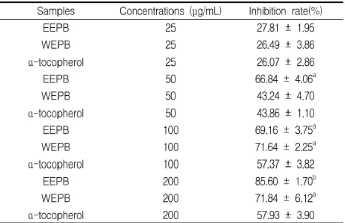
Table 2 shows the inhibitory property of PB extracts on FeCl
2-ascorbic acid-induced lipid peroxidation in rat liver homogenate. At concentrations of 25-200 μg/mL, all the tested samples showed a dose-dependent inhibition of lipid peroxidation. At the 200 μg/mL concentration, EEPB (85.60%, p < 0.001) and WEPB (71.84%, p < 0.05) exhibited significantly higher inhibition activities than did α-tocopherol (57.93%). The IC
50values of EEPB and WEPB were 31.24±2.00 and 45.70±2.72 μg/mL, respectively, which were lower than 98.96±4.02 μg/mL for α-tocopherol. Based on these results, EEPB displayed the highest anti-lipid peroxidation activity.
Table 2. Inhibitory effect on FeC
l2-ascorbic acid-induced lipid peroxidation
Samples Concentrations (μg/mL) Inhibition rate(%)
EEPB 25 27.81 ± 1.95
WEPB 25 26.49 ± 3.86
α-tocopherol 25 26.07 ± 2.86
EEPB 50 66.84 ± 4.06a
WEPB 50 43.24 ± 4.70
α-tocopherol 50 43.86 ± 1.10
EEPB 100 69.16 ± 3.75a
WEPB 100 71.64 ± 2.25a
α-tocopherol 100 57.37 ± 3.82
EEPB 200 85.60 ± 1.70b
WEPB 200 71.84 ± 6.12a
α-tocopherol 200 57.93 ± 3.90
a Values are mean ±SD of three replicates. a p <0.05, b p <0.001 vs. α-tocopherol group.
Table 3 shows the IC
50values of PB extracts for ROS and RNS, including hydroxyl radical, superoxide anion, nitric oxide and peroxynitrite. The hydroxyl radical scavenging activity of PB extracts was tested using the fluorescent probe dichlorodihydrofluorescein diacetate (DCFH-DA) after exposure to H
2O
2and FeSO
4, and was assessed by monitoring
the fluorescent intensity of DCF. In this assay system, H
2O
2reacts with FeSO
4to produce hydroxyl radical, which oxidizes DCFH to fluorescent product DCF. As shown in Table 3, caffeic acid, a well-known antioxidant compound, revealed the highest scavenging activity (IC
50= 1.48 μg/mL), followed by EEPB (IC
50= 10.08 μg/mL), WEPB (IC
50= 14.37 μg/mL) and α -tocopherol (IC
50= 17.65 μg/mL). In addition, to determine the scavenging activity of PB extracts on superoxide anions, we used a hypoxanthine-xanthine oxidase system and measured the level of NBT reduction. In this assay, EEPB (IC
50= 1.43 μ g/mL) exhibited the strongest activity, followed by WEPB (IC
50= 1.61 μg/mL)and caffeic acid (IC
50= 2.00 μg/mL), whereas ascorbic acid (IC
50> 200 μg/mL) was ineffective in scavenging the superoxide anions. It has been reported that peroxynitrite is formed from a reaction of superoxide anion with nitric oxide, which can promote atherosclerosis through LDL oxidative modification and endothelial cell damage
22,23). To further investigate the antiradical ability of PB extracts, we evaluated the RNS scavenging activities using nitric oxide and peroxynitrite generating systems. In the nitric oxide scavenging assay, we monitored the level of triazolofluorescein, which is the product of the reaction between DAF-2 and NO donor SIN-1. As shown in Table 3, the nitric oxide scavenging activity of EEPB (IC
50= 2.02 μg/mL) and WEPB (IC
50= 2.20 μ g/mL) was slightly higher than that of ascorbic acid (IC
50= 2.54 μg/mL), but lower than that of caffeic acid (IC
50= 0.38 μ g/mL). Furthermore, the peroxynitrite scavenging activity of PB extracts was assessed by monitoring the level of DHR 123 oxidation by native peroxynitrite. In this assay, the peroxynitrite scavenging capacities of EEPB (IC
50= 1.70 μ g/mL) and WEPB (IC
50= 2.14 μg/mL) were comparable to that of the peroxynitrite scavenger, penicilamine (IC
50= 0.85 μ g/mL). EEPB and WEPB displayed strong scavenging activities in the ROS and RNS scavenging tests, suggesting that PB extractsmay function as an efficient protector against ROS- or RNS-induced oxidative damage in biological systems.
Table 3. Scavenging effects on ROS and RNS
Samples ·OH O2- ·NO ONOO-
EEPB 10.08 ± 0.29 1.43 ± 0.03 2.02 ± 0.01 1.70 ± 0.03 WEPB 14.37 ± 0.42 1.61 ± 0.09 2.20 ± 0.02 2.14 ± 0.07 Ascorbic acidb 17.65 ± 0.53 > 200 2.54 ± 0.05
Caffeic acidb 1.48 ± 0.07 2.00 ± 0.01 0.38 ± 0.02
Penicillamineb 0.85 ± 0.02
Results are expressed as IC50values, where each value represents the mean ± SD of three replicates. b Used as a positive control
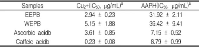
To confirm the possible antioxidant activity of PB
extracts, we measured anti-LDL oxidation effect using copper-
and AAPH (a free radical generator)-mediated LDL oxidation
model systems. Table 4 shows the inhibitory effect of PB extracts on human LDL oxidation. In the copper-mediated LDL oxidation assay system, caffeic acid, the positive control, revealed the highest antioxidant property (IC
50= 0.23 μg/mL), followed by EEPB (IC
50= 2.94 μg/mL), ascorbic acid (IC
50= 3.61 μg/mL) and WEPB (IC
50= 5.15 μg/mL). In the AAPH-mediated LDL oxidation model system, EEPB (IC
50= 31.92 μg/mL) and WEPB (IC
50= 39.42 μg/mL) exhibited a relatively low antioxidant ability compared to that of ascorbic acid (IC
50= 7.15 μg/mL) and caffeic acid (IC
50= 8.79 μg/mL).
The results of both LDL oxidation tests suggest that PB extracts may be more effective to prevent the human LDL oxidation through the chelating action of metal ions, rather than the direct removal ability of free radicals.
To further confirm the anti-LDL oxidation ability of PB extracts against copper-induced LDL oxidation, we used REM assay. Fig. 1 shows the effect of PB extracts and positive controls on copper-mediated LDL oxidation. When human LDL was incubated with 10 μM CuSO
4, the gel electrophoretic mobility of LDL was markedly increased (lane 2). In contrast, EEPB exhibited an inhibitory effect of 80.77% at 5 μg/mL (lane 5), which was higher than the 77.69% of 20 μg/mL ascorbic acid (lane 10) and the 76.92% of 10 μg/mL BHT (lane 11), but inferior to the 100% of 10 μM EDTA (lane 9). WEPB, however, revealed a relatively weak protective activity of 57.69% (lane 8). From the REM assay results, EEPB effectively suppressed the electrophoretic mobility during exposure of human LDL to copper ions.
In conclusion, PB extracts contained a high total phenolic content and exhibited strong antiradical and antioxidant activities in the various free radical generating systems.
Compared to WEPB, EEPB exhibited more potent activities on free radicals, lipid peroxidation and copper-induced human LDL oxidation.
These results suggest that PB branch extracts may protect against ROS- or RNS-involved diseases, including atherosclerosis. Therefore, further research is required to isolate the active components involved in the antioxidant activity, and to ascertain the antiatherosclerotic properties of the isolated compounds derived from PB branches.
Table 4. Inhibitory effects on LDL oxidation induced by copper or AAPH
Samples Cu2+(IC50, μg/mL)a AAPH(IC50, μg/mL)a
EEPB 2.94 ± 0.23 31.92 ± 2.11
WEPB 5.15 ± 1.88 39.42 ± 9.41
Ascorbic acidb 3.61 ± 0.85 7.15 ± 0.52
Caffeic acidb 0.23 ± 0.08 8.79 ± 0.99
a The level of LDL oxidation was measured by TBARS assay and the results are expressed as mean ± SD of three separate experiments. b Used as a positive control.