Analysis of Genes Expressed during Pepper-Phytophthora capsici Interaction using EST Technology
Dongyoung Kim
1, Jong-Hwan Lee
1,2,3and Woobong Choi
1,2,3*
1Department of Biomaterial Control, Dongeui University, Busan 614-714, Korea
2Department of Biotechnology and Bioengineering, Dongeui University, Busan 614-714, Korea
3Blue-Bio Industry RIC, Dongeui University, Busan 614-714, Korea
Received September 2, 2014 /Revised October 29, 2014 /Accepted November 13, 2014
Pepper, consumed as a typical spice food around world, is mainly cultivated in warm countries, in- cluding Korea, China, and Mexico. Phytophthora capsici is a pathogen on several economically im- portant crops, including pepper. The oomycete attacks the roots, stems, leaves, and fruit of the host plants. To understand the molecular mechanisms underlying development of the disease, the genes expressed during pepper-P. capsici interaction were explored by analyzing expressed sequence tags (ESTs). A cDNA library was constructed from total RNA extracted from pepper leaves challenged with P. capsici for three days, resulting in an early stage of symptom development for comparable interaction. A comprehensive analysis of single-pass sequencing of 5,760 randomly selected cDNA clones extracted 5,148 high-quality entries for contig assembly, which generated 2,990 unigenes. A ho- mology search of the unigenes with BLASTX resulted in 2,409 matches, of which 606 showed classi- fied functional catalogs.
Key words :
Expressed sequence tag (EST), pepper, Phytophthora capsici
*Corresponding author
*Tel : +82-51-890-2279, Fax : +82-51-890-2632
*E-mail : [email protected]
This is an Open-Access article distributed under the terms of the Creative Commons Attribution Non-Commercial License (http://creativecommons.org/licenses/by-nc/3.0) which permits unrestricted non-commercial use, distribution, and reproduction in any medium, provided the original work is properly cited.
Journal of Life Science 2014 Vol. 24. No. 11. 1187~1192 DOI : http://dx.doi.org/10.5352/JLS.2014.24.11.1187
Introduction
Phytophthora blight of pepper (Capsicum annuum L.), caused by Phytophthora capsici, is one of the most destructive diseases in pepper production worldwide [10]. This Phytophthora disease has been responsible for major pro- duction losses of pepper [5]. The pathogen infects all parts of pepper including roots, stems, leaves, and fruits. The most common symptoms associated with the disease are wilting and a root and crown rot characterized by a dark brown stem lesion extending upward from the soil line [2]. The ad- vancing lesion eventually girdles the main stem and kills the whole plant [3]. The control methods were limited due to the difficulty in dealing with this soil-borne disease.
Controlling of this disease has mainly depended on chemical treatment, cultural methods, and crop rotation [10].
However, the frequent appearance of fungicide resistant iso- lates has reduced the effectiveness of chemical treatment [9].
Use of resistant pepper cultivars has been considered as the most profound way for protecting peppers from the Phytophthora blight. In Korea, however, many pepper culti- vars susceptible to the disease have been cultivated in- tensively recently. The rational application of resistance in pepper protection could be achieved from the compre- hensive understanding on the defense mechanisms during the interactions between pepper and P. capsici. And the knowledge of genes involved in pathogenicity may contrib- ute to the development of novel strategies to control this devastating disease.
The pepper-P. capsici pathosystem is more complex com-
paring to other host-pathogen systems since P. capsici can
infect virtually every part of the host plant. Host infection
by virulent P. capsici can occur through stomata or by direct
penetration into the epidermis of plant surface. The infected
tissue can be heavily colonized by the pathogen in 24 hr
after inoculation [21]. Until now, there is no pathogenicity
gene characterized from P. capsici. On the other hand, a num-
ber of genes associated in defense have been cloned from
host plant pepper and characterized. Pathogenicity related
genes such as beta-1,3-glucanase [4, 12], chitinase [8, 11, 22],
PR-1 protein, Thionins [20], pepper lipid transfer protein
genes (LTP) [13], Sesquiterpene cyclase [17], and Hydroxy-
methylglutaryl-CoA reductase genes [7] have been studied
in their expression profile during pepper-P. capsici inter- actions.
The analysis of expressed sequence tags (ESTs) can pro- vide a global scope of information on the genes expressed at certain conditions. EST technique was first introduced for human brain research [1]. Recently, ESTs are available with plants in response to a variety of conditions [6, 28, 30] and phytopathogens [14, 16, 19, 24, 26, 27]. ESTs identified from plant-pathogen interactions as Rice-Magnaporthe grisea [18, 25], Pine-Heterobasidium [15], Wheat-Fusarium [23], Wheat head-Giberella zeae [19], and Plant-nematode [29] have of- fered an efficient way to identify genes involved in plant defense and pathogenicity in genome scale.
In this study, we describe the results of EST analysis per- formed to explore the genes associated in pepper-P. capsici interactions. A comparable interaction was chosen to identi- fy the defense-related genes from host plant and the patho- gen genes necessary for survival in the host.
Materials and Methods Plant, Pathogen, and Inoculation
Peppers (Capsicum annuum L.) of cultivar Bukang were grown in growth chambers under day-night illumination at 27
oC. A virulent isolate PC1 of P. capsici was used in this study. The Oomycete was grown on oatmeal agar in the dark for 7 days at 25
oC and exposed to fluorescent light for 7 days at 25
oC for sporangial production. Sporangia were collected with sterile deionized water and incubated at 5
oC for 1h to release zoospores. Zoospore suspension was ad- justed to the concentration of 10
5/ml with sterile deionized water and sprayed to 6 week old pepper. The pepper plants after inoculation were placed in moist chambers at 22
oC for 72 hr and the lesion areas were sampled for RNA preparation.
Construction of cDNA library
The pepper-P. capsici cDNA library, named KS13, was constructed from pepper leaves infected with virulent isolate of P. capsici. Total RNA was isolated using Trizole (Invitro- gen, USA) following the manufacturer instructions. Poly(A)+
RNA was purified through oligo cellulose chromatography and used for cDNA synthesis and cloning with cDNA li- brary system (Stratagene, USA). The cDNA products were size selected and inserted into the Uni-Zap XR vector. After in vivo excision, individual colonies were transferred to 96
well plates for further process.
EST sequencing
Plasmid DNA from cDNA clones was purified using a modified alkali lysis procedure in 96 well system (Qiagen, Germany). Prior to sequencing, all plasmids were checked for concentration. Sequencing of the cDNA clones was per- formed from the 5’ end with T3 universal primer using BigDye terminator kit (Perkin Elmer, USA) and the ABI 3700 DNA sequencer (Applied Biosystems, USA).
Data processing
Raw sequence data were treated in processing including vector trimming, quality screen, contig assembly, unique gene extraction and representative clone selection using the software stackPACK (STACK clustering system). In quality screen, sequences with over 100 bp after vector removal were used for further process. Non-redundant EST se- quences (unigenes) from contig assembly were used for searching against GenBank protein database using BLASTX algorithm in local computing system. Sequences with homo- logs in known database with function were classified into functional categories following the MIPS system (www.mips.
biochem.mpg.de). All sequences were deposited at GenBank (accession number 60489608~60494726).
Results
Constructon of cDNA library from pepper-P. capsici interactions
A cDNA library was constructed using mRNA isolated from pepper (cultivar Bukang) leaves harvested at 72 hr post-inoculation with virulent P. capsici isolate PC1. The average insert size was determined to be 1.2 kb approx- imately based on the agarose gel analysis of inserts that were PCR amplified using universal primers with randomly se- lected cDNA clones.
EST sequencing and contig assembly
Single pass, 5’-end sequences of 5,760 cDNA clones were
generated using the T3 primer resulting 4,867 high quality
ESTs. Redundant ESTs were grouped into 2,990 non- re-
dundant genes (unigenes) including 2,049 singletons (single
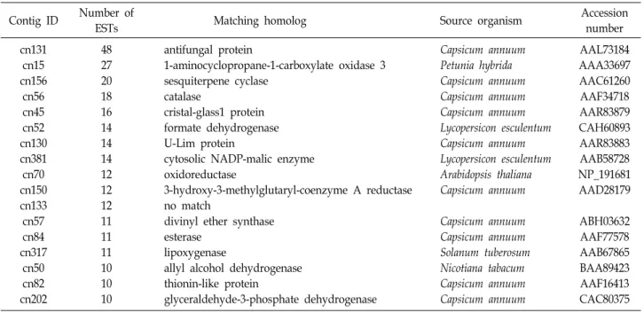
copy sequences) and 941 contigs (Table 1). There were 17
contigs with 10 or more ESTs, which were considered as
highly expressed transcripts (Table 2). The most redundant
Table 1. Primary report on EST sequencing of pepper-P.
capsici interaction
Items Number
Total sequenced Reliable sequenced Contigs assembled Singletons Unigenes Known unigene
Functionally classified unigenes Functionally unclassified unigenes Unknown unigene
5,760 4,867 941 2,049 2,990 2,409 606 1,803 581
Table 2. Most redundant genes showing significant similarity to known protein database
Contig ID Number of
ESTs Matching homolog Source organism Accession
number cn131
cn15 cn156
cn56 cn45 cn52 cn130 cn381 cn70 cn150 cn133 cn57 cn84 cn317
cn50 cn82 cn202
48 27 20 18 16 14 14 14 12 12 12 11 11 11 10 10 10
antifungal protein
1-aminocyclopropane-1-carboxylate oxidase 3 sesquiterpene cyclase
catalase
cristal-glass1 protein formate dehydrogenase U-Lim protein
cytosolic NADP-malic enzyme oxidoreductase
3-hydroxy-3-methylglutaryl-coenzyme A reductase no match
divinyl ether synthase esterase
lipoxygenase
allyl alcohol dehydrogenase thionin-like protein
glyceraldehyde-3-phosphate dehydrogenase
Capsicum annuum Petunia hybrida Capsicum annuum Capsicum annuum Capsicum annuum Lycopersicon esculentum Capsicum annuum Lycopersicon esculentum Arabidopsis thaliana Capsicum annuum
Capsicum annuum Capsicum annuum Solanum tuberosum Nicotiana tabacum Capsicum annuum Capsicum annuum
AAL73184 AAA33697 AAC61260 AAF34718 AAR83879 CAH60893 AAR83883 AAB58728 NP_191681 AAD28179
ABH03632 AAF77578 AAB67865 BAA89423 AAF16413 CAC80375 0
500 1000 1500 2000 2500
1 2 3 4 5 6 7 8 9 10 11 12 14 16 18 20 27 48
2049
564
18879 35 20 15 10 3 3 3 3 2 2 1 1 1 1
Number of ESTs
Number of ESTs in contig
Fig. 1. Redundancy of 4,867 high quality ESTs expressed during peppr-P. capsici compatible interaction.
gene, antifungal protein gene, was assembled with 48 ESTs.
Additionally, genes of oxidase (27 ESTs), sesquiterpene cy- clase (20), catalse (18), formate dehydrogenase (14), oxidor- eductase (12), divinyl ether synthase (11), esterase (11), lip- oxygenase (11), allyl alcohol dehydrogenase (10), thionin like protein (10), and glyceraldehydes-3- phosphate dehydro- gense (10) were also abundantly expressed. ESTs of 42.1%
(2,049/4,867) were deteced only once in the data set (Fig.
1).
Functional categorizing
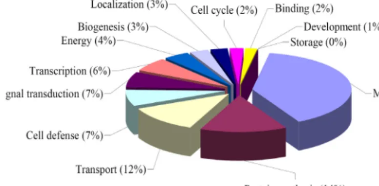
The data set of 2,990 unigenes was queried for homology search against public database by using BLASTX program with a threshold E-value of <10
-10. A number of 606 unigenes among the 2,409 encoding putative protein sequences were assigned in functional description by MIPS system (Fig. 2).
Major genes belong to housekeeping roles such as metabo- lism (39%), protein synthesis (14%), and transport (12%).
Also many genes were assigned to cell defense (7%), signal transduction (7%), transcription (6%) and energy (4%).
Proteins belonging to function of biogenesis (3%), local- ization (3%), cell cycle (2%), and development (1%) were detected.
Discussion
Phytophthora blight of pepper caused by P. capsici is con-
sidered as a novel system for studying host-pathogen inter-
action due to the great economic importance and special as-
pects of pathogen as included in oomycetes. However, stud-
Fig. 2. Categorization on the 606 unigenes from KS13 cDNA library constructed from peppr-P. capsici compatible in- teraction with classified functions.
ies of the interaction have been barely conducted in genome wide scale. Here we report EST analysis revealing the genes involved in the interaction between pepper and P. capsici.
EST analysis has been a powerful tool for identifying large numbers of genes that are involved in certain conditions and also is a rapid and relatively efficient way to discover genes from interesting status of organism in study. EST based se- quencing approaches are efficient comparing to whole ge- nome sequencing in terms of gene discovery. EST analysis as a genomics based approach provides an efficient means of gene discovery in studying the plant microbe pathogen interactions like pepper-P. capsici interactions.
Randomly selected 5,760 cDNA clones were sequenced in 5’-ends, from which more information on genes could be achieved comparing to 3’-ends. About 15% of EST sequences were discarded for next process due to their low quality in base calling. Large numbers of genes were identified by ex- amining 4,867 ESTs from cDNA library prepared from the comparable pepper-P. capsici interaction to provide insight into the molecular mechanisms of the plant defense and pathogenicity. Unnormalized library was chosen to explore the condition-dependent gene expression profiling during the interaction. Unigenes of 2,990 including 941 contigs and 2,049 singletons were assembled from 4,867 ESTs.
Gene of antifungal protein was most abundantly ex- pressed in the condition of comparable pepper-P. capsici interaction. Also high expression was detected in the genes encoding proteins of oxidase, sesquiterpene cyclase, catalse, formate dehydrogenase, oxidoreductase, divinyl ether syn- thase, esterase, lipoxygenase, allyl alcohol dehydrogenase, thionin like protein, and glyceraldehydes-3-phosphate dehydrogense. Considering the functions of abundantly ex- pressed genes during the contact of pepper and P. capsici, the interaction involves highly dynamic status in the life cy- cles of host and pathogen. Deeper dissection on the putative
function of the genes involved could suggest clearer clues to understand the feature of the interaction.
Among the 2,990 unigenes queried in BLASTX program, 2,409(80.6%) showed matches in non-redundant database of GenBank (Fig. 1). The high portion of matched genes in data- base suggests the rich status of gene information of GenBank in plant genome area. However, most of matched genes, 1,803 from 2,409, showed homology with unclassified pro- teins, which indicated more work on functional studies of genes is necessary. The matched 606 unigenes showing clas- sified functions were catalogued into 13 groups including metabolism, energy, storage, and cell cycle et al. Genes in- volved in ubiquitous metabolic pathways, protein synthesis, transport were prevalent. And also a high portion of genes were assigned to cell defense, signal transduction, tran- scription and energy. Proteins belonging to function of bio- genesis, localization, cell cycle and development were rela- tively often detected. Genes involved in cell division, struc- ture, differentiation were poorly represented in this library based on the functional classification. The normalized pro- portion of each gene group is not suggested since no com- parative set was assigned in this analysis.
As more ESTs from diverse plant-pathogen interactions become available, comparative analysis on the genes asso- ciated in pathosystems will be possible in deep sense.
Additionally, the value of this data will be fully appreciated with further analysis using functional genomics approaches such as microarray analysis and directed gene knockout mu- tant analysis. A better understanding of the molecular mech- anisms involved in pepper-P. capsici should lead to novel approaches to manage the important plant disease.
Acknowledgments
This research was supported by Dongeui University
Research Grant 2014AA221. Blue-Bio industry RIC (RIC 08-06-07) under Ministry of Knowledge Economy and Busan City contributed for experimental equipments.
References
1. Adams, M. D., Kelley, J. M., Gocayne, J. D., Dubnick, M., Polymeropoulos, M. H., Xiao, H., Merril, C. R., Wu, A., Olde, B. and Moreno, R. F. et al. 1991. Complementary DNA sequencing: expressed sequence tags and human genome project. Science 252, 1651-1656.
2. Black, L. L., Green, S. K., Hartman, G. L. and Poulos, J. M.
1991. Pepper diseases: a field guide. Asian vegetable research and development center, Tainan, Taiwan. AVRDC Publication 91, 347.
3. Bowers, J. H. and Mitchell, D. J. 1991. Relationship between inoculum level of Phytophthora capsici and mortality of peppers. Phytopathology 81, 178-184.
4. Egea, C., Dickinson, M. J., Candela, M. and Candela, M.
E. 1999. Beta-1,3-glucanase isoenzymes and genes in re- sistant and susceptible pepper (Capsicum annuum) cultivars infected with Phytophthora capsici. Physiol Plantarum 107, 312-318.
5. Erwin, D. C. and Rebeiro, O. K. 1996. Phytophthora, Diseases Worldwide. APS Press, St. Paul. 562 pp.
6. Gyorgyey, J., Vaubert, D., Jimenez-Zurdo, J. I., Charon, C., Troussard, L., Kondorosi, A. and Kondorosi, E. 2000.
Analysis of Medicago truncatula nodule expressed sequence tags. Mol Plant Microbe Interact 13, 62-71.
7. Ha, S. H., Kim, J. B., Hwang, Y. S. and Lee, S. W. 2003.
Molecular characterization of three 3-hydroxy-3-methyl- glutaryl-CoA reductase genes including pathogen-induced Hmg2 from pepper (Capsicum annuum). Biochim Biophys Acta 1625, 253-260.
8. Hong, J. K., Jung, H. W., Kim, Y. J. and Hwang, B. K. 2000.
Pepper gene encoding a basic class II chitinase is inducible by pathogen and ethephon. Plant Sci 159, 39-49.
9. Hwang, B. K. 2001. Cytology, physiology and molecular ge- netics of resistance to Phytophthora blight in pepper plants.
Plant Pathol J 17, 9-21.
10. Hwang, B. K. and Kim, C. H. 1995. Phytophthora blight of pepper and its control in Korea. Plant Dis 79, 221-227.
11. Jongedijk, E., Tigelaar, H., Van Roekel, J. S. C., Bres- Vloemans, S. A., Dekker, I., Van Den Elzen, P. J. M., Cornelissen, B. J. C. and Melchers, L. S. 1995. Synergistic activity of chitinases and beta-1,3-glucanases enhances fun- gal resistance in transgenic tomato plants. Euphytica 85, 173-180.
12. Jung, H. W. and Hwang, B. K. 2000. Pepper gene encoding a basic beta-1,3-glucanase is differentially expressed in pep- per tissues upon pathogen infection and ethephon or methyl jasmonate treatment. Plant Sci 156, 23-34.
13. Jung, H. W., Kim, W. and Hwang, B. K. 2003. Three patho- gen-induced genes encoding lipid transfer protein from pepper are differentially activated by pathogens, abiotic,
and environmental stresses. Plant Cell Environ 26, 915-928.
14. Kamoun, S., Hraber, P., Sobral, B., Nuss, D. and Govers, F. 1999. Initial assessment of gene diversity for the Oomycete pathogen Phytophthora infestans based on ex- pressed sequences. Fungal Genet Biol 28, 94-106.
15. Karlsson, M., Olson, A. and Stenlid, J. 2003. Expressed se- quences from the basidiomycetous tree pathogen Heterobasi- dion annosum during early infection of scots pine. Fungal Genet Biol 39, 51-59.
16. Keon, J., Bailey, A. and Hargreaves, J. 2000. A group of ex- pressed cDNA sequences from the wheat fungal leaf blotch pathogen, Mycosphaerella graminicola (Septoria tritici). Fungal Genet Biol 29, 118-133.
17. Kim, J. B., Lee, S. G., Ha, S. H., Lee, M. C., Ye, W. H., Lee, J. Y., Lee, S. W., Kim, J. B., Cho, K. J. and Hwang, Y. S.
2001. Molecular cloning and characterization of sesqui- terpene cyclase cDNAs from pepper plant infected with Phytophthora capsici. Agric Chem Biotechnol 44, 59-64.
18. Kim, S., Ahn, I. P. and Lee, Y. H. 2001. Analysis of genes expressed during rice-Magnaporthe grisea interactions. Mol Plant Microbe In 14, 1340-1346.
19. Kruger, W. M., Pritsch, C., Chao, S. and Muehlbauer, G.
J. 2002. Functional and comparative bioinformatic analysis of expressed genes from wheat spikes infected with Fusarium graminearum. Mol Plant Microbe Interact 15, 421-427.
20. Lee, S. C., Hong, J. K., Kim, Y. J. and Hwang, B. K. 2000.
Pepper gene encoding thionin is differentially induced by pathogens, ethylene and methyl jasmonate. Physiol Mol Plant Pathol 56, 207-216.
21. Lee, Y. K., Hong, J. K., Hippe-Sanwald, S. and Hwang, B.
K. 2000. Histological and ultrastructural comparison of com- patible, incompatible and DL-b-amino-n-butyric acid-in- duced resistance responses of pepper stems to Phytophthora capsici. Physiol Mol Plant Pathol 57, 269-280.
22. Lee, Y. K., Hippe-Sanwald, S., Jung, H. W., Hong, J. K., Hause, B. and Hwang, B. K. 2000. In situ localization of chiti- nase mRNA and protein in compatible and incompatible interactions of pepper stems with Phytophthora capsici.
Physiol Mol Plant Pathol 57, 111-121.
23. Pritsch, C., Vance, C. P., Bushnell, W. R., Somers, D. A., Hohn, T. M. and Muehlbauer, G. J. 2001. Systemic ex- pression of defense response genes in wheat spikes as a re- sponse to Fusarium graminearum infection. Physiol Mol Plant Pathol 58, 1-12.
24. Qutob, D., Hraber, P. T., Sobral, B. W. S. and Gijzen, M.
2000. Comparative analysis of expressed sequences in Phytophthora sojae. Plant Physiol 123, 243-253.
25. Rauyaree, P., Choi, W., Fang, E., Blackmon, B. and Dean, R. A. 2001. Genes expressed during early stages of rice in- fection with the rice blast fungus Magnaporthe grisea. Mol Plant Pathol 2, 347-354.
26. Soanes, D. M., Skinner, W., Keon, J., Hargreaves, J. and Talbot, N. J. 2002. Genomics of phytopathogenic fungi and the development of bioinformatic resources. Mol Plant Microbe Interact 15, 421-427.
27. Thomas, S. W., Rasmussen, S. W., Glaring, M. A., Rouster,
초록:EST기법을 이용한 고추와 고추역병균간의 상호작용에서 발현되는 유전자들의 분석 김동영
1․이종환
1,2,3․최우봉
1,2,3*
(
1동의대학교 바이오물질제어학과,
2동의대학교 생명공학과,
3동의대학교 블루바이오소재개발 RIC센터)
고추는 한국, 중국, 멕시코를 포함한 온대 및 아열대 지역을 중심으로 전세계적으로 전형적인 향신료로 식용되 고 있으며 그 생산량 및 사용량은 해마다 증가하는 추세에 있다. 고추역병균인 Phytophthora capsici는 고추의 생산 에 있어, 질적, 양적으로 많은 피해를 야기하는 식물병원균으로 알려져 있다. 난균강에 속하는 이 병원균은 기주 식물의 뿌리, 줄기, 잎과 함께 과실에 이르기까지 식물체 전체를 가해한다. 고추역병의 발병을 분자수중에서 이해 하기 위해서는, 발병과정에서 발현되는 유전자에 대한 연구분석이 필수적이며, 이를 위해 최근 개발되어 응용되고 있는 발현서열표지(expressed sequence tags, ESTs)의 분석을 시도하였다. 고추역병균을 접종한후 3일째 발병초 기의 고추잎으로부터 추출한 total RNA를 이용하여 고추-고추역병균 발병초기 cDNA library를 구축하였다. 이 cDNA library에서 무작위로 선발된 5,760 clone에 대하여 말단 염기서열 분석을 수행하여 5,148개의 양질의 염기 서열을 확보하고 contig assembly에 적용한 결과, 2,990개의 unigenes을 확보하였다. 이들 2,990개의 unigenes에 대한 BLASTX를 이용한 상동성 분석결과, 2,409개가 기존에 알려진 서열과 matching을 보였으며, 이중 606개가 기능적으로 구분되었다.
J. A., Christiansen, S. K. and Oliver, R. P. 2001. Gene identi- fication in the obligate fungal pathogen Blumeria graminis by expressed sequence tag analysis. Fungal Genet Biol 33, 195-211.
28. White, J. A., Todd, J., Newman, T., Focks, N., Girke, T., de Ilarduya, O. M., Jaworski, J. G., Ohlrogge, J. B. and Benning, C. 2000. A new set of Arabidopsis expressed sequence tags
from developing seeds. The metabolic pathway from carbo- hydrates to seed oil. Plant Physiol 124, 1582-1594.
29. Williamson, V. M. and Gleason, C. A. 2003. Plant-nematode interactions. Curr Opin Plant Biol 6, 1-7.
30. Yamamoto, K. and Sasaki, T. 1997. Large scale EST sequenc- ing in rice. Plant Mol Biol 35, 135-144.