Combined Treatment of Activin A and Heparin Binding-EGF (HB-EGF) Enhances In Vitro Production of Bovine Embryos
Se-woong Kim1, Yeon-Gil Jung2, Jong-im Park3 and Sangho Roh1,†
1Seoul National University School of Dentistry, Seoul 110-744, Korea
2ET Biotech Ltd, Jangsu 597-851, Korea
3Dept. of Theriogenology, Konkuk University, Seoul 143-701, Korea
ABSTRACT
This study was carried out to investigate the effects of tissue inhibitor of matalloproteinase-1 (TIMP-1), Activin A and Heparin binding epidermal growth factor (HB-EGF) on in vitro production of bovine embryos. In experiment 1, presumptive zygotes were cultured in the medium supplemented with TIMP-1 (0.5 μg/ml), Activin A (100 ng/ml), or HB-EGF (100 ng/ml) at 39 ℃ in a humidified atmosphere of 5% (v/v) CO2, 5% (v/v) O2 and 90% (v/v) N2. In experiment 2, TIMP-1 + HB-EGF or Activin A + HB-EGF combinations were supplemented in the culture medium.
The developmental rate to blastocysts, hatching rate and total cell numbers of the blastocysts were evaluated in both experiments. The embryos cultured in medium without growth factor supplementation was used as control group. In experiment 1, the embryos cultured in medium supplemented with TIMP-1 and Activin A showed significantly higher developmental rate to blastocysts than those cultured with HB-EGF and control (36.9%, 34.1%, 21.2% and 23.1%, respectively) (P<0.0001). However, the hatching rate of blastocyst was significantly higher in embryos with HB-EGF than those with TIMP-1, Actvin A and Control groups (84.4%, 58.8%, 51.4% and 49.3%, respectively) (P<0.001).
Total cell number per blastocyst was also significantly higher in embryos with HB-EGF group (174.3±2.5) than those with TIMP-1, Activin A (149.7 and 150.0, respectively) (P<0.05) and Control (119.0) (P<0.001). In experiment 2, embryos cultured with combined treatment of Activin A and HB-EGF resulted in significantly higher rates of blastocysts formation (48.0%), hatching rate (89.7%) and total cell number in blastocyst (182.3±2.1) than those with TIMP-1 and HB-EGF combination group (32.0%, P<0.001; 76.6%, P<0.05; 165.7±4.2, P<0.001, respectively). Our data demonstrate that in vitro production of bovine embryos could be improved by combined supplementation of Activin A and HB-EGF in culture medium.
(Key words: HB-EGF, Activin A, TIMP-1, in vitro production (IVP), bovine embryo)
* This study was supported by a grant from the National Research Foundation of Korea (NRF-2006-2004042) and the Technology Development Program for Agriculture and Forestry, Ministry of Agriculture, Food and Rural Affairs (MAFRA; 111160-04), Republic of Korea.
†Correspondence : sangho@snu.ac.kr
INTRODUCTION
To improve embryo quality, various growth factors known to be secreted from the reproductive tract in early embryonic period have been added to in vitro culture medium (Park et al., 2010; Vansteenbrugge et al., 1997; Lim et al., 2006), although their expression or secretion patterns showed spatial and temporal manners in reproductive tissues in vivo (Rowzee et al., 2008). Some of these factors are also known to be produced by early preimplantation embryo itself in paracrine/
autocrine manners (Gandolfi, 1994). Tissue inhibitor of metallo- proteinase (TIMP-1) is secreted from bovine oviduct cells and granulosa cells (Satoh et al., 1994; Gerena and Killian, 1990)
and Activin A is secreted from oviductal epithelial cells (Lu et al., 1993), whereas Heparin binding epidermal growth factor (HB-EGF) is known to be secreted solely in the luminal epithelium at the site of blastocyst apposition in mouse (Das et al., 1994). It is thought that HB-EGF may be expressed peri- implantation (blastocyst hatching and attachment to endometrial cells) stage, and Activin A and TIMP-1 are expressed during pre-implantation stage.
TIMP-1, showing embryotrophic activity, can be obtained from bovine oviduct conditioned medium (BOCM) and the mRNA transcript for bovine TIMP-1 is also detectable at the preimplantation embryos such as morula and blastocyst, although it is still unclear how TIMP-1 can improves the development
of bovine embryos (Vansteenbrugge et al., 1996). Activin A is one of the factors involved in maternal-embryonic interactions, and is known to act on embryogenesis and organogenesis in various species (Jones et al., 2002). Activin subunits and the activin receptor mRNA are expressed in oocytes and in em- bryos from zygote to morula in mice (Yoshioka et al., 1998).
Activins are secreted from the epithelial cells in oviduct and endometrium, and are present predominantly during the estrous cycle and pre-implantation phase (Jones et al., 2002). HB-EGF is identified as an early messenger of implantation. HB-EGF is also associated with the trophectoderm cell surface and its presence is coordinated with the competence of murine blasto- cyst attachment (Carson et al., 1993).
However, reciprocal action of growth factors secreted from reproductive tissues during pre- and peri-implantation period is unclear, although each growth factor are identified to support early embryonic development in vitro. The present study was performed to investigate the effects of Activin A, HB-EGF and TIMP-1, and their combinations on bovine embryos in a che- mically defined culture condition to improve the efficiency of in vitro produced (IVP) embryos in cattle.
MATERIALS AND METHODS
1. Chemicals
All inorganic and organic compounds were purchased from Sigma-Aldrich Korea (Yong-in, Korea) unless indicated in the text.
2. Oocyte Recovery and In Vitro Maturation (IVM)
Korean native Hanwoo cattle ovaries were collected at a local slaughterhouse and transported to the laboratory within 2∼3 h in saline at 25∼35℃. Cumulus-oocyte complexes (COCs) were recovered by aspiration of 3 to 8 mm follicles using an 18 gauge hypodermic needle attached to a 10 ml disposable syringe.
After washing three times in HEPES-buffered Tyrode’s solu- tion, the COCs that were enclosed by more than three layers of compact cumulus cells and an evenly granulated ooplasm were selected for IVM. Selected COCs were cultured in NuncTM 4-well culture dishes(Nunc, Roskilde, Denmark) containing 500 μL of IVMD101 medium (Research Institute for the Functional Peptides, Yamagata, Japan) supplemented with 10 μg/ml follicle- stimulating hormone (FSH)-P (Folltrophin-V; Vertrepharm,
London, UK) and 10% fetal bovine serum (FBS; Gibco-BRL, NY, USA) under warmed andgas-equilibrated mineral oil for 20∼22 hat 38.5℃, 5% CO2.
3. Sperm Preparation and In Vitro Fertilization (IVF)
After IVM, the matured oocytes were washed three times washing in IVF100 medium (Research Institute for the Functional Peptides, Yamagata, Japan), and placed into 45 μl drops of IVF100 medium under mineral oil. A frozen semen straw from the HanWoo cattle was rapidly thawed in a 38℃ water bath and the semen was diluted with Tyrode’s albumin lactate pyruvate (TALP) solution and washed twice in a same medium by centrifugation at 503 × g for 5 min. The final sperm pellet was resuspended in IVF100 medium and the number of spermatozoa was counted using a hemocytometer then adjusted to 1.0 × 10⁷/ml by further dilution. A 5 μl aliquot of the sperm suspension were introduced to a 45 μl droplet of IVF100 me- dium containing matured oocytes. Incubation was carried out at 38.5℃ in a humidified atmosphere of 5% CO2 in air for 6 h.
4. In Vitro Culture (IVC)
At the end of the insemination period, groups of 10 oocytes were stripped free from cumulus cells, and transferred into 50 μl drops of modified potassium simplex optimized medium containing 70.2 µM myo-inositol and 1 mM GlcNAc (mKSOM/
aa) supplemented with 20% RD (RPMI1640 + DMEM, 1:1 v/v) which was described by Momozawa and Fukuda (2011). This medium was used as Control. The incubation was conducted at 38.5°C under the 5% CO2, 5% O2 and 90% N2 humidified atmosphere for 7 to 9 days. Following 24 h of culture, the presumptive zygotes which did not undergo cleavage were removed and at this time, the IVC medium was replaced with fresh medium according to each experimental group. Total cell number of Day 7 blastocyst was assessed by nuclear staining with 2 μg/ml of Hoechst 33342 in IVC medium for 10 min.
Then, embryos were placed between a slide and a coverslip and nuclei were counted under UV light using an inverted microscope. Hatching rate of the blastocyst was evaluated on Day 9 of IVC.
5. Experimental Design
In Experiment 1, the effect of TIMP-1 (0.5 μg/ml), Activin A (100 ng/ml) and HB-EGF (100 ng/ml) on IVC from cleavage to blastocyst formation and hatching was evaluated. The con-
centration of these factors in the medium was determined in the preliminary experiment of the present study. Two-cell stage embryos were submitted to each culture condition (control, Activin A, TIMP-1 and HB-EGF supplementation), and then cultured for 7 days for the evaluation of blastocyst formation and total cell number, or cultured for 9 days for counting hatching of Day 7 blastocysts. In Experiment 2, the effect of combined treatments (Activin A + HB-EGF or TIMP-1 + HB- EGF) on IVC was evaluated.
6. Statistical Analysis
All the experiments of treated group were repeated 3∼4 times. Results subjected to statistical analyses were expressed as mean ± SD. Data were subjected to one-way ANOVA (PRISM software version 4.0; GraphPad, San Diego, USA). Difference at P<0.05 was considered significant.
RESULTS
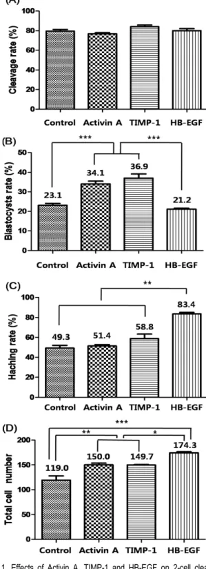
In Experiment 1, first cleavage was not affected by growth factor supplementation (control, 79.5%; Activin A, 76.9%; TIMP- 1, 84.0%; HB-EGF, 80.1%, Fig. 1A). However, the percentage of embryos developed to the blastocyst stage in Activin A (34.1%) and TIMP-1 (36.9%) groups were significantly higher than in Control (23.1%) and HB-EGF (21.2%) groups (P<
0.0001, Fig. 1B). the rate of blastocyst hatching was signifi- cantly higher (P<0.001) in embryos cultured with HB-EGF (83.4%) group than in all other experimental groups (Control, 49.3%; Activin A, 51.4%; TIMP-1, 58.8%, Fig. 1C). The mean cell number of blastocysts cultured with HB-EGF on Day 7 (174.3 ± 2.5) were significantly higher than that of other groups (Control, 119.0 ± 3.1; Activin A, 150.0 ± 2.6; TIMP-1, 149.7
± 1.9; P<0.001, Fig. 1D). In addition, total cell numbers in embryos cultured with Activin A or TIMP-1 groups were also significantly higher than those in control groups (P<0.05).
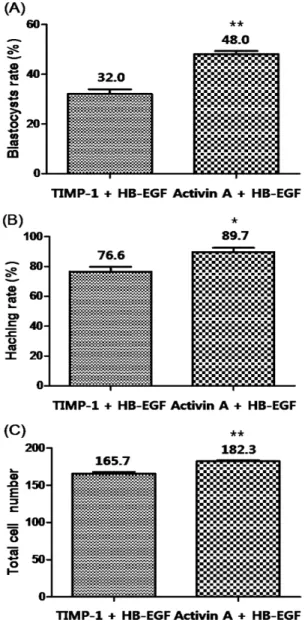
In Experiment 2, combination of Activin A and HB-EGF showed significantly increased blastocyst development (48.0%, P<0.001) and hatching (89.7%, P<0.05) than TIMP-1 + HB- EGF counterpart (32.0% and 76.6%, respectively, Fig. 2A, B).
The mean cell numbers in Day 7 blastocysts was significantly higher in Activin A + HB-EGF (182.3 ± 2.1) than the number in TIMP-1 + HB-EGF (165.7 ± 4.2, P<0.001, Fig. 2C).
DISCUSSION
Fig. 1. Effects of Activin A, TIMP-1 and HB-EGF on 2-cell cleavage (A), blastocyst formation from 2-cell embryos on Day 7 (B), hatching per total blastocysts (C) and total cell number in the blastocyst (D). Different letters indicate significant differences (***P<0.0001; **P<0.001 and *P<0.05). Data were expressed as mean ± SD from 3∼4 replicates.
Fig. 2. Effect of combined treatment of ‘Activin A and HB-EGF’
or ‘TIMP-1 and HB-EGF’ on blastocyst formation from 2-cell embryos on Day 7 (A), hatching per total blastocysts (B) and total cell number in the blastocyst (C). Different letters indicate significant differences (**P<0.001 and *P<0.05).
Data were expressed as mean ± SD from 3∼4 replicates.
The present investigation showed that combination of growth factors, Activin A and HB-EGF, which expressed in the repro- ductive tissue at different period of embryonic stages (early and late pre-implantation embryonic stages, respectively) can improve IVP of bovine embryos. In Experimental 1, Activin A and TIMP-1 significantly increased blastocyst formation and total cell number, whereas they did not influence blastocyst hatching. In contrast, HB-EGF improved hatching rate and
blastocyst cell number when compared with the other groups, although it did not affect blastocyst development. The result implies that each factor has different role (s) for bovine embryo development in vitro.
TIMP-1 is an inhibitor of matrix metalloproteinases (MMPs) in human (Moore and Crocker, 2012). TIMP-1 promotes the prehatched development of bovine embryos (Hwang et al., 2000), and MMP-2 is known as the key regulator of trophoblast invasion during early pregnancy period (Seval et al., 2004).
TIMP-1 also acts as a growth factor in various cells (reviewed by Denhardt et al., 1994). It was previously reported that trans- cripts for MMPs and TIMPs are expressed at various stages during mouse preimplantation embryo development (Brenner et al., 1989). Although major function of TIMP-1 is invasion, Satoh et al. (1994) suggested that TIMP-1 is one of the major factors for improving embryo development in cattle. On the other hand, Vansteenbrugge et al. (1997) suggested that TIMP- 1 do not play an important role in sustaining bovine embryo development, and also do not influence blastocyst quality in terms of total number of cells per embryo when the embryo was cultured in BOCM which contains TIMP-1. In the present study, TIMP-1 supplementation increased blastocyst formation and total blastocyst cell number except for hatching and this may be caused by promoting actions of cell migration, prolife- ration and cell-to-cell interaction (Wiley et al., 1992).
In recent studies, it has been reported that the expression of Activin A decreased in the ooplasm after maturation, and then increased after fertilization (Silva et al., 2003). Activin A is also known to be expressed in bovine oviducts (Gandolfi et al., 1995). These findings suggest an active role of Activin A in early embryonic development in mammals. Park et al. (2010) suggested that Activin A supports blastocyst formation and hatching when Activin A is supplemented at second half culture period in two-step culture system. However, Trigal et al. (2011) reported that although Activin A supplementation in second half culture stage support blastocyst formation, it increased apo- ptotic cell population and also decreased trophoblastic cells. In contrary, supplementation of Activin A in first half culture stage did not enhance blastocyst formation whereas the factor suppor- ted blastocyst hatching. In present study, Activin A signifi- cantly elevated blastocyst development and total cell number of the blastocyst. However, it did not affect hatching rate.
Therefore, role of Activin A in this experiment seems to be similar to the role of TIMP-1 in our experiment.
HB-EGF is expressed on the surface of trophoblasts and is related with acquisition of attachment competence for implan- tation and blastocyst hatching (reviewed by Jade Lim and Dey, 2009; Carson et al., 1993). The supportive role of EGF-related growth factors including HB-EGF for blastocyst hatching is caused by plasminogen activator in the blastocyst (reviewed by Paria and Dey, 1990). Previous report suggests the possibility of a ligand-receptor signaling of the EGF family at the initia- tion of implantation process in the mouse (Paria et al., 1993).
EGF receptor (EGF-R) is expressed in the trophectoderm of a blastocyst, and this expression is tightly regulated by the status of maternal steroid hormones at the time of implantation (Paria et al., 1993). However, effect of exogenous HB-EGF on bovine embryo itself is unknown yet. Our data show that HB-EGF support hatching, not blastocyst formation. This is in agreement with previous reports mentioned above and also implies that HB-EGF affect to bovine embryos after blastocyst formation.
Since our data demonstrated that HB-EGF supported blasto- cyst hatching, and Activin A and TIMP-1 improved blastocyst formation and its cell number increase, combined treatment of HB-EGF and Activin A or TIMP-1 was performed as the next step.
In Experiment 2, combination of Activin A and HB-EGF showed significantly higher blastocyst formation, hatching and total cell number in the blastocyst than TIMP-1 and HB-EGF combination group. The result indicates that embryonic quality was improved by synergetic effect of Activin A and HB-EGF.
Activin A might affect at the early stage as it is known that it induces fast embryonic cell cycle especially at 4∼8 cell stage bovine embryos (Yoshioka et al., 2000). On the other hand, HB-EGF could improve embryo quality in late stage blastocysts because expression of integrin αν and β3 subunits reached peak levels in the outgrowth stage embryo and HB-EGF support this (Lim et al., 2006). However, TIMP-1 and HB-EGF com- bination showed less synergic effect than Activin A and HB- EGF although it is known that EGF mediates TIMP-1 expre- ssion in cultured bovine trophoblasts (Dilly et al., 2010).
In conclusion, the present study suggests combined treatment of Activin A and HB-EGF for IVP of bovine embryos.
REFERENCES
Bavister BD. 1995. Culture of preimplantation embryos: facts and artifacts. Hum. Reprod. Update. 1: 91-148.
Brenner CA, Alder RR, Rappolee DA, Pedersen RA and Werb Z. 1989. Genes for extracellular matrix-degrading metallo- proteinase and their inhibitor, TIMP, are expressed during early mammalian development. Genes Dev. 3: 848-859.
Carson DD, Tang JP and Julian J. 1993. Heparan sulfate pro- teoglycan (perlecan) expression by mouse embryos during acquisition of attachment competence. Dev. Biol. 155: 97- 106.
Das SK, Wang XN, Paria BC, Damm D, Abraham JA, Klags- brun M, Andrews GK and Dey SK. 1994. Heparin-binding EGF-like growth factor gene is induced in the mouse uterus temporally by the blastocyst solely at the site of its appo- sition: a possible ligand for interaction with blastocyst EGF- receptor in implantation. Development 120: 1071-1083.
Denhardt DT, Rajan S and Walther SE. 1994. Structure-function studies of mouse tissue inhibitor of metalloproteinase-1. Ann.
NY. Acad. Sci. 732: 65-75.
Dilly M, Hambruch N, Haeger JD and Pfarrer C. 2010. Epi- dermal growth factor (EGF) induces motility and upregulates MMP-9 and TIMP-1 in bovine trophoblast cells. Mol. Re- prod. Dev. 77: 622-629.
Gandolfi F. 1994. Autocrine, paracrine and environmental fac- tors influencing embryonic development from zygote to blas- tocyst. Theriogenology 41: 95-100.
Gerena RL and Killian GJ. 1990. Electrophoretic characteri- zation of proteins in oviduct fluid of cows during the estrous cycle. J. Exp. Zool. 256: 113-120.
Hwang W, Kim H, Lee E, Lim J, Roh S, Shin T, Hwang K and Lee B. 2000. Purification and embryotropic roles of tissue inhibitor of metalloproteinase-1 in development of
“HanWoo” oocytes co-cultured with bovine oviduct epithelial cells. J. Vet. Med. Sci. 62: 1-5.
Jade Lim HJ and Dey SK. 2009. HB-EGF: a unique mediator of embryo-uterine interactions during implantation. Exp. Cell Res. 315: 619-626.
Jones RL, Salamonsen LA and Findlay JK. 2002. Potential roles for endometrial inhibins, activins and follistatin during human embryo implantation and early pregnancy. Trends Endocrinol. Metab. 13: 144-150.
Lim JJ, Lee DR, Song HS, Kim KS, Yoon TK, Gye MC and Kim MK. 2006. Heparin-binding epidermal growth factor (HB-EGF) may improve embryonic development and implan- tation by increasing vitronectin receptor (integrin ανβ3) expression in peri-implantation mouse embryos. J. Assist.
Reprod. Gen. 23: 111-119.
Lu RZ, Matsuyama S, Nishihara M and Takahashi M. 1993.
Developmental expression of activin/inhibin beta A, beta B, and alpha subunits, and activin receptor-IIB genes in preim- plantation mouse embryos. Biol. Reprod. 49: 1163-1169.
Momozawa K and Fukuda Y. 2011. Establishment of an advan- ced chemically defined medium for early embryos derived from in vitro matured and fertilized bovine oocytes. J. Re- prod. Dev. 57: 681-689.
Moore CS and Crocker SJ. 2012. An alternate perspective on the roles of TIMPs and MMPs in pathology. Am. J. Pathol.
180: 12-16.
Paria BC and Dey SK. 1990. Preimplantation embryo develop- ment in vitro: cooperative interactions among embryos and role of growth factors. Proc. Natl. Acad. Sci. USA 87:
4756-4760.
Paria BC, Huet-Hudson YM and Das SK. 1993. Blastocyst’s state of activity determines the “window” of implantation in the receptive mouse uterus. Dev. Biol. 90: 10159-10162.
Park JE, Oh HJ, Hong SG, Jang G, Kim MK and Lee BC.
2010. Effects of Activin A on the in vitro development and mRNA expression of bovine embryos cultured in chemically- defined two-step culture medium. Reprod. Dom. Anim. 45:
585-593.
Rowzee AM, Lazzarino DA, Rota L, Sun Z and Wood TL.
2008. IGF ligand and receptor regulation of mammary de- velopment. J. Mammary Gland Biol. Neoplasia 13: 361-370.
Satoh T, Kobayashi K, Yamashita S, Kikuchi M, Sendai Y and Hishi H. 1994. Tissue inhibitor of metalloproteinases (TIMP- 1) produced by granulosa and oviduct cells enhances in vitro development of bovine embryo. Biol. Reprod. 50: 835-844.
Scenna FN, Edwards JL, Rohrbach NR, Hockett ME, Saxton AM and Schrick FN. 2004. Detrimental effects of prosta- glandin F2α on preimplantation bovine embryos. Prostag- landins Other Lipid Mediat. 73: 215-226.
Scenna FN, Hockett ME, Townsa TM, Saxton AM, Rohrbach NR, Wehrman ME and Schrick FN. 2005. Influence of a prostaglandin synthesis inhibitor administered at embryo transfer on pregnancy rates of recipient cows. Prostaglan- dins Other Lipid Mediat. 78: 38-45.
Seval Y, Akkoyunlu G, Demir R and Asar M. 2004. Distribu- tion pattern of matrix metalloproteinase MMP-2 and MMP-9 and their inhibitors (TIMP-1 and TIMP-2) in the human decidua during early pregnancy. Acta. Histochem. 5: 353-
362.
Silva CC, Groome NP and Knight PG. 2003. Immunohistoche- mical localization of inhibin/activin alpha, betaA and betaB subunits and follistatin in bovine oocytes during in vitro maturation and fertilization. Reproduction 125: 33-42.
Trigal B, Gómez E, Díez C, Caamaño JN, Martín D, Carrocera S and Muñoz M. 2011. In vitro development of bovine em- bryos cultured with activin A. Theriogenology 75: 584-588.
van Soom A, Ysebaert MT and de Kruif A. 1997, Relationship between timing of development, morula morphology, and cell allocation to inner cell mass and trophectoderm in in vitro-produced bovine embryos. Mol. Reprod. Dev. 47: 47- 56.
Vansteenbrugge A, Van Langendonckt A, Donnay I, Massip A and Dessy F. 1996. Effect of high molecular weight factors present in bovine oviduct-conditioned medium on in vitro bovine embryo development. Theriogenology 46:631-641.
Vansteenbrugge A, Van Langendonckt A, Massip A and Dessy F. 1997. Effect of estrus-associated glycoprotein and tissue inhibitor of metalloproteinase-1 secreted by oviduct cells on in vitro bovine embryo development. Mol. Reprod. Dev.
46: 527-534.
Wilcox AJ, Weinberg CR, O’Connor JF, Baird DD, Schlatterer JP, Canfield RE, Armstrong EG and Nisula BC. 1988. Inci- dence of early loss of pregnancy. New Engl. J. Med. 319:
189-194.
Wiley LM, Wu JX, HArari I and Adamson ED. 1992. Epider- mal growth factor receptor mRNA and protein increase after four-cell preimplantation stage in murine development. Dev.
Biol. 149: 247-260.
Yamashita S, Abe H, Itoh T, Satoh T and Hoshi H. 1999. A serum-free culture system for efficient in vitro production of bovine blastocysts with improved viability after freezing and thawing. Cytotechnology 31: 121-129.
Yoshioka K, Suzuki C and Iwamura S. 2000. Effects of activin A and follistatin on developmental kinetics of bovine em- bryos: cinematographic analysis in a chemically defined medium. J. Reprod. Fertil. 118: 119-125.
Yoshioka K, Takata M, Taniguchi T, Yamanaka H, Sekikawa K. 1998. Differential expression of activin subunits, activin receptors and follistatin genes in bovine oocytes and preim- plantation embryos. Reprod. Fertil. Dev. 10: 293-298.
(Received: 2014. 4. 26/ Reviewed: 2014. 4. 28/ Accepted: 2014. 6. 16)