Received September 3, 2019 /Revised September 19, 2019 /Accepted September 23, 2019
Hepatic lipid accumulation and insulin resistance increases in patients with non-alcoholic fatty liver disease. Piperine is a major compound found in black pepper (Piper nigrum) and long pepper (P. lon-
gum). Piperine has been used in fine chemical for its anti-cancer, anti-obesity, anti-diabetic, anti-in-flammatory and anti-oxidant properties. However, the signaling-based mechanism of piperine and its role as an inhibitor of lipogenesis and insulin resistance in human hepatocyte cells remains ill-defined.
In the present study, we explored the effects of piperine on lipid accumulation and insulin resistance, and explored the potential underlying molecular mechanisms in palmitate-treated HepG2 cells.
Piperine treatment resulted in a significant reduction of triglyceride content. Furthermore, piperine treatment decreased palmitate-treated intracellular lipid deposition by inhibiting the lipogenic target genes, sterol-regulatory-element-binding protein 1c (SREBP-1c) and fatty acid synthase (FAS); whereas the expression of carnitine palmitoyl transferase (CPT-1) and phosphorylation of acetyl coenzyme A carboxylase (ACC) gene involved in fatty acid oxidation was increased. Moreover, piperine also in- hibited the phosphorylation of insulin receptor substrate (IRS)-1 (Ser307). Piperine treatment modu- lated palmitate-treated lipid accumulation and insulin resistance in HepG2 cells with concomitant re- duction of lipogenic target genes, such as SREBP-1 and FAS, and induction of CPT-1-ACC and phos- phorylation of IRS-1 (Tyr632)-Akt pathways. Therefore, piperine represents a promising treatment for the prevention of lipid accumulation and insulin resistance.
Key words : Insulin resistance, lipid accumulation, palmitate, piperine
*Corresponding author
*Tel : +82-51-510-2814, Fax : +82-51-518-2821
*E-mail : [email protected]
This is an Open-Access article distributed under the terms of the Creative Commons Attribution Non-Commercial License (http://creativecommons.org/licenses/by-nc/3.0) which permits unrestricted non-commercial use, distribution, and reproduction in any medium, provided the original work is properly cited.
Introduction
Obesity and associated conditions, such as type 2 dia- betes, coronary heart disease, and nonalcoholic fatty liver disease (NAFLD) are presently a global health problem [6, 28]. Hepatic steatosis is a metabolic disorder that is an early form of NAFLD, and is defined by an excessive amount of hepatic lipid accumulation. Palmitate (C16:0), are the most plentiful free fatty acids in liver triglycerides of patients with NAFLD, that express excess accumulation of cytosolic lipid and activate lipogenic genes in the liver tissue [17]. Palmitate also can affect insulin resistance in insulin target tissues, both in vitro and in vivo [15, 22, 34, 44]. Treatment with pal- mitate and lipogenic transcription factors; such as sterol-reg-
ulatory-element-binding protein 1c (SREBP-1c), plays a role in lipid accumulation by upregulating many lipogenic genes.
This lipogenic gene expression is related to fatty acid syn- thase (FAS) activity and leads to triglyceride synthesis [22].
In addition, acetyl coenzyme A carboxylase (ACC) inactiva- tion suppresses malonyl-CoA synthesis and, depresses carni- tine palmitoyl transferase (CPT-1) and hyper-activates fatty acid oxidation simultaneously [27]. Insulin signaling is ini- tiated by insulin that interacts and activates the insulin re- ceptor (IR), inducing auto-phosphorylation of IR’s core ty- rosine residues [35]. Moreover, tyrosine residues of the in- sulin receptor substrate (IRS) are subsequently phosphory- lated by the IR kinase, which activates downstream of the PI3K-Akt signaling pathway transduction.
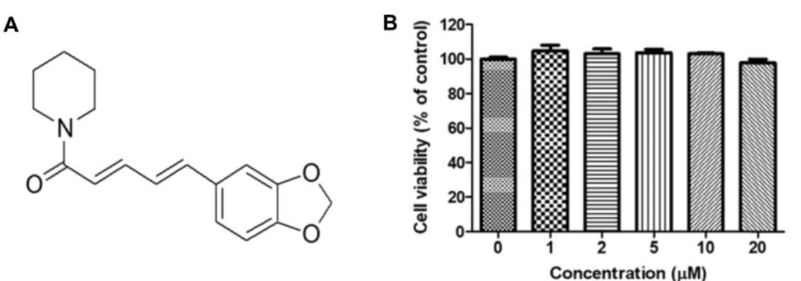
Piperine (1-piperoylpiperidine) (Fig. 1A), one of active al-
kaloid constituents in black pepper (Piper nigrum), long pep-
per (P. longum), and other Piper species (family: Piperaceae),
is used as an effective remedy for gonorrhea, tuberculosis,
menstrual pain, sleeping problems, respiratory-tract in-
fections, chronic gut-related pain and several arthritic con-
ditions [38]. Piperine affects various biological activities, in-
A B
Fig. 1. Chemical structure of piperine (A) and cell viability of piperine on HepG2 cells (B). Cells (1×10⁴ cells/well) were preincubated using various concentrations (up to 20 μM) of piperine for 24 hr. Cell viability was determined using the EZ-Cytox assay and expressed as the percentage of absorbance values relative to the control group. Data shown represents mean ± S.E.M.
of triplicate experiments.
cluding anti-cancer [26, 36], anti-angiogenesis [14], anti- oxidant [29], anti-diabetic [3, 4], improved metabolic syn- drome [13], and anti-inflammatory effects [39]. Previous re- search on piperine investigated the effects of hepatic stea- tosis and insulin resistance on high-fat diet-induced mice [23]. In spite of many previous studies, the underlying mo- lecular mechanism by which piperine inhibits palmitate- treated lipid accumulation and insulin resistance has cur- rently not been. Therefore, we explored the molecular mech- anism of piperine on lipid accumulation and insulin resist- ance in palmitate-treated HepG2 cells.
Materials and Methods
Materials
Piperine, sodium palmitate and Oil red O staining sol- ution were obtained from Sigma Aldrich, St. Louis, USA.
PVDF membrane was obtained from Millipore Corp. (Billelica, MA, Germany) and the enhanced chemiluminescence de- tection system was obtained from Amersham Life Sciences, Inc. (Buckinghamshire, UK). Antibodies targeted toward SREBP-1c, FAS, p-ACC (Ser79), CPT-1, p-IRS (Ser307), p-IRS (Try632), IRS-1, p-Akt (Ser473), Akt, and β-actin were ob- tained from Santa Cruz Biotechnology (Santa Cruz, CA, USA). Serum triglyceride content was determined enzymati- cally using commercial kits (Bio-Clinical System, Gyeonggi- do, Korea). All other chemicals were of the highest purity available from Junsei Chemical Co. (Tokyo, Japan).
Cell culture and treatments
Human HepG2 cell lines were obtained from American Type Culture Collection (Manassas, VA, USA). The cells were
cultured in Dulbecco’s Modified Eagle Media (Welgene, Daegu, Korea) accompanied with 10% heat-inactivated fetal bovine serum, 100 mg/ml penicillin-streptomycin, and 0.25
μg/ml amphotericin B in an atmosphere of 5% CO
2. Piperine was dissolved in 100% DMSO. The final concentration of DMSO did not exceed 0.1%.
Cell viability assay
Cell viability was determined by the EZ-Cytox assay.
Briefly, HepG2 cells were seeded in a 96-well plate at a den- sity of
1×10⁴cells/well, and incubated 37℃ for 24 hr. Media was replaced with fresh Dulbecco’s Modified Eagle Media containing piperine (up to 20
μM) and incubated for 24 hr.
After incubation, 10
μl of EZ-Cytox solution was added to each well, and cells were incubated for an additional 2-4 hr. The absorbance of each well was measured at 450 nm using the ELISA reader (TECAN, Salzburg, Austria). The per- cent inhibition due to piperine was obtained according to the formula: inhibition (%) = [(OD (sample) - OD (control)) / (OD (normal) - OD (control))] ×100. All assays were per- formed in triplicate and then averaged.
Preparation and treatment with sodium palmitate in HepG2 cells
Palmitate was conjugated with fatty acid-free BSA (Gen
DEPOT, Barker, TX, USA) using a previously reported meth-
od [32]. Briefly, 250 mM stock solution was made by alter-
nated heating and vortexing 69.6 mg of sodium palmitate
dissolved in 1 ml of 0.1 N sodium hydroxide at 70℃. After
dissolution of palmitate, the stock solution was immediately
added to serum-free DMEM (containing 5% fatty acid-free
BSA), to obtain 0.5 mM palmitate solution. The cells were
10 min. Cells were soaked in 60% isopropanol for 5 min, stained with Oil red O solution for 1 hr, and rinsed with ddH
2O several times to remove excess stain. Pictures were taken using a microscope (Motic, CA, USA).
Measurements of intracellular triglyceride content Intracellular triglyceride content were measured using en- zymatic colorimetric assay kits after lysis of the HepG2 cells with 1% Triton X-100 in PBS. Protein concentrations were determined using the BCA method with BSA as the standard.
Intracellular triglyceride levels were normalized to de- termine cellular protein content.
Western blotting
Western blotting was performed as described previously [19]. The cells were harvested, washed twice with ice-cold PBS and lysed in buffer (50 mM Tris-HCl, pH 8.0, 120 mM NaCl, 0.5% NP-40 supplemented with protease and phos- phatase inhibitors (1
μg/ml leupeptin, 1
μg/ml pepstatin, 1
μg/ml aprotinin, 1 mM phenylmethylsulphonyl fluoride, 0.1 mM sodium orthovanadate, and 50 mM sodium fluoride) for 1 hr on ice, vortexing every 5 min. Lysates were centri- fuged at 12,000 rpm for 10 min to remove insoluble materials.
Equal amounts of protein were separated on 8-10% SDS- PAGE gels. The separated proteins were subsequently trans- ferred onto PVDF membranes via electro-blotting for 2 hr at 80 V. The membranes were blocked in a 5% non-fat milk solution in TBS with 0.5% Tween-20; and incubated with pri- mary antibodies overnight at 4℃. The membranes were washed and incubated for 2 hr at room temperature with HRP-linked secondary antibodies. Pre-stained blue protein markers (Bio-Rad, Hercules, CA, USA) were used for molec- ular weight determination.
Statistical analysis
Statistical significance was analyzed using the one-way analysis of variance to determine differences within treat- ments followed by the Bonferroni test (GraphPad Prism 5
in palmitate-treated HepG2 cells
To evaluate the cytotoxic effects of piperine on HepG2 cells, the cells were treated with several concentrations of piperine (0-20
μM) for 24 hr. Cell viability was not affected up to 20
μM after 24 hr, indicating that the tested concen- trations of piperine did not affect cell growth and viability (Fig. 1B). Therefore, subsequent experiments were conducted using up to 20
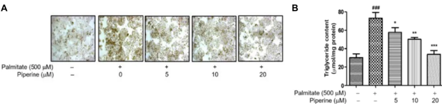
μM piperine. To examine whether piperine inhibited intracellular lipid content in palmitate-treated HepG2 cells, Oil red O staining and an intracellular trigly- ceride content assay were performed. The cells were in- cubated in 500
μM palmitate and several concentrations of piperine for 24 hr. As shown in Fig. 2, palmitate-treated HepG2 cells presented higher Oil red O staining than control cells (Fig. 2A), indicating higher triglyceride content (Fig.
2B). These results indicated that piperine treatment may sup- press palmitate-treated triglyceride content in HepG2 cells.
Piperine down-regulated SREBP-1c and FAS and up-regulated CPT-1 and phosphorylation of ACC in palmitate-treated HepG2 cells
SREBP-1c is an important transcription factor known to regulate the expression of lipid forming enzymes in the hep- atic lipogenic pathway [37]; and plays a crucial role in the pathogenesis of NAFLD [2]. In fatty acid synthesis, ace- tyl-CoA is the initiator and is involved in the addition of two carbon units that elongate the fatty acid chain. ACC mediates the conversion of acetyl-CoA to malonyl-CoA;
which is an essential rate-limiting step in lipogenesis [8]. FAS
utilized acetyl-CoA, malonyl-CoA, and NADPH, and length-
ens the fatty acid chains to generate palmitate [21]. There-
fore, we investigated the effects of piperine on several lipo-
genic genes, including SREBP-1c, CPT-1, FAS, and ACC, and
their resulting effects on lipogenesis and lipolysis in HepG2
cells using western blotting. As shown in Fig. 3A, palmitate
enhanced the expression of SREBP-1c and FAS; however, the
expression of SREBP-1c and FAS was significantly decreased
in the palmitate-treated HepG2 cells in a dose-dependent
A B
Fig. 2. Effects of piperine on palmitate-treated lipid content. HepG2 cells were left untreated or treated with piperine for 24 hr.
Then, cells were used for Oil red O staining (A) and intracellular triglyceride assay (B). Values are mean ± S.E.M. of three experiments; ###p<0.001 versus that of untreated cells; *p<0.05, **p<0.01, and ***p<0.001 versus that of the palmitate-treated cells.
A B C
D E
Fig. 3. Piperine suppressed lipid accumulation while enhancing fatty acid oxidation in palmitate-treated HepG2 cells. HepG2 cells were treated with piperine (5 or 20 μM) in serum free DMEM for 2 hr and changed to palmitate (500 μM) conjugated with fatty acid free-BSA for 24 hr. (A) Western blot was performed to detect SREBP-1c, FAS, p-ACC (Ser79), ACC, and CPT-1 protein levels. Levels were normalized to β-actin. (B-E) The protein levels of SREBP-1c, FAS, p-ACC (Ser79), ACC, and CPT-1 were quantified using CS analyzer software. A representation of three experiments that yielded similar results. One-fac- tor ANOVA: #p<0.05 versus vehicle treated controls; *p<0.05, **p<0.01, and ***p<0.001 versus palmitate-treated cells. Bars indicate standard errors of means (S.E.M.).
manner upon piperine treatment (Fig. 3B, Fig. 3C).
Moreover, we observed the phosphorylation of ACC (Ser79) in the palmitate-treated HepG2 cells were down- regulated compared into control group (Fig. 3E). On the oth- er hand, the expression of total ACC increased in the palmi- tate treatment. Next, we scrutinized whether piperine pro- moted fatty acid degradation via β-oxidation. We found that palmitate notably decreased the protein level of CPT-1 in HepG2 cells, while the protein level increase of CPT-1 was dose-dependent on piperine levels (Fig. 3C). Our results in- dicate that piperine activates the expression of CPT-1 and ACC phosphorylation, and selectively regulates the ex- pression of lipid metabolism-related proteins. Therefore, the
regulation of lipid regulatory proteins may facilitate the in- hibitory effects of piperine on lipid accumulation.
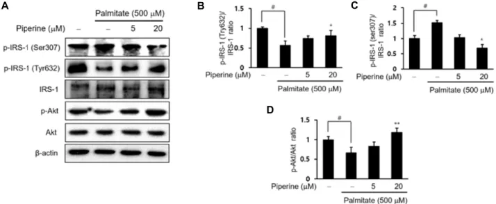
Piperine improved palmitate-treated insulin resistance through up-regulated p-IRS-1/p-Akt signaling
To elucidate the prospective mechanism underlying the
piperine ameliorating insulin resistance, the key proteins in-
volved in insulin signaling pathway in the palmitate-treated
HepG2 cells was investigated (Fig. 4). We first examined the
changes in the phosphorylation of IRS-1 in palmitate-treated
HepG2 cells to assess insulin resistance. IRS-1 was either
stimulated or inhibited by the phosphorylation of specific
tyrosine or serine resides [11]. Therefore, we confirmed that
A B C
D
Fig. 4. Piperine attenuated palmitate-treated insulin resistance in HepG2 cells. HepG2 cells were treated with piperine (5 or 20 μM) in serum free DMEM for 2 hr and palmitate (500 μM) conjugated with fatty acid free-BSA for 24 hr. (A) Western blot was performed to detect p-IRS-1 (Ser307), p-IRS-1 (Tyr632), IRS-1, p-Akt, and Akt protein levels. Levels were normalized to β-actin. (B-D) The protein levels of p-IRS-1 (Ser307), p-IRS-1 (Tyr632), IRS-1, p-Akt, and Akt were quantified using CS analyzer software. A representation of three experiments that yielded similar results. One-factor ANOVA: #p<0.05 versus vehicle treated controls; *p<0.05 and **p<0.01 versus palmitate-treated cells. Bars indicate standard errors of means (S.E.M.).
examined the protein’s expressions associated with insulin signaling Akt. As shown in Fig. 4D, the phosphorylation of Akt (Ser473) decreased in palmitate-treated HepG2 cell, but piperine treatment significantly alleviated the levels of Akt phosphorylation. Our findings demonstrate that piperine could increase the phosphorylated Akt and IRS-1 (Tyr632) phosphorylation, and improve insulin signaling in palmi- tate-treated HepG2 cells. Our results suggest that piperine is a modulator of palmitate-treated lipid accumulation and insulin resistance. We did not observe differences in the total amount of IRS and Akt protein (Fig. 4).
Discussion
Here, we demonstrated that piperine treatment alleviated lipid accumulation and insulin resistance induced by palmi- tate-treated HepG2 cells. As an active alkaloidal phenolic component of black pepper and long pepper [38], piperine
can induce lipid accumulation in a variety of cell types in- cluding, cardiomyocytes, NIT-1 pancreatic β-cells, human adipose-derived stem cells, C2C12 myotubes, and L6 fibro- blasts [1, 5, 26, 40, 42]. We investigated the inhibitory mecha- nism of piperine against hepatic lipid accumulation through improved insulin resistance using palmitate-treated HepG2 cells. Oil red O stains are well-known markers to detect in- tracellular lipid accumulation [12]. Consequently, our study clarified that piperine treatment suppresses palmitate-treat- ed lipid accumulation and concomitantly enhances insulin signaling (Fig. 3, Fig. 4). Our results suggest that treatment with piperine plays a beneficial role in preventing intra- cellular lipid accumulation in palmitate-treated HepG2 cells.
SREBP-1c is a transcription factor that plays a role in the
biosynthesis of cholesterol, fatty acid, and triglyceride [16,
18]. SREBP-1c functions through key regulators (FAS and
ACC) involved in fatty acid biosynthesis that mediate fatty
acid biosynthesis [24]. ACC is the critical rate-limiting en-
Fig. 5. Possible mechanism against lipid accumulation and in- sulin resistance of piperine.
zyme for malonyl-CoA synthesis that acts as a key substrate for fatty acid biosynthesis and is a potent inhibitor of fatty acid oxidation [45]. FAS is a major lipogenic enzyme in mammals; its concentration is carefully regulated by the nu- tritional and hormonal status of lipogenic tissues, such as adipose tissues and liver [7].
We examined the potential role of SREBP-1c in the lipid accumulation reduction of piperine. We found that piperine suppressed the expression of SREBP-1c, FAS, and ACC in palmitate-treated HepG2 cells. Moreover, upregulation of SREBP-1c significantly suppressed the alleviative effect of pi- perine against palmitate-mediated cellular insulin resistance and lipid accumulation in hepatocytes in vitro. Our results propose that SREBP-1c and its downstream target genes (including FAS and ACC) contributed to piperine-exhibited suppression of lipid accumulation and insulin resistance (Fig. 3). CPT-1 is an important rate-limiting enzyme that is involved in fatty acid oxidation [41], which can increase the ability of fatty acids to enter the mitochondria. Thus, it is reasonable to speculate that piperine alleviates lipid metabo- lism disorders by activating the ACC phosphorylation (Ser79)- CPT-1 pathway.
Palmitate-treated insulin resistance has been reported in other insulin sensitive cells, such as 3T3-L1 adipocytes [31]
and skeletal muscle cells [9]. Together with these results, our results suggest that palmitate inhibits insulin signal trans- duction at the level of phosphorylation of IRS (Tyr632) and Akt, regardless of cell type. Our results support in vitro find- ings that palmitate triggers insulin resistance in hepatocytes, in which phosphorylation of IRS (Tyr632) and Akt decreases [30, 43].
In summary, piperine treatment is beneficial in reducing lipid contents in palmitate-treated HepG2 cells. Moreover, piperine affects the lipid metabolic regulatory system by ac- tivating the expression of SREBP-1c, FAS, and ACC and changing the activation of the phosphorylation of ACC (Ser79) and CPT-1; an enzyme involved in lipid metabolism regulation that promotes catabolism of fuel storage. In addi- tion, we found that piperine treatment restored the levels of IRS (Tyr632) and Akt phosphorylation that were reduced by palmitate. The decline of hepatic lipid accumulation and insulin resistance by piperine might also contribute to its efficacy in suppressing hepatic metabolic disease (Fig. 5).
This study helps to understand the mechanisms of insulin resistance, and provides a new therapeutic target for the treatment of obesity and metabolic disorders. Further re-
search should focus on animal experimentation to confirm the metabolism-modulation activity of piperine in vivo.
Acknowledgements
This work was carried out with the support of "Cooper- ative Research Program for Agriculture Science & Technology Development (Project No. PJ006522132013)" Rural Development Administration, Republic of Korea. This research was sup- ported by Bio & Medical Technology Development Program of the National Research Foundation of Korea (NRF) funded by the Ministry of Science, ICT & Future Planning (NRF-2015M3A9B8029074).
References
1. Adrian, L., Lenski, M., Todter, K., Heeren, J., Bohm, M. and Laufs, U. 2017. AMPK prevents palmitic acid-induced apop- tosis and lipid accumulation in cardiomyocytes. Lipids 52, 737-750.
2. Ahmed, M. H. and Byrne, C. D. 2007. Modulation of sterol regulatory element binding proteins (SREBPs) as potential treatments for non-alcoholic fatty liver disease (NAFLD).
Drug Discov. Today 12, 740-747.
3. Arcaro, C. A., Gutierres, V. O., Assis, R. P., Moreira, T. F., Costa, P. I., Baviera, A. M. and Brunetti, I. L. 2014. Piperine, a natural bioenhancer, nullifies the antidiabetic and anti- oxidant activities of curcumin in streptozotocin-diabetic rats. PloS One 9, e113993.
4. Atal, S., Agrawal, R. P., Vyas, S., Phadnis, P. and Rai, N.
2012. Evaluation of the effect of piperine per se on blood glucose level in alloxan-induced diabetic mice. Acta. Pol.
Pharm. 69, 965-969.
5. Atshaves, B. P., Storey, S. M., Petrescu, A., Greenberg, C.
C., Lyuksyutova, O. I., Smith, R. 3rd. and Schroeder, F. 2002.
Expression of fatty acid binding proteins inhibits lipid accu-
Biol. Chem. 273, 29164-29171.
8. Brownsey, R. W., Boone, A. N., Elliott, J. E., Kulpa, J. E.
and Lee, W. M. 2006. Regulation of acetyl-CoA carboxylase.
Biochem. Soc. Trans. 34, 223-227.
9. Chavez, J. A. and Summers, S. A. 2003. Characterizing the effects of saturated fatty acids on insulin signaling and ce- ramide and diacylglycerol accumulation in 3T3-L1 adipo- cytes and C2C12 myotubes. Arch. Biochem. Biophys. 419, 101- 109.
10. Choi, S., Choi, Y., Choi, Y., Kim, S., Jang, J. and Park, T.
2013. Piperine reverses high fat diet-induced hepatic stea- tosis and insulin resistance in mice. Food Chem. 141, 3627- 3635.
11. Copps, K. D. and White, M. F. 2012. Regulation of insulin sensitivity by serine/threonine phosphorylation of insulin receptor substrate proteins IRS1 and IRS2. Diabetologia 55, 2565-2582.
12. Dihingia, A., Bordoloi, J., Dutta, P., Kalita J. and Manna, P. 2018. Hexane-isopropanolic extract of tungrymbai, a north-east indian fermented soybean food prevents hepatic steatosis via regulating AMPK-mediated SREBP/FAS/
ACC/HMGCR and PPARα/CPT1A/UCP2 pathways. Sci.
Rep. 8, 10021.
13. Diwan, V., Poudyal, H. and Brown, L. 2013. Piperine attenu- ates cardiovascular, liver and metabolic changes in high car- bohydrate, high fat-fed rats. Cell Biochem. Biophys. 67, 297- 304.
14. Doucette, C. D., Hilchie, A. L., Liwski, R. and Hoskin, D.
W. 2013. Piperine, a dietary phytochemical, inhibits angiogenesis. J. Nutr. Biochem. 24, 231-239.
15. Gao, D. and Li, Y. 2017. Identification and preliminary struc- ture-activity relationships of 1-indanone derivatives as nov- el indoleamine-2,3-dioxygenase 1 (IDO1) inhibitors. Bioorg.
Med. Chem. 25, 3780-3791.
16. Goldstein, J. L., DeBose-Boyd, R. A. and Brown, M. S. 2006.
Protein sensors for membrane sterols. Cell 124, 35-46.
17. Gomez-Lechon, M. J., Donato, M. T., Martinez-Romero, A., Jimenez, N., Castell, J. V. and O'Connor, J. E. 2007. A human hepatocellular in vitro model to investigate steatosis. Chem.
Biol. Interact. 165, 106-116.
18. Guillet-Deniau, I., Pichard, A. L., Koné, A., Esnous, C., Nier- uchalski, M., Girard, J. and Prip-Buus, C. 2004. Glucose induces de novo lipogenesis in rat muscle satellite cells through a sterol-regulatory-element-binding-protein-1c-dependent pathway. J. Cell Sci. 117, 1937-1944.
19. Habib, A., Creminon, C., Frobert, Y., Grassi, J., Pradelles, P. and Maclouf, J. 1993. Demonstration of an inducible cy-
synthase and liver triglyceride metabolism: housekeeper or messenger? Biochim. Biophys. Acta 1821, 747-753.
22. Jung, T. W., Choi, H. Y., Lee, S. Y., Hong, H. C., Yang, S.
J., Yoo, H. J., Youn, B. S., Baik, S. H. and Choi, K. M. 2013.
Salsalate and adiponectin improve palmitate-induced in- sulin resistance via inhibition of selenoprotein P through the AMPK- FOXO1alpha pathway. PloS One 8, e66529.
23. Jwa, H., Choi, Y., Park, U. H., Um, S. J., Yoon, S. K. and Park, T. 2012. Piperine, an LXRα antagonist, protects against hepatic steatosis and improves insulin signaling in mice fed a high-fat diet. Biochem. Pharmacol. 84, 1501-1510.
24. Li, J., Ding, L., Song, B., Xiao, X., Qi, M., Yang, Q., Yang, Q., Tang, X., Wang, Z. and Yang, L. 2016. Emodin improves lipid and glucose metabolism in high fat diet-induced obese mice through regulating SREBP pathway. Eur. J. Pharmacol.
770, 99-109.
25. Lin, W. C., Shih, P. H., Wang, W., Wu, C. H., Hsia, S. M., Wang, H. J., Hwang, P. A., Wang, C. Y., Chen, S. H. and Kuo, Y. T. 2015. Inhibitory effects of high stability fucox- anthin on palmitic acid-induced lipid accumulation in hu- man adipose-derived stem cells through modulation of long non-coding RNA. Food Funct. 6, 2215-2223.
26. Lin, Y., Xu, J., Liao, H., Li, L. and Pan, L. 2014. Piperine induces apoptosis of lung cancer A549 cells via p53-depend- ent mitochondrial signaling pathway. Tumour Biol. 35, 3305- 3310.
27. McGarry, J. D. and Brown, N. F. 1997. The mitochondrial carnitine palmitoyltransferase system. From concept to mo- lecular analysis. Eur. J. Biochem. 244, 1-14.
28. Michelotti, G. A., Machado, M. V. and Diehl, A. M. 2013.
NAFLD, NASH and liver cancer. Nat. Rev. Gastroenterol.
Hepatol. 10, 656-665.
29. Mittal, R. and Gupta, R. L. 2000. In vitro antioxidant activity of piperine. Method. Find. Exp.Clin. Pharmacol. 22, 271-274.
30. Nakamura, S., Takamura, T., Matsuzawa-Nagata, N., Takayama, H., Misu, H., Noda, H., Nabemoto, S., Kurita, S., Ota, T., Ando, H., Miyamoto, K. and Kaneko, S. 2009. Palmitate in- duces insulin resistance in H4IIEC3 hepatocytes through re- active oxygen species produced by mitochondria. J. Biol.
Chem. 284, 14809-14818.
31. Nguyen, M. T., Satoh, H., Favelyukis, S., Babendure, J. L., Imamura, T., Sbodio, J. I., Zalevsky, J., Dahiyat, B. I., Chi, N. W. and Olefsky, J. M. 2005. JNK and tumor necrosis fac- tor-alpha mediate free fatty acid-induced insulin resistance in 3T3-L1 adipocytes. J. Biol. Chem. 280, 35361-35371.
32. Park, J. Y., Kim, Y., Im, J. A. and Lee, H. 2015. Oligonol suppresses lipid accumulation and improves insulin resist-
초록:Palmitate처리된 인간 간세포주 HepG2 세포에서 piperine의 지질 축적과 인슐린 저항성 기전 에 대한 연구
정희진
1․방은진
2․정성호
2․김병무
2․정해영
1,2*
(1부산대학교 장수생명과학기술연구원, 2부산대학교 약학대학 약학과)
간의 지질 축적과 인슐린 저항성은 비알콜성 지방간 환자에게서 증가한다. Piperine은 후추(Piper nigrum)와 필 발(인도산 후추, P. longum)의 주요 성분으로 항암, 항비만, 항 당뇨병, 항염증 및 항산화 등의 생리활성이 보고되 었다. 그러나 piperine의 인간 간세포 HepG2 세포에서 지질 축적과 인슐린 저항성의 억제제로서의 연구는 보고된 바가 없다. 본 연구의 목적은 지질 축적 및 인슐린 저항성에 대한 piperine의 효과를 palmitate처리된 HepG2 세포 에서 잠재적인 분자 기전을 밝히는 것이다. 그 결과 piperine처리군은 지질 함량을 감소시켰고, 지방 형성 표적 유전자인 SREBP-1c와 FAS의 발현을 억제함으로써 palmitate처리된 세포내 지질 축적을 감소시켰다. 게다가 pi- perine처리군은 지방산 산화에 관련된 CPT-1과 인산화된 ACC 및 인산화된 IRS-1 (Tyr632)와 Akt의 레벨을 증가 시켰다. 또한, piperine처리군은 인산화된 IRS-1 (Ser307)의 레벨을 감소시켰다. 결론적으로 palmitate처리된 HepG2 세포에서 piperine은 SREBP-1와 FAS발현의 감소 및 CPT-1과 ACC 인산화의 증가 및 인산화된 IRS-1 (Try632)와 Akt 신호전달 경로를 조절함으로써 지질 축적 및 인슐린 저항성을 개선함을 확인하였다. 따라서 pi- perine의 지질 축적 및 인슐린 저항성을 예방하는 약물로써 가능성이 제시되었다.
ance in a palmitate-induced in HepG2 hepatocytes as a cel- lular steatosis model. BMC Complement Altern. Med. 15, 185.
33. Rauscher, F. M., Sanders, R. A. and Watkins, J. B. 3rd. 2000.
Effects of piperine on antioxidant pathways in tissues from normal and streptozotocin-induced diabetic rats. J. Biochem.
Mol. Toxicol. 14, 329-334.
34. Reddy, J. K. and Rao, M. S. 2006. Lipid metabolism and liver inflammation. II. Fatty liver disease and fatty acid oxidation. Am. J. Physiol. Gastrointest. Liver Physiol. 290, G852-858.
35. Saltiel, A. R. and Kahn, C. R. 2001. Insulin signalling and the regulation of glucose and lipid metabolism. Nature 414, 799-806.
36. Selvendiran, K., Banu, S. M. and Sakthisekaran, D. 2004.
Protective effect of piperine on benzo(a)pyrene-induced lung carcinogenesis in Swiss albino mice. Clin. Chim. Acta 350, 73-78.
37. Shimano, H., Yahagi, N., Amemiya-Kudo, M., Hasty, A. H., Osuga, J., Tamura, Y., Shionoiri, F., Iizuka, Y., Ohashi, K., Harada, K., Gotoda, T., Ishibashi, S. and Yamada, N. 1999.
Sterol regulatory element-binding protein-1 as a key tran- scription factor for nutritional induction of lipogenic en- zyme genes. J. Biol. Chem. 274, 35832-35839.
38. Srinivasan, K. 2007. Black pepper and its pungent princi- ple-piperine: a review of diverse physiological effects. Crit.
Rev. Food Sci. Nutr. 47, 735-748.
39. Umar, S., Golam Sarwar, A. H., Umar, K., Ahmad, N., Sajad, M., Ahmad, S., Katiyar, C. K. and Khan, H. A. 2013. Piperine ameliorates oxidative stress, inflammation and histological
outcome in collagen induced arthritis. Cell Immunol. 284, 51-59.
40. Wang, D., Tian, M., Qi, Y., Chen, G., Xu, L., Zou, X., Wang, K., Dong, H. and Lu, F. 2015. Jinlida granule inhibits palmitic acid induced-intracellular lipid accumulation and enhances autophagy in NIT-1 pancreatic beta cells through AMPK activation. J. Ethnopharmacol. 161, 99-107.
41. Wolfgang, M. J. and Lane, M. D. 2011. Hypothalamic ma- lonyl-CoA and CPT1c in the treatment of obesity. FEBS J.
278, 552-558.
42. Yang, M., Wei, D., Mo, C., Zhang, J., Wang, X., Han, X., Wang, Z. and Xiao, H. 2013. Saturated fatty acid palmi- tate-induced insulin resistance is accompanied with my- otube loss and the impaired expression of health benefit my- okine genes in C2C12 myotubes. Lipids Health Dis. 12, 104.
43. Yokoyama, K., Tatsumi, Y., Hayashi, K., Goto, H., Ishikawa, T. and Wakusawa, S. 2017. Effects of ursodeoxycholic acid and insulin on palmitate-induced ROS production and down-regulation of PI3K/Akt signaling activity. Biol. Pharm.
Bull. 40, 2001-2004.
44. Yu, X. X., Murray, S. F., Pandey, S. K., Booten, S. L., Bao, D., Song, X. Z., Kelly, S., Chen, S., McKay, R., Monia, B.
P. and Bhanot, S. 2005. Antisense oligonucleotide reduction of DGAT2 expression improves hepatic steatosis and hyper- lipidemia in obese mice. Hepatology 42, 362-371.
45. Zhang, Y., Liu, X., Han, L., Gao, X., Liu, E. and Wang, T.
2013. Regulation of lipid and glucose homeostasis by mango tree leaf extract is mediated by AMPK and PI3K/AKT sig- naling pathways. Food Chem. 141, 2896-2905.